
Digital Poster Session
Molecular Imaging: Hyperpolarization and Molecular Imaging
Molecular Imaging
3000 -3014 Hyperpolarization and Molecular Imaging - Hyperpolarization 1
3015 -3028 Hyperpolarization and Molecular Imaging - Hyperpolarization 2
3029 -3042 Hyperpolarization and Molecular Imaging - Hyperpolarization 3
3043 -3056 Hyperpolarization and Molecular Imaging - Molecular Imaging: Technical
3057 -3070 Hyperpolarization and Molecular Imaging - Molecular Imaging: Applications 1
3071 -3085 Hyperpolarization and Molecular Imaging - Molecular Imaging: Applications 2
3000.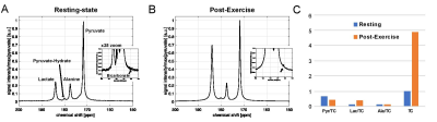 |
Effects of Anaerobic Exercise in Skeletal Muscle Measured by Hyperpolarized [1-13C]Pyruvate in Humans
Jae Mo Park1,2,3, Edward P Hackett2, Crystal E Harrison2, Galen D Reed4, Avneesh Chhabra1, and Craig R Malloy2
1Radiology, University of Texas Southwestern Medical Center, Dallas, TX, United States, 2Advanced Imaging Research Center, University of Texas Southwestern Medical Center, Dallas, TX, United States, 3Electrical Engineering, University of Texas Dallas, Richardson, TX, United States, 4GE Healthcare, GE Healthcare, Dallas, TX, United States
Direct studies of lactate and pyruvate metabolism by biopsy or vascular cannulation during or after exercise is undesirable. The fraction of pyruvate that is oxidized to CO2 vs. reduced to lactate in skeletal muscle was studied in healthy human subjects using hyperpolarized [1-13C]pyruvate at rest and after toe-lift exercise. There was a 2.5x fold increase in lactate production but negligible oxidation of pyruvate in the Krebs cycle. Hyperpolarization methods offer a novel approach to studying lactate and pyruvate metabolism in human skeletal muscle that is readily acceptable to subjects.
|
|
3001.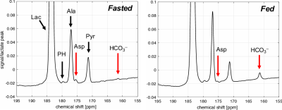 |
Hyperpolarized [1-13C] Lactate as an Agent for Measuring Pyruvate Carboxylase Activity
Jun Chen1, Edward Hackett1, and Jae Mo Park2,3
1AIRC, UT Southwestern Medical Center at Dallas, Dallas, TX, United States, 2UT Southwestern Medical Center at Dallas, Dallas, TX, United States, 3Electrical Engineering, University of Texas at Dallas, Richardson, TX, United States
[1-13C] lactate was studied as hyperpolarized substrate to measure hepatic pyruvate carboxylase activity in vivo with fed and fasted conditions. Besides pyruvate, alanine and bicarbonate, 13C-aspartate could be detected from hyperpolarized lactate. Although the intracellular pyruvate pool size is tightly regulated, we found that lactate conversion to pyruvate and alanine is not saturated when we increased the concentration of hyperpolarized lactate from 30 to 60 mM, whereas the bicarbonate production was saturated. This study demonstrates the utility of hyperpolarized lactate to detect pyruvate carboxylase activity in vivo, and suggests that the metabolite ratio analysis should consider saturable enzyme activities.
|
|
3002.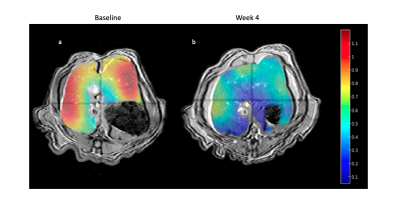 |
Hyperpolarized [1-13C]pyruvate MR spectroscopic imaging to detect metabolic changes in liver in a high-fat diet rat model of NASH and diabetes
Joao Piraquive Agudelo1, Shubhangi Agarwal1, Ting Sun1,2, Robert Bok1, Aras Mattis3,4, Jacquelyn Maher5,6, John Kurhanewicz1, Cornelius Von Morze7, and Michael Ohliger1,8
1Radiology and Biomedical Imaging, University of California San Francisco, San francisco, CA, United States, 2Peking Union Medical College Hospital, Peking, China, 3Department of Pathology, University of California San Francisco, San francisco, CA, United States, 4Liver center, University of California San Francisco, San Francisco, CA, United States, 5Department of Medicine, Division of Gastroenterology, University of California San Francisco, San francisco, CA, United States, 6Liver center, University of California San Francisco, San francisco, CA, United States, 7Biomedical Magnetic Resonance Laboratory, Washington University School of Medicine, St. Louis, MO, United States, 8Liver center, University of California San Francisco, San Franscisco, CA, United States
The present study was focused on using HP 13C MRSI to detect noninvasively metabolic changes in diabetic liver rats fed with a high-fat diet. Planned treatment is 20 weeks to induce NASH, but we present interim data at 4 weeks of treatment. Liver fat signal fraction was significantly increased. In animals, in which the fat and body weight increased, lactate/pyruvate ratio was significantly decreased. This might be explained by a stimulation of gluconeogenesis by high levels of fatty acids in the liver. Long-term monitoring allows us a better understanding of the metabolic changes in the progression from NAFLD diabetic rats.
|
|
3003.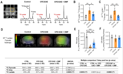 |
Hyperpolarized 13C MRSI of pyruvate and urea can detect immunomodulatory responses to dimethyl fumarate therapy in a model of multiple sclerosis
Caroline Guglielmetti1,2, Christian Cordano3, Chloe Najac4, Ari Green3, and Myriam M Chaumeil1,2
1Department of Physical Therapy and Rehabilitation Science, University of California San Francisco, San Francisco, CA, United States, 2Department of Radiology and Biomedical Imaging, University of California San Francisco, San Francisco, CA, United States, 3Department of Neurology, University of California San Francisco, San Francisco, CA, United States, 4Department of Radiology, C.J. Gorter Center for High Field MRI, Leiden University Medical Center, Leiden, Netherlands
We used hyperpolarized 13C magnetic resonance spectroscopic imaging (HP 13C MRSI) and T1-MRI to assess dimethyl fumarate (DMF) response in a model of multiple sclerosis (MS). Gadolinium-enhanced T1-MRI showed blood-brain-barrier breakdown, regardless of DMF treatment. In contrast, DMF therapy prevented an increase compared to untreated MS animals in HP 13C lactate, HP 13C lactate-to-pyruvate and HP 13C lactate-to-(pyruvate/urea) ratios. HP 13C MRSI findings were further correlated to pyruvate dehydrogenase activity and pro-inflammatory macrophages. Altogether, we demonstrated that HP 13C MRSI has potential to monitor the effect of immunomodulatory therapies within the central nervous system.
|
|
3004.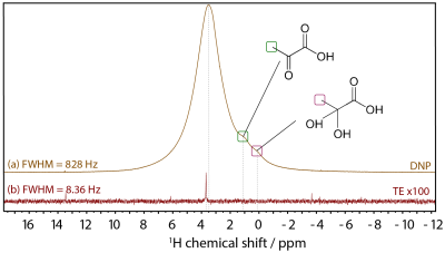 |
Boosting the sensitivity of MRI: 10,000 Tesla equivalent water proton signal
Arthur C. Pinon1, Andrea Capozzi1, and Jan Henrik Ardenkjær-Larsen1
1Department of Health Technology, Technical University of Denmark, Kongens Lyngby, Denmark
In recent years, hyperpolarization of water protons via dissolution Dynamic Nuclear Polarization has attracted increasing interest in the magnetic resonance community. Hyperpolarized water may provide an alternative to Gd-based contrast agents, and it may report on chemical and biochemical reactions, and proton exchange. However, hyperpolarizing water protons is challenging, since the presence of radicals is the main source of relaxation during dissolution and transfer to the MRI system. In this work, we report water signals otherwise requiring >10,000 T at room temperature by employing UV-generated labile radicals, opening up for novel MRI applications.
|
|
3005. |
Hyperpolarized MRI Evaluation of Streptozotocin Injection in the Fasted or Postprandial State: A Metabolic and Functional Study of Diabetes
Dragana Savic1, Vicky Ball1, Carolyn Carr1, Lisa Heather1, and Damian J Tyler1
1University of Oxford, Oxford, United Kingdom
Heart failure in diabetes is the leading cause of mortality in patients. Many studies show cardiac dysfunction in the STZ-induced diabetes model, however variability in cardiac metabolism and function is observed due to differences in the method of model induction. This study investigated both a high and a low dose STZ-induced diabetic model, with STZ injected in both the fasted and postprandial states. Different metabolic and functional severities were observed; with the high-dose STZ-induced diabetic model injected in the fasted state resulting in the most severe form of diabetes with remodelling of both cardiac function and metabolism.
|
|
3006.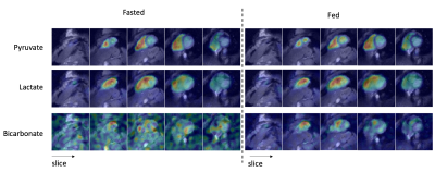 |
Investigating cardiac metabolism between the fasted and fed state using hyperpolarized [1-13C]pyruvate MRI
Shuyu Tang1, Jing Liu1, Robert Bok1, Karen Ordovas1, Jeremy Gordon1, James Slater1, M. Roselle Abraham1, and Peder Larson1
1University of California, San Francisco, San Francisco, CA, United States
In this work, we investigated the influence of a fed versus fasted state on human heart metabolism using hyperpolarized [1-13C]pyruvate MRI in order to develop protocols for human studies of heart disease. Good image quality was obtained in all volunteers, and approximately 40-50% increases in the lactate and bicarbonate signals were observed in the fed state. Distinct metabolic patterns are observed between the left and right ventricle and warrant further investigation.
|
|
3007.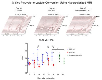 |
Imaging the metabolic evolution of glioblastoma throughout tumor development, regression and relapse
Travis Salzillo1,2, Joy Gumin3, Jaehyuk Lee1, Frederick Lang3, and Pratip Bhattacharya1
1Cancer Systems Imaging, MD Anderson Cancer Center, Houston, TX, United States, 2The University of Texas MD Anderson Cancer Center UTHealth Graduate School of Biomedical Sciences, Houston, TX, United States, 3Neurosurgery, MD Anderson Cancer Center, Houston, TX, United States
Glioblastoma metabolism was interrogated at several time-points during tumor development, regression following radiotherapy, and eventual recurrence. Pyruvate-to-lactate conversion measured with hyperpolarized magnetic resonance imaging (MRI) was compared with tumor volume measured with conventional MRI to determine which was more sensitive at detecting tumor progression. These results were correlated with ex vivo measurements of global metabolism using nuclear magnetic resonance spectroscopy as well as the expression of relevant proteins such as LDH-A, ALT, HIF-1a, MCT1, and MCT4 using immunohistochemistry. A comprehensive analysis of metabolic trajectories throughout the entirety of tumor evolution will be presented.
|
|
3008.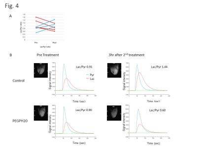 |
Molecular imaging assessment of changes in tumor microenvironment in response to PEGPH20 treatment.
Yu Saida1, Tomohiro Seki1, Yasunori Otowa1, Shun Kishimoto1, Jisook Lee2, Kazutoshi Yamamoto1, Nallathamby Devasahayam1, Jeffery R. Brender1, and Murali C. Krishna1
1National Cancer Institute, Bethesda, MD, United States, 2Halozyme, San Diego, CA, United States
PEGylated human hyaluronidase (PEGPH20) enzymatically deplete tumor hyaluronan, and allows re-expansion of intra-tumor vasculature to increase the delivery of therapeutic molecules. The purpose of this study was to monitor physiologic and metabolic changes in pancreatic adenocarcinoma xenograft in response to PEGPH20 treatment by using multi-modal imaging methods including hyperpolarized 13C-MRI with [1-13C] pyruvate, EPRI, DCE-MRI, and photoacoustic imaging. Significantly decreased glycolysis, increased intratumor pO2, improved perfusion, and increased oxyhemoglobin saturation as well as increased blood volume, were observed after PEGPH20 treatment. The results validate the utility of these imaging methods to non-invasively monitor the PEGPH20-induced changes in the tumor microenvironment.
|
|
3009.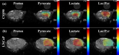 |
Association of prostate cancer androgen sensitivity with dynamic metabolic imaging of hyperpolarized [1-13C]pyruvate
Aditya Jhajharia1, Dexue Fu2, Maninder Singh1, Shu Wang2, Ian Qian2, Aidan Kennedy2, Mohummad M. Siddiqui2,3,4, and Dirk Mayer1,3
1Diagnostic Radiology and Nuclear Medicine, University of Maryland School of Medicine, Baltimore, MD, United States, 2Department of Surgery, Division of Urology, Baltimore, MD, United States, 3Greenebaum Cancer Center, University of Maryland School of Medicine, Baltimore, MD, United States, 4The Veterans Health Administration Research and Development Service, Baltimore, MD, United States
Metabolic signatures of androgen-dependent (LNCaP) and androgen-independent (CSS90) prostate tumors were investigated using hyperpolarized [1-13C]pyruvate imaging. Higher pyruvate-to-lactate conversion in CSS90 were confirmed by higher glycolysis in CSS90 measured by extracellular acidification rate of the tumor slices. Treatment with MDV3100 lead to higher oxidative phosphorylation measured by oxygen consumption rate. Initial in vivo experiments suggested a reduced pyruvate-to-lactate conversion in LNCaP tumors after treatment, potentially a marker for positive response to therapy. These findings demonstrated that conversion of hyperpolarized pyruvate to lactate is a useful biomarker to both characterize tumor androgen sensitivity and potentially to evaluate response to therapy.
|
|
3010.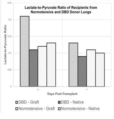 |
Differentiating Between DBD and Normotensive Syngeneic Grafts in a Rat Lung Transplant Model Using HP [1-13C] Pyruvate MRI
Sarmad Siddiqui1, Federico Sertic1,2, Mehrdad Pourfathi1, Jon Snow1, Ian Duncan1, Stephen J Kadlecek1, and Rahim R. Rizi1
1Radiology, University of Pennsylvania, Philadelphia, PA, United States, 2Surgery, University of Pennsylvania, Philadelphia, PA, United States
Our previous study in a rat lung transplant model using HP [1-13C]-pyruvate MRI demonstrated that an increased lactate-to-pyruvate ratio (LtP) precedes lung rejection. However, most lungs in a clinical setting are obtained after donor brain death (DBD), which is associated with increased ischemia-reperfusion injury and risk of acute lung rejection. In this study, we transplanted DBD-lungs into syngeneic rats and compared the metabolism of DBD-donor lungs to our previous normotensive-donor model. The LtP of the DBD-graft is approximately two-fold higher than that of the other lungs, suggesting that HP [1-13C]-pyruvate imaging can be used to identify grafts with ischemia-reperfusion injury.
|
|
3011.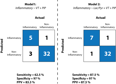 |
Hyperpolarized 13C MRI Improves Sensivity of Pulmonary Inflammation Detection in an Acid Aspiration Rat Model of ARDS
Mehrdad Pourfathi1, Hooman Hamedani1,2, Michael Rosalino1, Stephen J Kadlecek1, Yi Xin1,2, Ian Duncan1, Maurizio Cereda1,3, Sarmad Siddiqui1, Harrilla Profka1, Luis Loza1, Faraz Amzajerdian 1,2, Tahmina Achekzai1, Kai Ruppert1,
Federico Sertic1,4, Ryan Baron1, Jon Snow1, Yiwen Qian1,2, Gabriel Unger1, Shampa Chatterjee5, and Rahim R. Rizi1
1Radiology, University of Pennsylvania, Philadelphia, PA, United States, 2Bioengineering, University of Pennsylvania, Philadelphia, PA, United States, 3Anesthesiology and Critical Care, University of Pennsylvania, Philadelphia, PA, United States, 4Surgery, University of Pennsylvania, Philadelphia, PA, United States, 5Physiology, University of Pennsylvania, Philadelphia, PA, United States
Elevated Hyperpolarized (HP) lactate-to-pyruvate ratio has previously been linked to pulmonary inflammation. In this study, we used a logistic regression model to assess the added value of HP lactate-to-pyruvate ratio as a predictive marker of lung inflammation. Our data suggests, the combination of metabolic and functional information improves both sensitivity of detecting lung inflammation than functional information alone.
|
|
3012.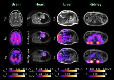 |
Safety, Tolerability and Reproducibility of HP13C-MRI in Normal, Healthy Human Subjects
Robert Bok1, Hsin-Yu Chen1, Jeremy Gordon1, Adam Autry1, Shuyu Tang1, Brian Chung1, James Slater1, Peder E.Z. Larson1, Yan Li1, Mark van Criekinge1, Priscilla Chan1, Romelyn Delos Santos1, Evelyn Escobar1, Kimberly Okamoto1,
Mary Frost1, John Kurhanewicz1, and Daniel B. Vigneron1
1Radiology & Biomedical Imaging, University of California San Francisco, San Francisco, CA, United States
HP 13C-MRI with 13C-Pyruvate was performed in normal, healthy volunteers over four major body regions and organs, including head/brain, chest/heart, abdomen/liver, and pelvis/kidneys. Many individuals received multiple HP 13C injections and a variety of acquisition sequence. Pyruvate-to-lactate conversion rate measurements were reproducible across days and coil configurations. All injections and imaging were very well tolerated with no adverse effects observed in over 70 scans.
|
|
3013.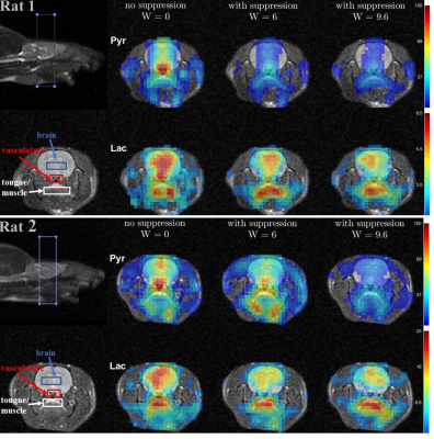 |
Flow-Suppressed Metabolic Imaging of Hyperpolarized [1-13C]Pyruvate in the Healthy Rat Brain In Vivo
Minjie Zhu1, Aditya Jhajharia1, and Dirk Mayer1
1Diagnostic Radiology & Nuclear Medicine, University of Maryland Baltimore, Baltimore, MD, United States
Dynamic metabolic imaging was used to investigate the origin of the lactate signal in rat brain after injection with hyperpolarized (HP) [1-13C] pyruvate. In order to suppress signal with vascular components, we applied a bipolar gradient pulse with different duration prior to data acquisition. The presented results provide further evidence that transport of HP pyruvate into the brain and its subsequent conversion to lactate is limited by blood-brain barrier transport and that the majority of the HP lactate observed in brain was indeed produced there.
|
|
3014. |
Distinguishing metabolic signals of liver tumors from surrounding liver cells using hyperpolarized 13C MRI and Gadoxetate
Shubhangi Agarwal1, Jeremy Gordon1, Natalie Korn1, Robert A Bok1, Cornelius von Morze2, Daniel B Vigneron1, John Kurhanewicz1, and Michael Ohliger1,3
1Department of Radiology and Biomedical Imaging, University of California, San Francisco, San Francisco, CA, United States, 2Biomedical Magnetic Resonance Laboratory, Washington University School of Medicine, St. Louis, MO, United States, 3Liver Center, University of California, San Francisco, San Francisco, CA, United States
Hyperpolarized (HP) 13C MRI is a promising technique that can assess the metabolic pathways in a variety of tumors non-invasively and in real time. In this study, we show the ability of a gadolinium-based contrast agent (gadoxetate) to selectively suppress the HP metabolic signal arising from normal hepatocytes by altering the relaxation rates of metabolites 13C pyruvate and 13C lactate in order to evaluate the metabolic profile of tumors within the liver.
|
 View the Poster
 Watch the Video
3015.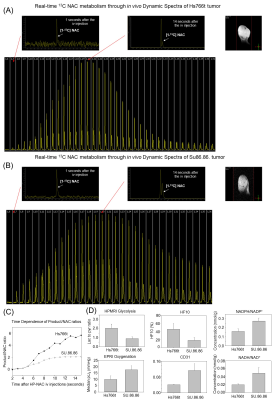 |
Real-time Monitoring of in vivo free radical scavengers through hyperpolarized [1-13C] N-acetyl cysteine
Kazutoshi Yamamoto1, Ana Opina2, Keita Saito3, Ronja M Malinowski4, Tomohiro Seki1, Nobu Oshima1, Deepak Sail2, Jeffrey R Brender3, Shun Kishimoto3, Nallathamby Devasahayam1, Jan H Ardenkjær-Larsen4, James B Mitchell5, Rolf E Swenson2,
and Murali C Krishna1
1National Cancer Institute, National Institutes of Health, Bethesda, MD, United States, 2Imaging Probe Development Center, National Heart, Lung, and Blood Institute, National Institutes of Health, Rockville, MD, United States, 3National Institutes of Health, Bethesda, MD, United States, 4Department of Electrical Engineering, Technical University of Denmark, Lyngby, Denmark, 5NCI/RBB, National Institutes of Health, Bethesda, MD, United States
N-acetyl cysteine (NAC) is a widely used therapeutic and involved to stimulate glutathione synthesis. Glutathione elevates detoxification and works directly as a free radical scavenger. Here, we synthesized and demonstrated [1-13C]NAC as a promising novel probe for hyperpolarized 13C MRI methodologies, which has limitations in available number of probes. In vivo hyperpolarized NAC was broadly distributed throughout the body. The chemical conversions into products were observed in pancreatic tumor xenografts, Hs766t, and SU.86.86, with various conversion efficiencies depending on metabolic characteristics and status. Hyperpolarized NAC can provide insights into redox status, metabolic profile, and enzymatic activities.
|
|
3016.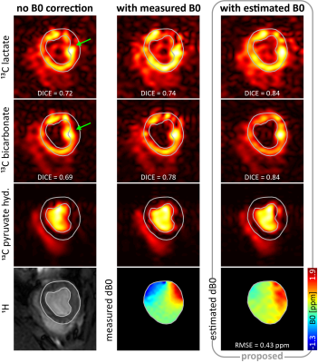 |
Joint Image and Field Map Estimation for Multi-Echo Hyperpolarized 13C Metabolic Imaging
Julia Traechtler1, Valery Vishnevskiy1, Maximilian Fuetterer1, Andreas Dounas1, and Sebastian Kozerke1
1Institute for Biomedical Engineering, University and ETH Zurich, Zurich, Switzerland
An advanced signal model-based reconstruction jointly estimating image and field map from multi-echo, multi-coil acquisition of hyperpolarized metabolic data was developed and validated using synthetic and in-vivo data. Relative to standard multi-echo reconstruction methods, reconstruction accuracy improved by up to 30% for synthetic data considering realistic noise levels and field map gradients. Geometric distortion correction resulted in less than 20% error. For in-vivo data, the average improvement was 15%. Depending on the direction of the field gradients present, multi-coil reconstruction was found to be beneficial for addressing signal folding issues.
|
|
3017.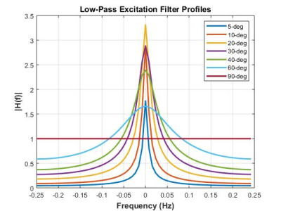 |
The filtering effect of signal excitations on hyperpolarized imaging agents
James A Bankson1, Keith A Michel1, Zhan Xu1, Collin J Harlan1, Gary Martinez 1, and Christopher M Walker1
1Department of Imaging Physics, The University of Texas MD Anderson Cancer Center, Houston, TX, United States
Imaging tumor metabolism using hyperpolarized (HP) pyruvate shows tremendous potential for new insight into disease and response to therapy, but these technically challenging measurements must be carefully designed to maximize accuracy and reproducibility. In this work, we explore the temporal filtering effects of signal excitations on observed HP MRI magnetization. The signal flow diagram and its associated transfer function confirm a low-pass filtering effect that can be controlled by the prescribed excitation angle and sampling interval. Low excitation angles promote temporal averaging but reduce sensitivity to higher frequency content. We explore the effects of signal excitation on quantification.
|
|
3018.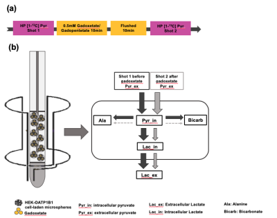 |
Selective hyperpolarized 13C metabolites suppression from a liver-specific gadolinium contrast agent using an NMR-compatible bioreactor
Ting Sun1,2, Renuka Sriram1, Kathy Giacomini1, James Chien1, Sook Wah Yee1, Shubhangi Agarwal1, Joao Piraquive Agudelo1, Cornelius Von Morze3, John Kurhanewicz1, Huadan Xue2, and Michael Ohliger1
1University of California, San Francisco, San Francisco, San Francisco, CA, United States, 2Peking Union Medical College Hospital, Peking Union Medical College and Chinese Academy of Medical Sciences, Beijing, China, 3Washington University, Seattle, WA, United States
This study demonstrated that hyperpolarized 13C metabolic signals arising from intracellular and extracellular 13C metabolites can be selectively suppressed by the intracellular liver-specific gadolinium contrast agent. This data further elucidates how targeted relaxation agents affect observed hyperpolarized 13C signals, expanding the way these agents can be used to distinguish between specific cell types in vivo.
|
|
3019.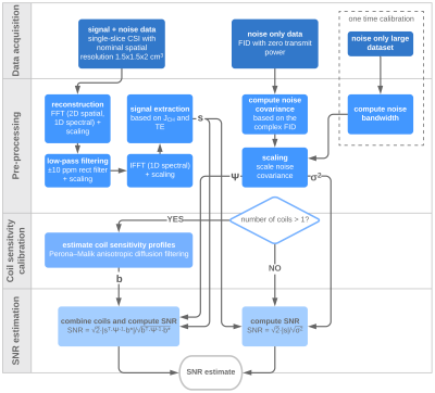 |
A new open-source quality assurance protocol for unbiased 13C coil comparison across sites
Rie B Olin1, Juan D Sánchez-Heredia1, Mary A McLean2, Christoffer Laustsen3, Adam E Hansen4, Lars G Hanson1,5, and Jan H Ardenkjær-Larsen1,6
1Department of Health Technology, Technical University of Denmark, Kgs. Lyngby, Denmark, 2Cancer Research UK Cambridge Institute, University of Cambridge, Cambridge, United Kingdom, 3MR Research Centre, Department of Clinical Medicine, Aarhus University, Aarhus, Denmark, 4Department of Clinical Physiology, Nuclear Medicine and PET, Rigshospitalet, Copenhagen, Denmark, 5Danish Research Centre for Magnetic Resonance, Centre for Functional and Diagnostic Imaging and Research, Copenhagen University Hospital Hvidovre, Hvidovre, Denmark, 6GE Healthcare, Brøndby, Denmark
We propose a new quality assurance (QA) protocol for signal to noise ratio (SNR) evaluation of 13C receive coils to both guide coil developments and assist the choice of the best suited coil for a given hyperpolarized 13C MR experiment. The protocol allows fair comparison of coils with different number of elements and comparison across scanner vendors. The protocol was used to test and compare seven different coils at three different MR sites, which provided insights into coil performance, trade-offs, and malfunctions. All data and source code are shared publicly online.
|
|
3020. |
Multi-compartment pH detection in healthy and tumour bearing mice using hyperpolarized deuterated [1,5-13C2]zymonic acid
Martin Grashei1, Sandra Sühnel1, Geoffrey Topping1, Elisabeth Bliemsrieder1, Stephan Düwel1, Christian Hundshammer1, and Franz Schilling1
1Department of Nuclear Medicine, Klinikum rechts der Isar, Technical University of Munich, Munich, Germany
Here, we demonstrate multi-compartment in vivo pH imaging in healthy and EL4-tumor bearing mice using deuterated hyperpolarized [1,5‑13C2, 3,6,6,6-D4]zymonic acid. Three pH-compartments of the healthy kidney (pHureter = 6.53±0.16, pHmedulla = 7.06±0.06 and pHcortex = 7.38±0.03) could be reliably detected using PRESS-voxel spectroscopy and chemical shift imaging. pH-imaging in mice bearing subcutaneous EL4-tumors allowed to identify one physiologic (pHphys = 7.39±0.05) and two acidic pH-compartments in the tumour (pHac,1 = 6.96±0.17, n=5), with the second pH-compartment being only present in subregions (pHac,2 = 6.62±0.10, n=5).
|
|
3021.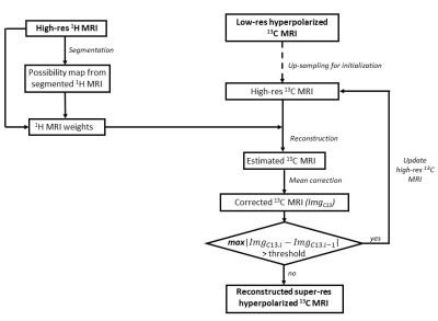 |
Super-Resolution Hyperpolarized 13C Imaging of Human Brain Using Patch-Based Algorithm
Junjie Ma1 and Jae Mo Park1,2,3
1Advanced Imaging Research Center, UT Southwestern Medical Center, Dallas, TX, United States, 2Radiology, UT Southwestern Medical Center, Dallas, TX, United States, 3Electrical Engineering, UT Dallas, Richardson, TX, United States
In this study, a patch-based super-resolution algorithm is proposed, which uses prior knowledge from high-resolution 1H MRI to guide the reconstruction of hyperpolarized 13C images. The algorithm was validated with simulation and phantom, and the results show enhanced spatial resolution, SNR and contrast as well as comparable quantification accuracy to the upsampled images from nearest-neighbor, bilinear and spline interpolation methods. Finally, the proposed algorithm was applied to metabolic imaging of human brain with hyperpolarized [1-13C]pyruvate injection, which significantly improve the image resolution, SNR and contrast while keeping the quantification accuracy.
|
|
3022.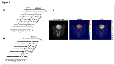 |
Imaging a tumor hallmark: hyperpolarized [U-13C, U-2H]-glucose and [1-13C]-alanine non-invasively monitor TERT expression in gliomas in vivo
Pavithra Viswanath1, Georgios Batsios1, Anne Marie Gillespie1, Peng Cao1, Peder E Z Larson1, Russell O Pieper2, and Sabrina M Ronen1
1Radiology and Biomedical Imaging, University of California San Francisco, San Francisco, CA, United States, 2Neurological Surgery, University of California San Francisco, San Francisco, CA, United States Poster Permission Withheld
Telomerase reverse transcriptase (TERT) expression is a fundamental hallmark of tumor proliferation, including in low-grade oligodendrogliomas. Inhibiting TERT expression is also a therapeutic strategy for gliomas. In this study, we show, for the first time, that metabolism of hyperpolarized [U-13C,U-2H]-glucose to 6-phosphogluconate via the pentose phosphate pathway and hyperpolarized [1-13C]-alanine flux to lactate can serve to non-invasively detect TERT expression in orthotopic low-grade oligodendrogliomas in vivo. Our imaging biomarkers will allow non-invasive, longitudinal detection of a molecular hallmark of tumor proliferation and have the potential to enhance imaging of glioma burden and response to emerging TERT therapies in the clinic.
|
|
3023.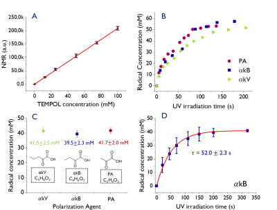 |
Radical-free and metal-free hyperpolarized MRI using endogenous pyruvate analogues
Claudia C Zanella1, Andrea Capozzi1, Hikari A I Yoshihara1, Alice Radaelli1, Lionel P Arn2,3, Rolf Gruetter1, and Jessica A M Bastiaansen2,3
1Laboratory for Functional and Metabolic Imaging (LIFMET), EPFL, Lausanne, Switzerland, 2Department of Diagnostic and Interventional Radiology, CHUV, Lausanne, Switzerland, 3Department of Diagnostic and Interventional Radiology, UNIL, Lausanne, Switzerland
Using nonpersistent radicals generated by UV-irradiation of endogenous metabolite precursors for dissolution DNP avoids the need for radical filtration and may potentially lengthen the measurement window of hyperpolarized MRI measurements. Here, the endogenous pyruvate-analogues alpha-ketobutyrate and alpha-ketovalerate were proposed as nonpersistent radical precursors. Radical yields were characterized along with their performance as polarizing agents for in vitro and in vivo dDNP experiments. A 13C-glucose liquid state polarization of 26.4% was attained using alpha-ketobutyrate-derived radical, while pyruvate-derived radical yielded 21.7% (compared to 18.9% reported with the persistent trityl radical). Alpha-ketobutyrate was used to hyperpolarize [1-13C]butyrate and measure cardiac metabolism in vivo.
|
|
3024.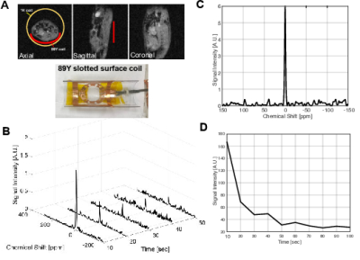 |
Feasibility of Hyperpolarized [89Y]-Labeled Substrates for In Vivo Studies
Jun Chen1, Richard Martin1, Lloyd Lumata2, Zoltan Kovacs1, and Jae Mo Park1,3
1AIRC, UT Southwestern Medical Center at Dallas, Dallas, TX, United States, 2Physics, University of Texas at Dallas, Richardson, TX, United States, 3Electrical Engineering, University of Texas at Dallas, Richardson, TX, United States
Yttrium-89 in the form of diamagnetic 89Y complexes is an attractive nucleus for the design of responsive magnetic resonance spectroscopy and imaging probes the 89Y NMR chemical shift is quite sensitive to the coordination environment of the Y3+ ion. 89Y has a great potential for in vivo hyperpolarized NMR due to its extremely long T1 relaxation time. In this study, we developed an RF coil for imaging 89Y signal in vivo, tested it with phantoms containing thermally polarized and hyperpolarized yttrium compounds, and demonstrated the feasibility of using hyperpolarized 89Y complexes for in vivo studies in a rat.
|
|
3025.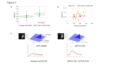 |
Detecting early response to immune checkpoint inhibitors by multimodal molecular imaging
Yu Saida1, Shun Kishimoto1, Yasunori Otowa1, Kazutoshi Yamamoto1, Jeffery R. Brender1, Nallathamby Devasahayam1, and Murali C. Krishna1
1National Cancer Institute, Bethesda, MD, United States
The purpose of this study is to detect physiologic and metabolic changes in tumor-bearing mouse model in response to immune checkpoint blockade therapy by using multi-modal imaging methods including hyperpolarized 13C-MRI with [1-13C] pyruvate, with [1,4-13C2] fumarate, and DCE-MRI. Lactate/Pyruvate ratio tended to decrease in αPD-L1 Ab + αCTLA-4 Ab treated tumor in mouse model although not statistically significant. Significantly increased Malate/Fumarate ratio and higher permeability were observed in treated tumor. We will investigate capability of detecting early response to immune checkpoint blockade therapy using multi-modal imaging.
|
|
3026.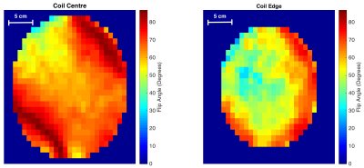 |
Rapid Multi-Point B1+ Calibration for Large Field-of-View 13C Imaging
Nadia D. Bragagnolo1,2, Benjamin J. Geraghty1,2, Casey Y. Lee1,2, Albert P. Chen3, and Charles H. Cunningham1,2
1Medical Biophysics, University of Toronto, Toronto, ON, Canada, 2Physical Sciences, Sunnybrook Research Institute, Toronto, ON, Canada, 3GE Healthcare Technologies, Toronto, ON, Canada
This project aims to minimize the B1+ field inhomogeneities that arise when using a flexible transmit coil system in multi-organ hyperpolarized 13C MRI. A method for rapid B1+ measurement of several locations immediately prior to image acquisition was developed to determine scaling factors for the power input of slices over a large field-of-view. These scaling factors ensure that the desired flip angle is achieved across multiple organs. The rapid B1+ measurement technique has been tested in in vivo scans, giving values accurate within ±1μT. This method will be used to minimize the variability in multi-organ thoracic and abdominal imaging.
|
|
3027.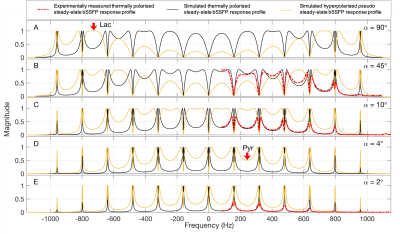 |
Fast 3D hyperpolarized 13C metabolic MRI at 7 T using spectrally selective bSSFP
Jason Graham Skinner1, Geoffrey Topping1, Irina Heid2, Maximilian Aigner1, Martin Grashei1, Christian Hundshammer1, Lukas Kritzner2, Frits Hendrik Anton van Heijster1, Tim Wartewig3,4, Erik Hameister3,4, Jürgen Ruland3,4,5,6, Rickmer Braren2, and Franz Schilling1
1Nuclear Medicine, Klinikum rechts der Isar, The Technical University of Munich, Munich, Germany, 2Institute of Radiology, Klinikum rechts der Isar, The Technical University of Munich, Munich, Germany, 3Institute for Clinical Chemistry and Pathobiochemistry, Klinikum rechts der Isar, The Technical University of Munich, Munich, Germany, 4TranslaTUM, Center for Translational Cancer Research, The Technical University of Munich, Munich, Germany, 5German Cancer Consortium (DKTK), Heidelberg, Germany, 6German Center for Infection Research (DZIF), Munich, Germany
A fast, spectrally selective 3D bSSFP sequence was developed for preclinical metabolic imaging of hyperpolarized 13C agents at 7 T. High spatiotemporal resolution 3D images of metabolism were produced in healthy mice and in two endogenous models of cancer: pancreatic ductal adenocarcinoma (PDAC) and T-cell lymphoma. Using 900 Hz FHWM pulses, 1.75 mm3 isotropic whole-body images were obtained with a scantime of 1.212 s per image. A significant difference between 3D bSSFP derived AUC ratios in healthy and high-glycolytic-phenotype T-cell lymphoma mice was found, and AUC ratios correlated with PET measurements. Potential tumour heterogeneity was detected in PDAC mice.
|
|
3028.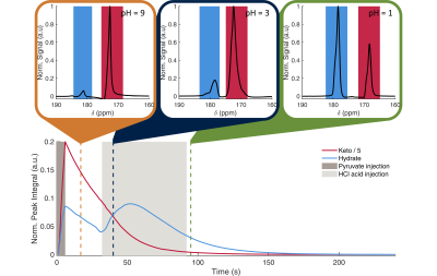 |
ROKET: a Robust Keto Enol Tautomerisation phantom for multi-site, multi-vendor hyperpolarized 13C studies
Liam A J Young1,2, Jack JJJ Miller1,3, Ladislav Valkovic1,4, Esben SS Hansen5, Mary A McLean6,7, Ferdia A Gallagher7, Christoffer Laustsen5, Damian J Tyler1,3, Christopher T Rodgers1,2, and Justin YC Lau1,3
1Oxford Centre for Clinical Magnetic Resonance Research (OCMR), University of Oxford, Oxford, United Kingdom, 2Wolfson Brain Imaging Centre, Department of Clinical Neurosciences, University of Cambridge, Cambridge, United Kingdom, 3Department of Anatomy and Physiology, University of Oxford, Oxford, United Kingdom, 4Department of Imaging Methods, Institute of Measurement Science, Slovak Academy of Sciences, Bratislava, Slovakia, 5Department of Clinical Medicine, the MR Research Centre, Aarhus University, Aarhus N, Denmark, 6CRUK Cambridge Institute, University of Cambridge, Cambridge, United Kingdom, 7Department of Radiology, University of Cambridge, Cambridge, United Kingdom
As dissolution dynamic nuclear polarization (d-DNP) hyperpolarised 13C magnetic resonance imaging progresses towards multi-centre clinical trials, a reproducible, scalable in vitro testing platform is required that allows quality assurance, multi-centre validation, and further technical development. We present a method to exploit the inherent pH dependence of the hydrate-to-keto ratio of pyruvic acid to generate a simple, inexpensive, and reproducible non-enzymatic dynamic phantom to address this need. The method was validated at three different sites with a variety of hardware produced by different vendors.
|
3029.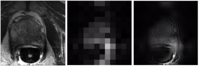 |
Di-chromatic Interpolation of Metabolic Imagery
Nicholas Dwork1, Jeremy Gordon1, Shuyu Tang1, Daniel O'Connor2, and Peder E.Z. Larson1
1Radiology and Biomedical Imaging, University of California in San Francisco, San Francisco, CA, United States, 2Mathematics and Statistics, University of San Francisco, San Francisco, CA, United States
We present a method for using a high-resolution proton image to inform the interpolation of a low-resolution metabolic image. An observer is able to better localize the metabolic activity in the interpolated imagery.
|
|
3030.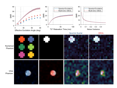 |
Comparison of Selective Excitation and IDEAL for Chemical Shift Imaging of Hyperpolarized [1-13C]Pyruvate
Keith A Michel1, Christopher M Walker1, Matthew E Merritt2, and James A Bankson1
1Imaging Physics, The University of Texas MD Anderson Cancer Center, Houston, TX, United States, 2Biochemistry and Molecular Biology, University of Florida, Gainesville, FL, United States
Imaging of hyperpolarized (HP) agents permits real-time non-invasive measurement of metabolic flux, however these experiments require specialized techniques for rapidly encoding chemical shift in a way that is sensitive metabolic signals that are often low SNR. The SNR performance of two chemical shift encoding methods commonly used for HP 13C MRI, IDEAL and spectral-spatial excitation pulses, were compared for a typical preclinical experiment imaging [1-13C]pyruvate at 7 T. The SNR of these two techniques were found to be equivalent in numerical simulation, while the SNR measured for IDEAL was ~30% greater than for spectral-spatial pulses in a thermal phantom experiment.
|
|
3031.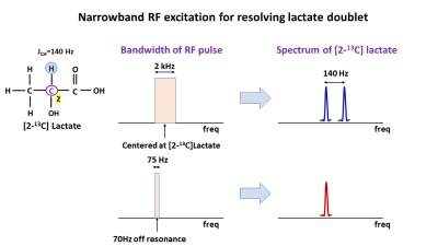 |
A fast, single-shot narrowband excitation method for resolving J-modulated artifacts in hyperpolarized [2-13C] Lactate imaging
Keshav Datta1 and Daniel Mark Spielman1
1Department of Radiology, Stanford University, Stanford, CA, United States
Magnetic resonance spectroscopic imaging of hyperpolarized [2-13C] Pyruvate and its metabolic products enables real-time assessment of in vivo glycolysis and oxidative phosphorylation simultaneously. However, significant technical difficulties arising from the widely dispersed spectrum and the J-coupling of the 13C labeled carbon with its chemically bonded proton limit its practical use for imaging. We propose a narrowband radiofrequency excitation based-method for eliminating the J-modulated artifacts in imaging [2-13C] Lactate. Results from simulations, phantoms and rat brain demonstrate the utility of this method, and highlight its potential role in the real-time simultaneous assessment of GLY and OXPHOS in vivo.
|
|
3032.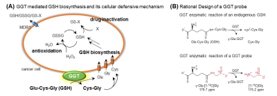 |
Novel Real-time Hyperpolarized 13C Metabolic Tracer Probes γ-Glutamyl Transferase Activities in vivo Tumor Xenografts
Tomohiro Seki1, Kazutoshi Yamamoto2, Nobu Oshima1, Marino Itoda3, Yohei Kondo3, Yutaro Saito3, Yoichi Takakusagi4, Shun Kishimoto1, Jeffrey R Brender1, Ronja M Malinowski5, Jan Henrik Ardenkjær-Larsen6, Hiroshi Nonaka3, Murali C Krishna1,
and Shinsuke Sando3
1National Institutes of Health, Bethesda, MD, United States, 2National Cancer Institute, National Institutes of Health, Bethesda, MD, United States, 3University of Tokyo, Bunkyo ku, Japan, 4Quantum Medical Science, Chiba, Japan, 5Technical University of Denmark, Lyngby, Denmark, 6Department of Electrical Engineering, Technical University of Denmark, Lyngby, Denmark
It is well known that dysregulation of γ-glutamyl transferase (GGT) activities in malignant cells leads to more aggressive phenotypes by producing reactive oxygen species. GGT is important for glutathione homeostasis, and has also been used as a diagnostic marker for various pathologies in the liver, biliary system, and pancreas. Here, for the first time, a novel hyperpolarized 13C probe, γ-Glu-[1-13C]Gly, was demonstrated in in vivo tumor xenografts, including human pancreatic ductal adenocarcinoma and ovarian adenocarcinoma, to detect real-time γ-glutamyl transferase activities as a prospective biomarker for monitoring the tumor progression and prognosis with/without various cancer therapeutic approaches.
|
|
3033.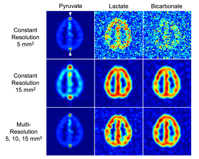 |
Simulation and Application of a Variable Resolution Hyperpolarized 13C MRI Approach for Improved Quantification of Brain Metabolism
Jasmine Y. Graham1,2, Peder E.Z. Larson2, Daniel B. Vigneron2, and Jeremy W. Gordon2
1Bioengineering, UC San Francisco, UC Berkeley, San Francisco, CA, United States, 2Radiology and Biomedical Imaging, UC San Francisco, San Francisco, CA, United States
Downstream metabolic products from hyperpolarized pyruvate, such as lactate and bicarbonate, are acquired with coarse resolution for adequate SNR. However, the injected pyruvate exhibits high signal and can be acquired with higher resolution. A simulation framework has been developed to perform multi-resolution acquisitions of hyperpolarized [1-13C]pyruvate, which analyzes SNR and kinetic rate fits for differing resolution schemes. Utilizing the multi-resolution acquisition, lactate and bicarbonate have respective SNR improvement of 3.3-fold and 5.7-fold compared to high resolution images. Kinetic fits for multi-resolution images accurately estimated ground truth values. This simulation framework performed and analyzed variable resolution acquisitions to improve metabolic quantification.
|
|
3034.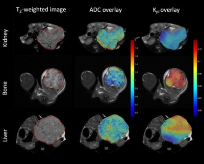 |
Hyperpolarized 13C MR imaging of metastatic prostate cancer murine models
Shubhangi Agarwal1, Jinny Sun1, Robert A Bok1, Romelyn Delos Santos1, Mark van Criekinge1, Rahul Aggarwal2, Daniel B Vigneron1, John Kurhanewicz1, and Renuka Sriram1
1Department of Radiology and Biomedical Imaging, University of California, San Francisco, San Francisco, CA, United States, 22Division of Hematology/Oncology, Department of Medicine, University of California, San Francisco, San Francisco, CA, United States
The study of prostate patient derived xenografts in kidney, bone and liver demonstrated the impact of microenvironment on metabolic characteristics of the metastatic prostate cancer and how it differs with respect to the host organ’s characteristics. Hyperpolarized 13C MRI was able to identify the modifications in the pyruvate metabolism between the kidney, bone and liver tumors before and after therapeutic intervention. This study demonstrates that Hyperpolarized 13C MRI can be used to monitor the real time changes in metabolic profile of prostate cancer and its metastases.
|
|
3035.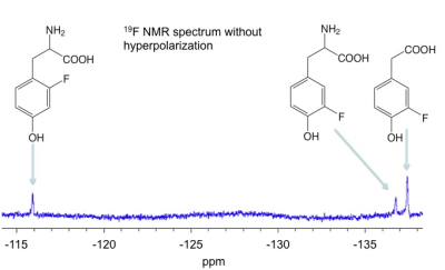 |
Simultaneous 19F hyperpolarization of aromatic molecules in aqueous solution
Frederike Euchner1, Christian Bruns1, Johannes Bernarding1, and Markus Plaumann1
1Institute of Biometrics and Medical Informatics, Otto-von-Guericke University Magdeburg, Magdeburg, Germany
Fluorinated aromatic substrates are of high interest in biomedical and pharmaceutical applications. To increase the low 19F MR signal intensity in aqueous solutions, the hyperpolarization technique photo-Chemical Induced Dynamic Nuclear Polarization (photo-CIDNP) can be used. Compounds such as 2-fluoro-tyrosine, 3-fluoro-tyrosine or 3-fluoro-4-hydroxyphenylacetic acid were hyperpolarized repeatable inside the detection field (7T). Strong 19F signal enhancements could be detected for aromatic systems, which were fluorinated in meta-position. The simultaneous hyperpolarization of different substrates allows e.g. the study of molecule-molecule interactions and their binding behavior.
|
|
3036.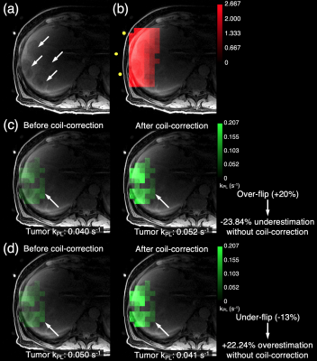 |
Development of Computational Methods for B1-Corrected Hyperpolarized 13C MRI Human Studies
Philip M Lee1, Hsin-Yu Chen2, Jeremy W Gordon2, Peder EZ Larson2, Mark Van Criekinge2, Michael A Ohliger2, Duan Xu2, John Kurhanewicz2, Robert A Bok2, Rahul Aggarwal3, Pamela N Munster3, and Daniel B Vigneron2
1Department of Radiology & Biomedical Imaging, University of California, San Francisco, Berkeley, CA, United States, 2Department of Radiology & Biomedical Imaging, University of California, San Francisco, San Francisco, CA, United States, 3Department of Medicine, University of California, San Francisco, San Francisco, CA, United States
The inhomogeneous B1 excitation profile of 13C surface transmit/receive coils results in a decreasing gradient of flip angles for voxels increasingly farther away from the coil. For accurately quantifying the pyruvate to lactate conversion rate (kPL), the flip angle needs to be corrected based on the B1 excitation profile. A voxel-wise B1 excitation field correction method for hyperpolarized 13C MRSI scans of human patients was developed and applied, yielding an improved quantitative accuracy of kPL values in metastatic cancer patients. The corrected kPL estimations agree with simulations where over-flip leads to an underestimation of kPL, whereas under-flip leads to overestimation.
|
|
3037.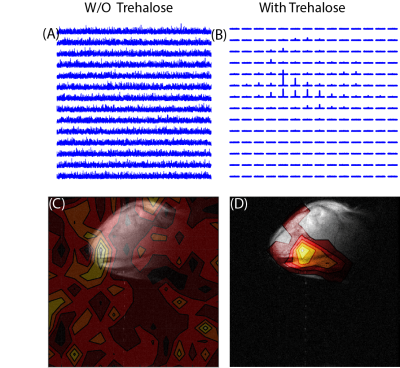 |
Trehalose as an alternative to glycerol as a glassing agent for in vivo DNP MRI
Jeffrey R. Brender1, Shun Kishimoto1, Gareth R. Eaton2, Sandra S. Eaton2, Yu Saida1, and Murali C. Krishna1
1Radiation Biology Branch, Center for Cancer Research, National Cancer Institute, National Institutes of Health, Bethesda, MD, MD, United States, 2Department of Chemistry & Biochemistry, University of Denver, Denver, CO, United States
Glassing agents are essential in DNP studies but have the potential to perturb the metabolic measurements that are being studied. Glycerol, the glassing agent of choice for in vivo DNP studies, is effective at reducing ice crystal formation during freezing but is rapidly metabolized, potentially altering the redox and ATP balance of the system. As a biologically inert alternative to glycerol, we show here that 15-20 wt % trehalose yields a glass that polarizes samples more rapidly than the commonly used 60% wt formulation of glycerol and yields similar polarization levels within clinically relevant timeframes.
|
|
3038. |
Towards Comprehensive Assessment of Lung Injury Using Multi-Modality Hyperpolarized MRI
Mehrdad Pourfathi1, Hooman Hamedani1,2, Yi Xin1,2, Stephen J Kadlecek1, Ian Duncan1, Maurizio Cereda1,3, Sarmad Siddiqui1, Harrilla Profka1, Luis Loza1, Faraz Amzajerdian 1,2, Tahmina Achekzai1, Kai Ruppert1, Michael Rosalino1,
Federico Sertic1,4, Ryan Baron1, Jon Snow1, Yiwen Qian1,2, Gabriel Unger1, Shampa Chatterjee5, and Rahim R. Rizi1
1Radiology, University of Pennsylvania, Philadelphia, PA, United States, 2Bioengineering, University of Pennsylvania, Philadelphia, PA, United States, 3Anesthesiology and Critical Care, University of Pennsylvania, Philadelphia, PA, United States, 4Surgery, University of Pennsylvania, Philadelphia, PA, United States, 5Physiology, University of Pennsylvania, Philadelphia, PA, United States
Hyperpolarized 13C and 129Xe MRI are novel imaging methods that have been used to characterize tissue metabolism and lung function, respectively. In this study, we demonstrate the use of these techniques in tandem in order to characterize lung injury in a porcine model of aspiration pneumonitis.
|
|
3039. |
Progression towards in-vivo detection of Signal Amplification by Reversible Exchange (SABRE) Hyperpolarisation
Aneurin J Kennerley1, Marie-Christine BD Labarthe1, Elizabeth J Fear1, Peter J Rayner1, and Simon B Duckett1
1Chemistry, University of York, York, United Kingdom
We demonstrate that signal amplification by reversible exchange (SABRE) presents as a promising and fast route to hyperpolarising a wide range of biologically relevant contrast agents, including nicotinamide (a precursor for NAD+ a mediator of metabolism) and pyruvate (for mapping anaerobic metabolism) for in-vivo preclinical magnetic resonance imaging and spectroscopic detection. Images recorded in a rat model at 7 Tesla (Bruker BioSpec 70/30) are presented. We highlight necessary chemical preparation steps to ensure that the SABRE agents are bio-compatible following exposure to high pressure para-hydrogen gas and the heavy metal Iridium catalyst required for SABRE hyperpolarisation.
|
|
3040.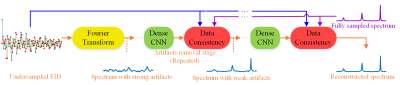 |
Deep Learning-based Fast Magnetic Resonance Spectroscopy
Xiaobo Qu1, Yihui Huang1, Hengfa Lu1, Tianyu Qiu1, Di Guo2, Tatiana Agback3, Vladislav Orekhov4, and Zhong Chen1
1Department of Electronic Science, Xiamen University, Xiamen, China, 2School of Computer and Information Engineering, Xiamen University of Technology, Xiamen, China, 3Department of Molecular Sciences, Swedish University of Agricultural Sciences, Uppsala, Sweden, 4Department of Chemistry and Molecular Biology, University of Gothenburg, Gothenburg, Sweden
Nuclear magnetic resonance (NMR) spectroscopy serves as an indispensable tool in chemistry and biology but often suffers from long experimental time. In this work, we present a proof-of-concept of application of deep learning and neural network for high-quality, reliable, and very fast NMR spectra reconstruction from limited experimental data. Experimental results show that the neural network training can be achieved using solely synthetic NMR signal with exponential functions, which lifts the prohibiting demand for a large volume of realistic training data usually required in the deep learning approach.
|
|
3041.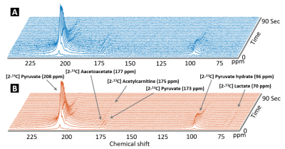 |
Quantification pipeline for in-vivo hyperpolarized 13C magnetic resonance spectroscopy in rats
Donghyun Hong1, Georgios Batsios1, Pavithra Viswanath1, Elavarasan Subramani1, Marina Radoul1, and Sabrina M. Ronen1
1Department of Radiology & Biomedical Imaging, University of California, San Francisco, San Francisco, CA, United States Although the hyperpolarized (HP) spectroscopy shows significant SNR enhancement, quantification of the HP in-vivo spectrum can sometimes be challenging due to low metabolite levels, rapid signal relaxation, short acquisition time, and signal overlap. This study describes a quantification pipeline which includes spectral denoising and automatic fitting using LCModel in order to monitor dynamic HP in-vivo brain metabolism. HP [2-13C] Pyruvate in-vivo data was acquired from healthy rats. Proposed quantification pipeline revealed low concentration metabolites which were not clearly visible before denoising and allowed for improved metabolic kinetic information. |
|
3042.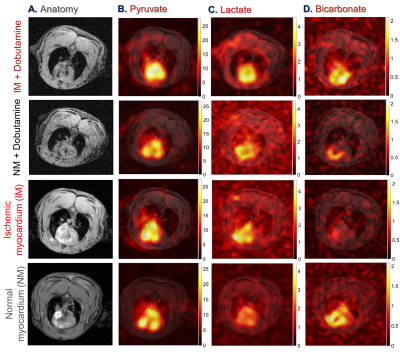 |
In vivo evaluation of metabolic response to dobutamine stimulation in ischemic rat hearts using hyperpolarized 13C-pyruvate
Gaurav Sharma1, Alexander Funk1, Xiaodong Wen1, Crystal Harrison1, Nesmine Maptue1, Craig R. Malloy1,2,3, A. Dean Sherry1,3,4, and Chalermchai Khemtong1,3
1Advanced Imaging Research Center, UT Southwestern Medical Center, Dallas, TX, United States, 2Internal Medicine, UT Southwestern Medical Center, Dallas, TX, United States, 3Radiology, UT Southwestern Medical Center, Dallas, TX, United States, 4Chemistry, The University of Texas at Dallas, Richardson, TX, United States
Heart failure due to ischemic coronary artery disease is a leading cause of morbidity and mortality. Metabolic assessment of myocardium could be important for assessing myocardial ischemia in patients. This study evaluated in vivo metabolism of hyperpolarized 13C-pyruvate in ischemia rat hearts under normal and elevated workload by dobutamine stimulation. Decreased production of bicarbonate was observed in hearts with ischemic myocardium compared to control hearts. Dobutamine stimulation lowered bicarbonate production in control hearts but did not change the appearance of bicarbonate signals in ischemic group.
|
3043. |
Methemoglobin Modulation as an Alternative Intravascular Contrast agent for T1 contrast Enhancement
Seong-Eun Kim1, J Scott McNally1, Matthew Alexander 1, Dennis L Parker1, and Ronald Day2
1UCAIR, Department of Radiology and Imaging Sciences, University of Utah, Salt Lake City, UT, United States, 2Department of Pediatrics, University of Utah, Salt Lake City, UT, United States There are increasing concerns about Gadolinium based contrasts safety. One potential alternative agent is endogenous intracellular methemoglobin(MrtHb), a paramagnetic molecule in small amounts in our blood cells. Intracellular levels of MetHb can be increased by exposing blood to nitric oxide or sodium nitrite. The previous in vitro studies suggest that an intracellular MetHb level less than 10% sufficiently increases the T1 signal of blood to be an effective contrast agent. The purpose of this work is to evaluate the change of T1 of blood according to a transient increase in intracellular MetHb in an in vivo animal model. |
|
3044. |
A novel RXDLXXL-coupled peptide-conjugated USPIO nanoparticles for integrin αvβ6-targeted MR molecular imaging
Dengfeng Li1, Xiaohong Ma1, Chengyan Dong2, and Xinming Zhao1
1Cancer Hospital, Chinese Academy of Medical Sciences and Peking Union Medical College, Beijing, China, 2GE Healthcare, Beijing, China
Integrin αvβ6 is considered a promising molecular imaging target because it is significantly up-regulated in various types of cancers and correlates with the survival time of patients. MR imaging has advantages of non-invasive, non-radioactive, high soft-tissue contrast, and multiparameter imaging. Therefore, this study synthesized an integrin αvβ6-targeted nanoparticle probe for MR molecular imaging. Our experiments showed that novel targeted nanoparticles can specifically bind with integrin αvβ6 overexpressed cells in vitro and in vivo, presenting potential applications in the fields of αvβ6-positive tumor targeted MR molecular imaging.
|
|
3045. |
Sub-5 nm Ultrafine Iron Oxide Nanoparticles for T1-Weighted MRI of Brain Tumors in Intracranial Mouse Model of Glioma
Yuancheng Li1, Yaolin Xu1, Kecheng Lei2, Bing Ji1, Seong Kang2, Keqiang Ye2, and Hui Mao1
1Radiology and Imaging Sciences, Emory University School of Medicine, Atlanta, GA, United States, 2Pathology and Laboratory Medicine, Emory University School of Medicine, Atlanta, GA, United States
Sub-5 nm ultrafine iron oxide nanoparticles (uIONP) is capable of entering intracranial U87 brain tumors in mice, leading to T1-contrast enhancement in the tumor similar to the clinically used gadolinium chelate contrast agents. The intra-tumoral accumulation of uIONP was supported by in vivo and ex vivo NIR imaging of NIR830-labeled uIONP and histological analysis. The developed uIONPs demonstrated faster clearance via kidney and liver comparing to conventional IONPs with larger sizes. Further development of uIONP-based MRI probes and drug carriers can provide a theranostic platform for precision medicine in brain tumors.
|
|
3046.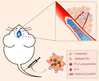 |
A novel ANG-BSA/Car/ICG MNPs integrated for targeting therapy of glioblastoma
Fan Lin1
1The First Affiliated Hospital of Shenzhen University, Shenzhen, China
Moat chemotherapeutic drugs failed to effectively treat glioblastoma (GBM) on account of the existence of blood-brain barrier, which play a role in GBM. In this study, we constructed an integrated nanoprobe based on albumin nanoparticles for targeted diagnosis and treatment of GBM. The nanoprobe consists of albumin-coated superparamagnetic iron oxide, Carmustine and indocyanine green to achieve bimodal imaging and drug delivery. And the surface-coupled Angiopep-2 can specifically bind to LRP, which is overexpressed in BBB and GBM cells. This novel targeting imaging and drug delivery system provides an efficient strategy for targeted therapy and intraoperative localization of GBM.
|
|
3047.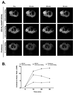 |
Effective MR Molecular Imaging of Prostate Cancer with an EDB-Fibronectin-Specific Contrast Agent at Reduced Doses
Nadia Ayat1, Sarah Roelle1, Songqi Gao1, Yajuan Li1, and Zheng-Rong Lu1
1Case Western Reserve University, Cleveland, OH, United States
This research highlights the potential of a targeted contrast agent specific to the extra-domain B (EDB-FN) to be utilized in the diagnosis of prostate cancer at reduced dosages. EDB-FN specific MR contrast agent MT218 produced robust tumor enhancement at dosages as low as 0.02 mmol Gd/kg.
|
|
3048.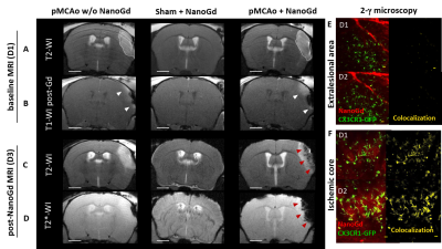 |
NanoGd, a novel hybrid multimodal nanoparticle designed for inflammation imaging: pre-clinical evaluation in ischemic stroke
Violaine Hubert1, Ines Hristovska2, Szilvia Karpati3, Frederic Lerouge3, Maelle Monteil4, Emmanuel Brun5, Naura Chounlamountri2, Chantal Watrin2, Fabien Chauveau6, Yves Berthezene7, Marc Lecouvey4, Stephane Parola3, Olivier Pascual2,
and Marlene Wiart1
1Université Lyon, CarMeN laboratory, Inserm U1060, Lyon, France, 2Université Lyon Institut Neuromyogène CNRS UMR 5310, INSERM U1217, Lyon, France, 3Université de Lyon, Ecole Normale Supérieure de Lyon, CNRS UMR 5182, Université Lyon 1, Laboratoire de Chimie, Lyon, France, 4Université Paris 13, Sorbonne Paris Cité, Laboratoire CSPBAT, CNRS UMR 7244, Paris, France, 5STROBE team, European Synchrotron Radiation Facility, Grenoble, France, 6Université Lyon, Lyon Neuroscience Research Center, CNRS UMR5292, Inserm U1028, Lyon, France, 7Université Lyon, CREATIS CNRS UMR 5220-INSERM U1206, Lyon, France
We here report the first quantitative study using MRI and intravital two-photon microscopy back-to-back to image inflammation in a mouse model of permanent middle cerebral artery occlusion, thanks to NanoGd, a new multimodal nanoparticle. Overall, our data suggest that NanoGd is safe and does not induce inflammation. NanoGd is phagocytosed by Macrophages/Microglia both in-vitro and in-vivo. In permanent ischemic stroke, NanoGd-enhanced MRI allows to visualize inflamed areas within the ischemic lesion and thus appears as a promising tool for the evaluation of anti-inflammatory treatments.
|
|
3049.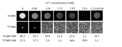 |
New class of biodegradable responsive phosphorus–containing contrast agent for 1H/31P MR
Natalia Ziółkowska1,2, Martin Hrubý3, Martin Vít1,4, Zdislava Pechrová3,5, and Daniel Jirák1,2
Video Permission Withheld
1Department of Computed Tomography, Magnetic Resonance and Clinical Experimental Spectroscopy, Institute for Clinical and Experimental Medicine, Prague, Czech Republic, 2First Faculty of Medicine, Institute of Biophysics and Informatics, Charles University, Prague, Czech Republic, 3Supramolecular polymer systems department, Institute of Macromolecular Chemistry, Prague, Czech Republic, 4Faculty of Mechatronics Informatics and Interdisciplinary Studies, Technical University of Liberec, Liberec, Czech Republic, 5Faculty of Sciences, Department of Physical and Macromolecular Chemistry, Charles University, Prague, Czech Republic
We present a new class of biodegradable responsive 31P phosphorus–containing contrast agent for dual 1H/31P MRI. Both 1H and 31P MR imaging modalities offer complementary information and can be easily combined at the same experiment. The switch to phosphorus MR signal can reflect biochemical changes in the organism. An implementation for 1H/31P MR at 4.7 T is presented and its properties are investigated.
|
|
3050.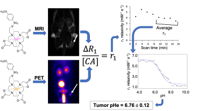 |
A PET/MRI contrast agent for measuring tumor extracellular pH
Alyssa C Pollard1,2, Aikaterini Kotrotsou2, Jorge de la Cerda2, F William Schuler2, Federica Pisaneschi2, Seth T Gammon2, and Mark D Pagel1,2
1Chemistry, Rice University, Houston, TX, United States, 2Cancer Systems Imaging, MD Anderson Cancer Center, Houston, TX, United States
Extracellular pH (pHe) in the tumor microenvironment is a biomarker that is used to assess tumor acidosis, which is caused by upregulated aerobic glycolysis in cancer cells. A PET/MRI contrast agent has developed consisting of a pH-responsive MRI co-agent, and a PET co-agent with matching pharmacokinetics to report on the concentration of the MRI co-agent. Using our simultaneous PET/MRI methodology, the pHe of a MIA PaCa-2 pancreatic tumor mouse model was found to be 6.76. These in vivo pH measurements can be used to detect a response to therapy or identify tumor vs. non-tumor tissue.
|
|
3051. |
In Vivo T2* Mapping of Hyperpolarized 13C-Labeled Metabolites Using a Metabolite-Selective Multi-Echo Spiral Imaging Sequence
Junjie Ma1, Edward P. Hackett1, and Jae Mo Park1,2,3
1Advanced Imaging Research Center, UT Southwestern Medical Center, Dallas, TX, United States, 2Radiology, UT Southwestern Medical Center, Dallas, TX, United States, 3Electrical Engineering, UT Dallas, Richardson, TX, United States
Multi-echo 13C spiral imaging sequence combined with a spectral-spatial radiofrequency pulse is implemented for sequential imaging of hyperpolarized [13C]bicarbonate, [1-13C]lactate and [1-13C]pyruvate. From the multi-echo 13C images of each metabolite in vivo T2* maps are generated. The pulse sequence was tested with a 13C-phantom and applied to measure T2*s of hyperpolarized [1-13C]pyruvate and the products in rat heart in vivo.
|
|
3052. |
Natural Abundance Deuterium (2H) MRI of the Brain
Joshua D Kaggie1, Mary McLean1, Rolf F Schulte2, Dimitri A Kessler1, Frances Henson3, Fiona J Gilbert1, Martin J Graves1, and Ferdia A Gallagher1
1Radiology, University of Cambridge, Cambridge, United Kingdom, 2GE Healthcare, Munich, Germany, 3Veterinary Medicine and Divisino of Trauma and Orthopaedic Surgery, University of Cambridge, Cambridge, United Kingdom
We demonstrate natural abundance deuterium imaging of the normal human brain in vivo, without the requirement of an injected deuterated molecule, and within a 10-minute chemical shift imaging acquisition.
|
|
3053. |
The Best Combination of Genes for Genetic Reporter of Cellular Level MR Imaging
Naoya Hayashi1,2, Junichi Hata2,3, Tetsu Yoshida2, Yawara Haga1,2, Hideyuki Okano2,4, Hirotaka James Okano3, and Akira Furukawa1
1Department of Radiological Sciences, Human Health Sciences, Tokyo Metropolitan University Graduate School, Tokyo, Japan, 2Laboratory for Marmoset Neural Architecture, Center for Brain Science, RIKEN, Saitama, Japan, 3Division of Regenerative Medicine, The Jikei University School of Medicine, Tokyo, Japan, 4Department of Physiology, Keio University School of Medicine, Tokyo, Japan
We examined which gene we should transduce for genetic reporter for Magnetic Resonance Imaging (MRI) by comparing T1 or T2 values of the cells followed by exposure to MnCl2 or Holo-transferrin. We identified the cell which has the shortest T1 or T2 values and it indicates that they can be useful genetic reporter for T1 Weighted Imaging (T1WI) or T2 Weighted Imaging (T2WI).
|
|
3054. |
Three compartment model approach to acquire kinetic information of reactive contrast agent
Asaduddin Muhammad1, Wonsik Jung2, Sangyong Jon2, and Sung-Hong Park1
1Bio and Brain Engineering, KAIST, Daejeon, Korea, Republic of, 2Biological Science, KAIST, Daejeon, Korea, Republic of
Common contrast enhanced MRI study relies on low retention and inert contrast. With the emerging trend of using paramagnetic metal for theragnostic study, few nanoparticles have been reported to have T1 enhancement property. In this research, we try to model the kinetics of such nanoparticles in mouse with A549 tumor. A novel three compartment model were introduced to explain the behavior of Mn-BRNP nanoparticles within mouse blood circulation system. This is the first study to explore reactive contrast agent with high retention, and successfully derived kinetic parameters that characterize the drug uptake behavior.
|
|
3055.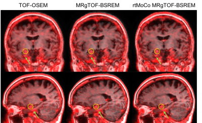 |
Rigid Motion Correction for High Resolution Brain PET/MR Imaging
Mohammad Mehdi Khalighi1, Mackenzie Carlson2, Matthew Spangler-Bickell3, Timothy Deller4, Kristen Wangerin4, Dan Rettmann4, Tim Skloss4, Brian Burns4, Dawn Holley1, Kim Halbert1, Tyler N Toueg5, Phillip S DiGiacomo1, Murat Aksoy6,
Julian Maclaren6, Roland Bammer6, Floris Jansen4, Fred T. Chin1, Michael Moseley1, Greg Zaharchuk1, Elizabeth Mormino5, and Michael Zeineh1
1Radiology, Stanford Univ., Stanford, CA, United States, 2Bioengineering, Stanford Univ., Stanford, CA, United States, 3Was with: Nuclear Medicine, IRCCS Ospedale San Raffaele, Milan, Italy, 4GE Healthcare, Waukesha, WI, United States, 5Neurology, Stanford Univ., Stanford, CA, United States, 6HobbitView, San Jose, CA, United States
In a brain PET/MR scan, the PET acquisition is done during the entire exam which typically lasts between 30-60 min. Most subjects, especially patients with a motion disorder or AD, exhibit some degree of motion which results in image blurring, quantitative errors due to mismatched attenuation correction, and mis-registered PET and MR images which limits the advantages of PET reconstruction using anatomical priors. In this study, we describe the benefits of an optical motion tracking technique, which has been recently introduced for PET/MR, to acquire high resolution isotropic PET/MR images.
|
|
3056. |
Effectiveness of fat suppression using a water-selective binomial-pulse excitation in Chemical Exchange Saturation Transfer (CEST) MRI
Yu Zhao1, Daniel F. Gochberg2, and Jianqi Li1
1Shanghai Key Laboratory of Magnetic Resonance, Shanghai, China, 2Vanderbilt University Institute of Imaging Science, Nashville, TN, United States
The purpose of this study was to characterize the contribution of fat to the CEST signal when using a water-selective binomial-pulse excitation and the effects of multiple fat peaks and B0 inhomogeneity. A CEST sequence with binomial-pulse excitation and a modified PRESS localization was applied to in vivo experiments to determine signal contributions of lipid resonances. Water excitation using a {1-3-3-1} pulse provided a broad signal suppression, which provided robustness against B0 inhomogeneity. Significant fat signal contributions to CEST imaging of hydroxyl and amine were unavoidable, while much smaller contamination was seen when imaging amide sites, limited by B0 inhomogeneity.
|
3057.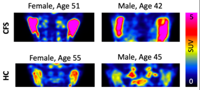 |
TSPO-PET/MRI Reveals Increased Neuroinflammation in Basal Ganglia of Chronic Fatigue Syndrome Patients
Mackenzie Leigh Carlson1, Jun-Hyung Park2, Tullia Lieb3, Bin Shen2, Marc Stevens2, Brian Mills2, Nicole Mouchawar2, Greg Zaharchuk2, Michael Zeineh2, and Michelle James2,4
1Bioengineering, Stanford University, Stanford, CA, United States, 2Radiology, Stanford University, Stanford, CA, United States, 3Medicine, Stanford University, Stanford, CA, United States, 4Neurology, Stanford University, Stanford, CA, United States
Myalgic encephalomyelitis/chronic fatigue syndrome (ME/CFS) is a debilitating disease affecting millions of people in the United States alone, but little is known about the underlying pathophysiology. We show that simultaneous TSPO-PET/MRI measurements using [11C]DPA-713, including SUV, SUVr, QSM, R2*, and volume can uncover new information about this disease. We find that the putamen has significantly higher TSPO-PET signal in ME/CFS subjects compared to healthy controls, indicating an elevated inflammatory response in this area. This finding corresponds to previous fMRI and diffusion imaging findings and may help with future diagnosis and tracking of this chronic, widespread disease.
|
|
3058.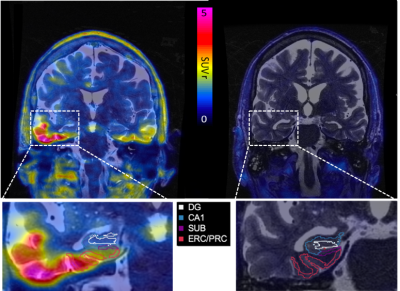 |
Hippocampal Subfield Tau Progression Identified using Simultaneous Tau-PET/MRI at 3T in Alzheimer’s Disease
Mackenzie Leigh Carlson1, Tyler Toueg2, Mehdi Khalighi3, Jessa Castillo3, Bin Shen3, Carmen Azevedo3, Phillip DiGiacomo1, Nicole Mouchawar3, Greg Zaharchuk3, Michelle James2,3, Elizabeth Mormino2, and Michael Zeineh3
1Bioengineering, Stanford University, Stanford, CA, United States, 2Neurology, Stanford University, Stanford, CA, United States, 3Radiology, Stanford University, Stanford, CA, United States
We found structural changes and tau protein accumulation within the hippocampus that can be simultaneously assessed at the subfield level in Alzheimer’s Disease. We found more separation among AD, mild cognitive impairment (MCI), and control groups when considering subfields than whole hippocampal SUVr. This study highlights the potential of our combined subfield analysis technique in disentangling pathologic derangements accompanying AD in the hippocampus and for providing more nuanced imaging-based measures for tracking and staging disease in vivo.
|
|
3059.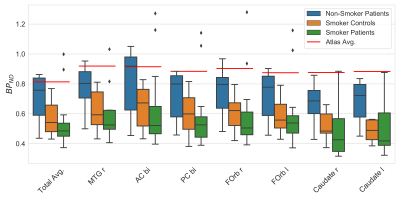 |
Construction of mGluR5 receptor based in vivo atlas using simultaneously acquired [11C]ABP688 PET / MR data
Nicolas Kaulen1,2, Claudia Régio Brambilla1,3,4, Ravichandran Rajkumar1,3,4, Shukti Ramkiran1,3, Linda Orth1,3, Hasan Sbaihat1, Markus Lang5, Elena Rota Kops1, Jürgen Scheins1, Bernd Neumaier5, Johannes Ermert5, Hans Herzog1, Karl-Josef Langen1,4,6,
Christoph Lerche1, N. J. Shah1,4,7,8, and Irene Neuner1,3,4
1Institute of Neuroscience and Medicine 4, INM-4, Forschungszentrum Jülich, Jülich, Germany, 2Faculty of Physics, Technical University Dortmund, Dortmund, Germany, 3Department of Psychiatry, Psychotherapy and Psychosomatics, RWTH Aachen University, Aachen, Germany, 4JARA – BRAIN – Translational Medicine, Aachen, Germany, 5Institute of Neuroscience and Medicine 5, INM-5, Forschungszentrum Jülich, Jülich, Germany, 6Department of Nuclear Medicine, RWTH Aachen University, Aachen, Germany, 7Institute of Neuroscience and Medicine 11, INM-4, Forschungszentrum Jülich, Jülich, Germany, 8Department of Neurology, RWTH Aachen University, Aachen, Germany
This project aims to create a standard space in vivo atlas of the mGluR5 distribution in the healthy brain using structural and metabolic information from simultaneously acquired [11C]ABP688-PET / MR data. The structural MRI scans served as an anatomical reference to create a parametric MNI space atlas of mGluR5 non-displaceable binding potential. Validation of the atlas was performed and global as well as local reductions of mGluR5 availability in healthy smokers and schizophrenic smokers that are in line with the results of previous studies could be shown. Preliminary investigation of schizophrenic non-smokers showed slight, though not conclusive, reductions of mGluR5.
|
|
3060.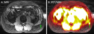 |
Sigma-1 Receptor PET-MRI for Imaging Nociceptive Sources of Low Back Pain
Daehyun Yoon1, Peter Cipriano1, and Sandip Biswal1
1Radiology, Stanford university, Stanford, CA, United States
Low back pain is a leading cause of job-related disability and missed workdays in the U.S. Unfortunately, the overall treatment effectiveness is significantly limited by our poor diagnostic capability, which fails to identify nociceptive sources in 80% to 90% of the cases. In this work, we introduce a PET/MRI approach for low back pain diagnosis using a novel radioligand to track the sigma-1 receptor, which is upregulated during nociceptive processes. Our early results demonstrate promise in achieving improved sensitivity and specificity to the local sources of low back pain.
|
|
3061.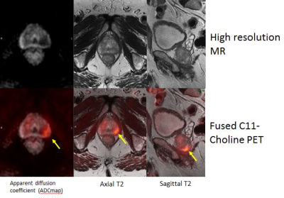 |
Hybrid C11 Choline PET/MRI in staging untreated high risk and very high risk prostate cancer patients at single institution: preliminary results
Kelly T Smith1, Akira Kawashima2, Mark D Tyson3, Erik P Castle4, Alvin Silva1, Yuxiang Zhou1, Michael C Roarke1, and Ming Yang1
1Radiology, Mayo Clinic Arizona, PHOENIX, AZ, United States, 2Radiology, Mayo Clinic Arizona, Phoenix, AZ, United States, 3Urology, Mayo Clinic Arizona, PHOENIX, AZ, United States, 4Urology, Mayo Clinic Arizona, Phoenix, AZ, United States
The most prevalent malignancy in US men is prostate cancer. Appropriate treatment regimen is dependent on accurate tumor staging. Pelvic multiparametric MR (mpMRI) is the established modality for tumor and nodal staging. C-11 choline is FDA approved in diagnosis of recurrent prostate cancer. Introduction of a hybrid time-of-flight PET/MR system affords the opportunity to perform a combined C-11 choline PET + mpMRI for prostate cancer staging. We feel this system would be beneficial in staging of treatment naïve high risk prostate cancer.
|
|
3062.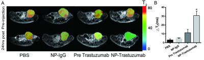 |
The utility of trastuzumab-based imaging agents for the development of theranostic for breast cancer
Xiaojun Luo1, Chunwu Zhou2, and Lizhi Xie3
1Department of Radiology, Wangjing Hospital of China Academy of Chinese Medical Sciences, Beijing, China, 2National Cancer Center/National Clinical Research Center for Cancer/Cancer Hospital, Chinese Academy of Medical Sciences and Peking Union Medical College, Beijing, China, 3GE healthcare, China, Beijing, China
Superparamagnetic iron oxide nanoparticles (SPIONs) has been applied in diagnosis of different cancers, however its potential in breast cancer has not been fully explored. In this study, we research into the value of SPIONs conjugated with trastuzumab and indocyanine green (ICG) in becoming an effective multifunctional imaging agent and a tool for photothermal therapy for HER2-positive breast cancer. It was found that the biodistribution of dual-modality imaging agents has been observed by MRI and fluorescence imaging, which could provide effective technical assistance for HER2-positive breast cancer treatment.
|
|
3063.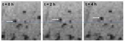 |
Magnetic Resonance Imaging of Single-Cell Dynamics in the Olfactory Bulb
Nikorn Pothayee1, Stephen Dodd1, Gary Zabow2, and Alan Koretsky1
1Laboratory of Functional and Molecular Imaging, National Institutes of Health, Bethesda, MD, United States, 2Magnetic Imaging Group, National Institute of Standards and Technology, Boulder, CO, United States
There is great potential if single cells can be tracked in vivo in the mammalian brain. Here, we demonstrate the applicability of microfabricated gold-coated iron particles for in vivo tracking of single-cell dynamics. These particles have a pure iron core as opposed to the more commonly used iron-oxide based particles. Following in situ labeling of neural precursor cells, we show that the migration of individual cells can be visualized in real-time.
|
|
3064.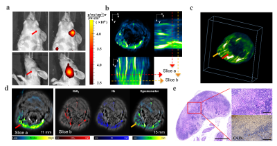 |
In vivo three-dimensional evaluation of hypoxia in nasopharyngeal carcinomas using FMT-CT and MSOT
Wenhui Huang1,2, Kun Wang2, Jie Tian2, and Shuixing Zhang1
1the First Affiliated Hospital,Jinan University, Guangdong, China, 2Institute of Automation, Chinese Academy of Sciences, Beijing, China
The accurate imaging hypoxia is especially vital for nasopharyngeal carcinoma (NPC) patients undergoing radiotherapy. Multimodality molecular imaging have great potential to acquire hypoxic imaging with high sensitivity and more accuracy. We propose a novel imaging strategy,which combined the hybrid fluorescence molecular tomography-computed tomography (FMT-CT) and the multispectral optoacoustic tomography (MSOT), for achieving three-dimensional (3D) quantitative evaluation of NPC hypoxia in small animal models.The results could not only detect the hypoxia in small sizes of NPCs and lymph nodes metatasis, but also visualize the heterogeneity of hypoxia in 3D.
|
|
3065.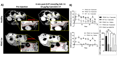 |
Imaging Zinc Dysregulation in Pancreatic Ductal Adenocarcinoma by Secretagogue-Stimulated Zinc Secretion MRI
Mozhdeh Sojoodi1, Ian Ramsay2, Eric Gale2, Peter Caravan2,3, Kenneth Tanabe1, and Veronica Clavijo Jordan 2
1Division of Surgical Oncology, Massachusetts General Hospital/Harvard Medical School, Boston, MA, United States, 2Athinoula A. Martinos Center for Biomedical Imaging, Massachusetts General Hospital/Harvard Medical School, Charlestown, MA, United States, 3Institute for Innovation in Imaging (i3), Massachusetts General Hospital/Harvard Medical School, Charlestown, MA, United States
Zinc homeostasis is markedly dysregulated in pancreatic ductal adenocarcinoma (PDAC), and this dysregulation can be probed by selecting a secretagogue to stimulate the secretion of zinc and functional components from the exocrine pancreas. Here, we introduce the use of caerulein, a surrogate cholecystokinin agonist of pancreatic exocrine secretion, and a Gd-based zinc probe to monitor exocrine function in the healthy and PDAC mouse pancreas by MRI. Our results indicate that zinc dysregulation involves an increase in zinc accumulation in malignant tissue mediated by upregulation of zinc import transporters, and hypersecretory capacity as reported by caerulein-stimulated zinc secretion MRI in vivo.
|
|
3066.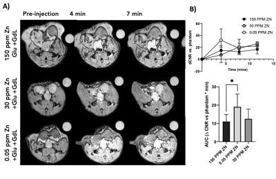 |
Molecular MRI and synchrotron radiation X-Ray fluorescence of zinc homeostasis in the healthy and malignant mouse prostate
Veronica Clavijo Jordan 1, Andre Martins2,3, Erica Dao4, Alia Al-Ebraheem4, Kalotina Geraki5, Xiaodong Wen3, Sara Chirayil3, Xiaojing Wang3, Mozhdeh Sojoodi6, Michael Farqhuarson4, and A.Dean Sherry3,7
1Athinoula A. Martinos Center for Biomedical Imaging, Massachusetts General Hospital/Harvard Medical School, Charlestown, MA, United States, 2Werner Siemens Imaging Center, Tuebingen, Germany, 3Advanced Imaging Research Center, University of Texas Southwestern Medical Center, Dallas, TX, United States, 4McMaster University, Hamilton, ON, Canada, 5Diamond Light Source, Harwell, United Kingdom, 6Division of Surgical Oncology, Massachusetts General Hospital/Harvard Medical School, Boston, MA, United States, 7Department of Chemistry, University of Texas at Dallas, Richardson, TX, United States
Zinc homeostasis is markedly dysregulated in prostate cancer (PCa), and this dysregulation can be detected with glucose-stimulated zinc secretion (GSZS) by MRI. Here we explore the use of GSZS MRI and synchrotron-radiation X-Ray fluorescence to interrogate the effect of dietary zinc on the healthy and malignant mouse prostate. Our results indicate that the lateral lobe of the healthy mouse is the only prostatic structure responsive to a variable zinc diet acting as the zinc “regulator” of the gland, and that in PCa this lobe no longer responds to changes in dietary zinc, highlighting the dysregulation of zinc transporters in PCa.
|
|
3067.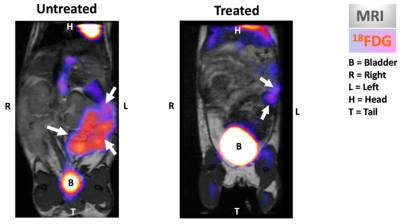 |
Quantitative PET/MRI of Immunotherapy Response in Preclinical Epithelial Ovarian Cancer
Marie-Laurence Tremblay1,2,3, Caitrin Sobey-Skelton1,2, Hailey Wyatt1,2, Victoria Gonzalez2, Andrea Nuschke1,4, Christa Davis1, Alecia MacKay5, Kim Bobbitt5, Andrea West2,5, Barbara Vanderhyden6,7, Genevieve Weir5, Marianne Stanford5, and Kimberly Brewer1,2
1Biomedical Translational Imaging Centre (BIOTIC), Halifax, NS, Canada, 2Dalhousie University, Halifax, NS, Canada, 3IWK Inc., Halifax, NS, Canada, 4Carleton University, Ottawa, ON, Canada, 5IMV Inc., Halifax, NS, Canada, 6Ottawa Hospital Research Institution, Ottawa, ON, Canada, 7University of Ottawa, Ottawa, ON, Canada
Epithelial ovarian cancers are highly aggressive tumor types making them a prime target for research using immunotherapies. Using simultaneous PET/MRI, we monitored an orthotopic ovarian cancer model to evaluate longitudinal tumor growth and metabolism in response to therapy while tracking immune cell subsets labeled with superparamagnetic iron oxide (SPIO). Treatment with the combination of immune therapies significantly decreased the volume of primary tumors and improved survival times. We quantified cytotoxic T lymphocytes (CTLs) and dendritic cells (DCs) recruited to the primary tumors and found treatment with the combination therapy increased recruitment of CTLs but resulted in decreased recruitment of DCs.
|
|
3068. |
In Vivo Tracking and Quantification of Murine Natural Killer Cells via Fluorine-19 MRI
Lawrence M Lechuga1, Matthew H Forsberg2, Kai D Ludwig1, Christian M Capitini2,3, and Sean B Fain1,4,5
1Medical Physics, University of Wisconsin, Madison, Madison, WI, United States, 2Pediatrics, University of Wisconsin, Madison, Madison, WI, United States, 3Carbone Comprehensive Cancer Center, University of Wisconsin, Madison, Madison, WI, United States, 4Biomedical Engineering, University of Wisconsin, Madison, Madison, WI, United States, 5Radiology, University of Wisconsin, Madison, Madison, WI, United States
Flourine-19 (19F) MRI has demonstrated the ability to monitor immune cell populations in vivo. Murine green fluorescent protein natural killer (NK) cells were labeled with perfluoropolyether (PFPE-red) and injected intratumorally into lymphoma-bearing mice. In this work, we demonstrate the ability to track and quantify NK cells up to 5 days in vivo, via 19F MRI. In vivo quantification indicated that 88% of the initially detected NK cells remained at the tumor site on day 5. Flow cytometry analysis provided validation that the cellular label remains colocalized within a significant portion of the NK cells at day 5.
|
|
3069.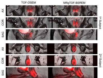 |
High Resolution Isotropic PET/MR imaging using Anatomical Priors for PET Reconstruction
Mohammad Mehdi Khalighi1, Timothy Deller2, Matthew Spangler-Bickell3, Kristen Wangerin2, Dawn Holley1, Kim Halbert1, Michael Zeineh1, Greg Zaharchuk1, Elizabeth Mormino4, Andrei Iagaru5, and Michael Moseley1
1Radiology, Stanford University, Stanford, CA, United States, 2PET/MR Engineering, GE Healthcare, Waukesha, WI, United States, 3Was with: Nuclear Medicine, IRCCS Ospedale San Raffaele, Milan, Italy, 4Neurology, Stanford University, Stanford, CA, United States, 5Nuclear Medicine, Stanford University, Stanford, CA, United States The current spatial resolution of PET images is ~4mm for whole body PET/MR. Anatomical MR images with higher resolution and superior SNR, have been used in PET reconstruction to improve the image quality and spatial resolution. However, these methods are generally vulnerable to mismatches between the anatomical image and the true activity distribution. To address this concern, we propose a feature based approach to incorporate anatomical priors in PET image reconstruction, where the location of a voxel or its neighbors doesn't play a direct role and both functional and anatomical images are used to construct the feature space. |
|
3070.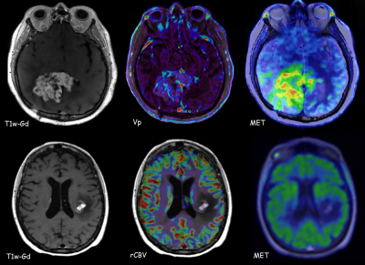 |
Spatial relationship between brain tumor regions assessed by PWI and 11C-Methionine using a fully hybrid PET/MRI system
Paola Scifo1, Maurizio Barbera2, Federico Fallanca1, Antonella Castellano2, Paola Mapelli1, Valentino Bettinardi1, Annarita Savi1, Riccardo Rigamonti1, Andrea Falini2, Luigi Gianolli1, Maria Picchio1,3, and Nicoletta Anzalone2
1Nuclear Medicine, IRCCS San Raffaele Scientific Institute, Milan, Italy, 2Neuroradiology and CERMAC, VIta-Salute University, IRCCS San Raffaele Scientific Institute, Milan, Italy, 3Vita-Salute San Raffaele University, Milan, Italy Poster Permission Withheld
Simultaneous PET/MRI offers a unique opportunity to investigate the relationship between concurrent phenomena measured by MET-PET and PWI-MR at the same physiological condition. Although 11C-Methionine uptake and perfusion are tightly coupled, it is not clear whether tumor regions (VOIs) defined by PWI and MET are overlapped and how the values of the corresponding parametric maps are distributed within each VOI. This preliminary work aims at studying the spatial relationship between PWI-MRI maps and MET-PET in brain tumors using a fully integrated PET/MRI system.
|
3071.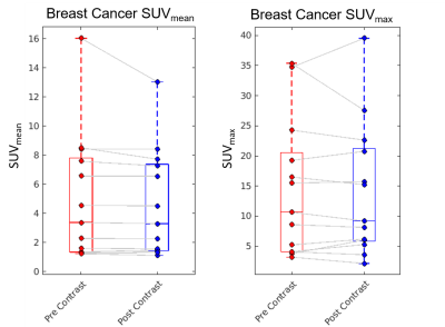 |
In Vivo Effect of Gadolinium-Based Contrast Agent on PET Quantification for Breast Imaging
Timothy J Allen1, Leah C Henze Bancroft2, Manoj Kumar2, Tyler Bradshaw2, Roberta M Strigel1,2,3, Alan McMillan1,2, and Amy M Fowler1,2,3
1Medical Physics, University of Wisconsin-Madison, Madison, WI, United States, 2Radiology, University of Wisconsin-Madison, Madison, WI, United States, 3Carbone Cancer Center, University of Wisconsin-Madison, Madison, WI, United States
The effect of gadolinium-based contrast agents (GBCAs) on dynamic PET quantification was investigated. Simultaneous PET/MRI was performed in patients with biopsy confirmed invasive breast cancer. Subjects received an injection of a GBCA as part of standard clinical dynamic contrast enhanced breast MRI exam. SUVmean and SUVmax were calculated for the biopsy-proven breast cancer, aorta, liver, and benign fibroglandular tissue prior to and following GBCA injection. No significant difference in SUVmean or SUVmax was measured suggesting GBCAs are unlikely to affect PET quantification in combined breast PET/MRI.
|
|
3072.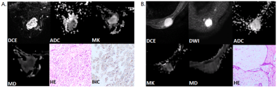 |
Assessment of Diffusion Kurtosis at 3.0T as an in vivo Imaging Marker for Breast Lesions Characterization
wan tang1, ting ouyang1, and yan lin1
1radiology, Second Affiliated Hospital, Shantou University Medical College, China, shantou, China
Breast multi-parametric magnetic resonance imaging (mp-MRI) is a well-established technique in breast cancer for tumor characterization, treatment strategies and post operative predicting, owing to its non-invasive nature of excellent soft-tissue contrast. Our preliminary work demonstrated that MK showed promise in characterizing breast lesions as well as predicting prognostic factors. However, due to the small samples in our previous research and few research relating to the correlation between MK and molecular subtypes, further study with larger cohort is needed to verify our initial results and explore more potential of MK in other clinical applications (assessing therapeutic response to MWA in BBLs).
|
|
3073.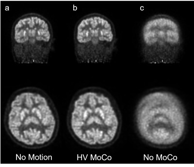 |
Evaluation of marker-based head motion tracking for prospective correction in MR and retrospective correction in PET for integrated PET/MR
Samuel Anthony Hurley1, Matthew G Spangler-Bickell2, Timothy Deller3, Timothy W Skloss3, Floris P Jansen3, Tyler J Bradshaw1, and Alan B McMillan1
1Radiology, University of Wisconsin, Madison, WI, United States, 2Nuclear Medicine Unit, IRCCS Ospedale San Raffaele, Milan, Italy, 3GE Healthcare, Waukesha, WI, United States
Motion during PET acquisition will result in blurring, which may reduce the diagnostic value of the images and cause inaccurate quantitation. Motion during an MR exam results in image replicates and ghosting along the phase encoding direction and other effects due to inconstant phase, rendering images non-diagnostic. In this work, we evaluate the ability of an optical motion tracking system to simultaneously enable prospective correction in MR and retrospective correction in PET of rigid body head motion.
|
|
3074.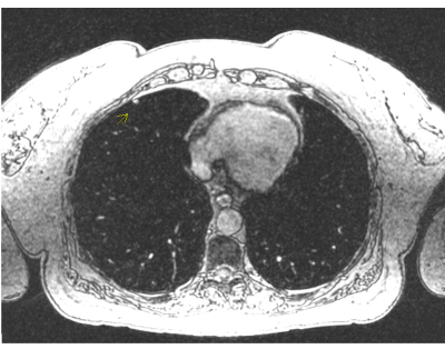 |
Comparing PET/MRI with PET/CT in detection of lung nodules 4mm or larger using a single-breath hold MR acquisition
Nevein Ibrahim1, Minnie Kieler1, Elizabeth A Sadowski1, Steve Cho1, Emmanuel Sampene1, Alan McMillan1,2, and Joanna E Kusmirek1
1Diagnostic radiology, University of Wisconsin School of Medicine and Public health, Madison, WI, United States, 2Medical physics, University of Wisconsin School of Medicine and Public health, Madison, WI, United States
One of the limitations of PET/MRI is poor evaluation of lung parenchyma with free breathing sequences. In this study, 41 patients with gynecological malignancies underwent PET/MRI with rapid single breath hold lung acquisition after PET/CT. The CT and MRI of the lungs were reviewed blindly for detection of pulmonary nodules 4mm or larger. The study showed that there was no significant difference between the CT and single breath hold MRI using Chi square test. Improved detection of small pulmonary nodules using single breath hold sequence would increase the feasibility of stand-alone PET/MR imaging for clinical staging in oncologic patients.
|
|
3075.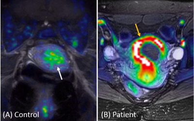 |
Early experience in identifying chronic pelvic pain generators by sigma-1 receptor PET/MRI
Daehyun Yoon1, Angela Fast2, Peter Cipriano1, Bin Shen1, Michelle James1, Deirdre Lum3, and Sandip Biswal1
1Radiology, Stanford university, Stanford, CA, United States, 2Diagnostic, Molecular and Interventional Radiology, The Mount Sinai Hospital, New York, NY, United States, 3Gynecology, Stanford university, Palo Alto, CA, United States
Chronic pelvic pain is a highly prevalent pain condition among women, but it is quite challenging to determine the exact cause of pain. MRI is currently the imaging modality of choice, but even the most common etiology is very difficult to identify. In this abstract, we introduce a novel PET/MRI approach for chronic pelvic pain diagnosis to achieve improved sensitivity by enabling image tracking of sigma-1 receptor, a chaperone protein modulating ion channels for activating nociceptive processes. Our early results present promising results in identifying potential pain generators of chronic pelvic pain, which could not be detected in MRI alone.
|
|
3076.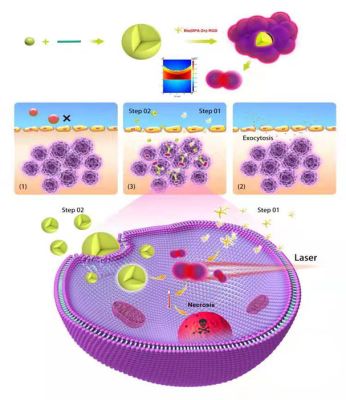 |
Orthotopic Brain Tumor Imaging and Therapy by Microenvironment-Responsive Gold Nanoclusters
Haiyan Gao1, Yan Bai1, Lijuan Chen1, and Meiyun Wang1
1Henan Provincial People's Hospital, Zhengzhou, China
Glioblastoma is one of the most lethal cancers with few available therapeutic options. Herein, we report a in situ self-assembled nanoplatform formed by RGD peptide-modified Bisulfite-ZincII-Dipicolylamine-Arg-Gly-Asp (Bis(DPA-Zn)-RGD) and ultrasmall Au-ICG nanoparticles. Taking advantage of RGD-mediated neovascular targeting, the Bis(DPA-Zn)-RGD selectively navigates to the tumor site, and then assembles into large nanoclusters with subsequently administered Au-ICG nanoclusters which can successfully cross the BBB due to ultrasmall particle size (~7 nm). This study demonstrates that such in situ self-assembly metal–organic nanosystems are ideal to overcome the BBB and BBTB, which promise a new way for orthotopic brain tumors imaging and therapy.
|
|
3077.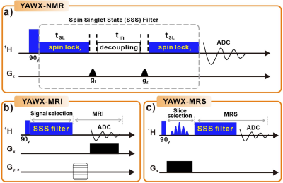 |
Molecular Targeted Magnetic Resonance Imaging and Spectroscopy
Jia-Xiang Xin1, Yi Li1, Jian-Qi Li1, Guang Yang1, Jia-Chen Wang1, Da-Xiu Wei1, and Ye-Feng Prof. Yao1
1East China Normal University, Shanghai, China
In this work, we report a novel method for obtaining an exact molecular targeted MRI and MRS. This method uses the nuclear spin singlet state to select the signals from a specific molecule. Several endogenetic molecules in living organism such as N-acetylaspartate and dopamine have been imaged and probed as the targeted molecules in the MRI and MRS experiments, demonstrating the unique molecular selectivity of the developed method.
|
|
3078.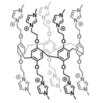 |
In-Vitro HyperCEST detection of decacationic pillar-5-arene
Camryn Jayne Newman1, Yurii Shepelytskyi2,3, Ashvin Fernando4, Paul T Cesana4, Tao Li5, Alanna Wade5, Vira Grynko3, Adriana M Mendieta4, Francis T Hane2,5, Brenton DeBoef4, and Mitchell S Albert2,5,6
1Biology, Lakehead University, Thunder Bay, ON, Canada, 2Thunder Bay Regional Research Institute, Thunder Bay, ON, Canada, 3Chemistry and Material Science, Lakehead University, Thunder Bay, ON, Canada, 4Chemistry, University of Rhode Island, Kingston, RI, United States, 5Chemistry, Lakehead University, Thunder Bay, ON, Canada, 6Northern Ontario School of Medicine, Thunder Bay, ON, Canada
Hyperpolarized (HP) 129Xe molecular imaging technology has been working towards the detection of biochemically inactive supramolecular cage-molecules within a living mammalian model. The issue originates from the non-specific natural biodistribution of the biosensor molecules, which makes it difficult to precisely localize them within a living mammalian body using HP 129Xe MRI. We have previously identified cyclodextrin-based pseudorotaxanes and benzene-appended CB6 as conjugatable scaffolds for xenon biosensors; in this work, we introduce a third class of conjugatable scaffolds, with the hyperCEST detection of pillar[5]arene, a potential precursor to a large variety of targeted molecular imaging probes.
|
|
3079.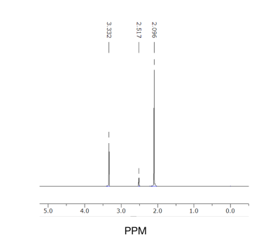 |
Evaluation of Cucurbit[6]uril Diffusion Across an Artificial Blood-Brain Barrier
Camryn Jayne Newman1, Yurii Shepelytskyi2,3, Michael Campbell2,4, Vira Grynko2,3, Francis Theodor Hane3,4, and Mitchell Scott Albert4,5
1Biology, Lakehead University, Thunder Bay, ON, Canada, 2Thunder Bay Regional Research Institute, Thunder Bay, ON, Canada, 3Chemistry and Material Science, Lakehead University, Thunder Bay, ON, Canada, 4Chemistry, Lakehead University, Thunder Bay, ON, Canada, 5Northern Ontario School of Medicine, Thunder Bay, ON, Canada
The selective nature of the blood-brain barrier (BBB) is highly regarded when evaluating the permeability of a molecule such as Cucurbit[6]uril (CB6). CB6 has previously demonstrated a strong hyperCEST effect, which makes it a valid candidate for detecting low concentrations of biomarkers of neurodegenerative pathologies. However, CB6 must be permeable to the BBB to use the hyperCEST technique for neurological biomarkers. Therefore, this work evaluates the permeability of CB6 across an artificial BBB based on diffusion. It was found that CB6 can cross the brain sphingomyelin powder (BSM) based artificial BBB.
|
|
3080.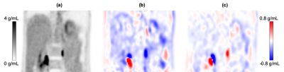 |
PET image quality improvement for PET/MR with low-attenuating AIR Technology coils
Timothy W. Deller1, Nicholas Mathew2, Samuel Hurley2, Chad Bobb1, and Alan McMillan2
1GE Healthcare, Waukesha, WI, United States, 2University of Wisconsin-Madison, Madison, WI, United States
Floating surface coils, such as the anterior array coil, are ignored in PET attenuation correction for PET/MR because their position is difficult to determine. This leads to quantitative bias in reconstructed PET images. This study investigates the potential to improve quantitative accuracy in PET when using a new lightweight "AIR Technology" anterior array coil, as compared to the traditional anterior array coil. Metrics of quantification accuracy and error variation were explored with a phantom and clinical study.
|
|
3081.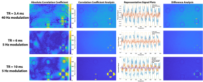 |
Detection of Superparamagnetic Iron Oxide Nanoparticles using Statistical Analysis of a Block Design Modulated bSSFP (BDM-bSSFP) Acquisition
Eugene Milshteyn1,2, Bastien Guerin1,2, and Lawrence L. Wald1,2,3
1Athinoula A. Martinos Center for Biomedical Imaging, Charlestown, MA, United States, 2Harvard Medical School, Boston, MA, United States, 3Harvard-MIT Division of Health Sciences and Technology, Cambridge, MA, United States
The current standard methods for magnetic resonance imaging of superparamagnetic iron oxide nanoparticles (SPIONs) can be subject to “1/f” physiological fluctuations that can confound pre/post comparisons. We present a method to combine Block Design contrast Modulations and statistical analysis with the bSSFP sequence (BDM-bSSFP) in order to mitigate this effect. To do this, we leverage the frequency response of the bSSFP sequence to the local fields created by SPIONs. We show the ability of this method to detect SPIONs down to 10 μg/mL through simulations and in-vitro acquisitions.
|
|
3082.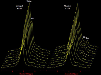 |
Monitoring Inflammation-associated Metabolic Alterations by Deuterium MRS
Vera Flocke1, Pascal Bouvain1, Sebastian Temme1, and Uli Flögel1
1Molecular Cardiology, Heinrich-Heine-University Düsseldorf, Düsseldorf, Germany There is increasing evidence that inflammatory diseases go along with substantial changes in intermediatory metabolism. Also it is known that metabolic syndrome is characterized by chronic low-grade inflammation in multiple organs[1,2]. In this context, the metabolism of glucose is of major interest, since in particular in the initial phase of an inflammation infiltrating immune cells mainly rely on glycolysis to cover their energy demand. The present study demonstrates that 2H MRI can be successfully used for monitoring metabolic alterations in an inflammatory environment with a switch to a more anaerobic glucose utilization in the initial phase of immune cell recruitment. |
|
3083.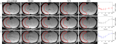 |
Timely Visualizing the Collaterals Formed during Acute Ischemic Stroke with Fe3O4 Nanoparticle-based MR Imaging Probe
Wang Ting1, Ma Lin1, and Lou Xin1
1Chinese PLA General Hospital, Beijing, China
An αvβ3-specific Fe3O4-RGD nanoprobe was prepared for MR imaging of the collaterals during AIC. The MCAO rat models were constructed for imaging studies on 7.0-T MRI. To show the binding specificity to the collaterals, the imaging results acquired with the Fe3O4-RGD nanoprobe, Fe3O4 mother nanoparticles and RGD blocking experiment. Cerebral ischemia-reperfusion studies were also performed to show the collateral dynamics upon reperfusion. The studies have the first-time enabled the direct observation of collaterals in a quasi-real time fashion and further disclosed that the antegrade flow upon reperfusion dominates the blood supply of primary ischemic tissue during early stage of infarction.
|
|
3084.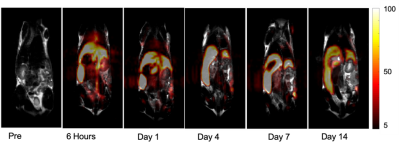 |
Assessment of Passive Tumor Targeting of a Novel PFCE-Loaded Nanoemulsion via 19F-MRI
Lawrence M Lechuga1, Alexa R Barres2, Montira Tangsangasaksri3, Paul Begovatz1, Sandro Mecozzi3,4, and Sean B Fain1,5,6
1Medical Physics, University of Wisconsin, Madison, Madison, WI, United States, 2Morgridge Institute for Research, Madison, WI, United States, 3Pharmacy, University of Wisconsin, Madison, Madison, WI, United States, 4Chemistry, University of Wisconsin, Madison, Madison, WI, United States, 5Biomedical Engineering, University of Wisconsin, Madison, Madison, WI, United States, 6Radiology, University of Wisconsin, Madison, Madison, WI, United States
Fluorine-19 MRI is a method that can utilize biocompatible perfluorocarbons to detect cancer. Implementation of a novel nanoemulsion has allowed for improved particle stability and PFC loading. Tumor-bearing mice were injected with perfluoro-[15-crown-5]-ether (PFCE) loaded nanoemulsion (NE) to explore the passive tumor targeting across 14 days. 19F MRI and subsequent ROI-image analysis suggested that there was a preferential retention of NE particles due to the enhanced permeability and retention (EPR) effect. This work demonstrates repeatable 19F imaging of a novel nanoemulsion that passively targets a fast growing tumor with improved stability and PFCE loading.
|
|
3085.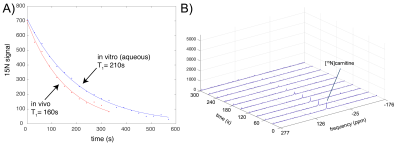 |
Hyperpolarization and 1H-based detection of [15N]carnitine, a new long-lived metabolic imaging probe
Cornelius von Morze1, James D Quirk1, John A Engelbach1, Galen D Reed2, Albert P Chen2, Craig R Malloy3, Joel R Garbow1, and Matthew E Merritt4
1Department of Radiology, Washington University, St Louis, MO, United States, 2GE Healthcare, Dallas, TX, United States, 3Advanced Imaging Research Center, UT Southwestern, Dallas, TX, United States, 4Department of Biochemistry and Molecular Cell Biology, University of Florida, Gainesville, Gainesville, FL, United States 13C hyperpolarization has opened the door for real-time MR metabolic imaging but remains limited by T1's of approximately one minute or less. 15N offers potentially much longer T1's, but suffers from poor intrinsic sensitivity and small chemical-shift separation. We have hyperpolarized L-[15N]carnitine-d9, a novel metabolic imaging probe, and found ultra-long T1's of 210s (in H2O) / 160s (in vivo). We also demonstrate successful 15N->1H hyperpolarization transfer for enhanced sensitivity of detection as well as larger chemical-shift separation among metabolites. The long signal lifetime and excellent safety profile of [15N]carnitine suggest great potential for metabolic imaging investigations and future clinical translation. |