
Digital Poster Session
Contrast Mechanisms: CEST, Magnetization Transfer and Relaxometry
Contrast Mechanisms
3086 -3101 CEST, Magnetization Transfer and Relaxometry - CEST: Methods & Applications
3102 -3115 CEST, Magnetization Transfer and Relaxometry - CEST Friends & Family
3116 -3131 CEST, Magnetization Transfer and Relaxometry - Diet CEST (No Glucose)
3132 -3146 CEST, Magnetization Transfer and Relaxometry - Magnetization Transfer Imaging
3147 -3162 CEST, Magnetization Transfer and Relaxometry - Relaxation
3163 -3178 CEST, Magnetization Transfer and Relaxometry - Contrast Mechanisms: Quantitation & Validation
Session Topic: CEST, Magnetization Transfer and Relaxometry
Session Sub-Topic: CEST: Methods & Applications
Digital Poster
Contrast Mechanisms
3086.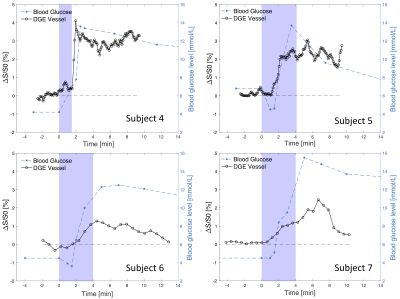 |
Optimization of glucose infusion protocol for glucoCEST imaging
Anina Seidemo1, Patrick M. Lehmann1, Anna Rydhög2, Ronnie Wirestam1, Xiang Xu3,4, Akansha A. Sehgal3,4, Yi Zhang5, Frederik Testud6, Pia C. Sundgren7,8, Peter C.M. van Zijl3,4, and Linda Knutsson1,3
1Department of Medical Radiation Physics, Lund University, Lund, Sweden, 2Centre for Medical Imaging and Physiology, Skåne University Hospital, Lund and Malmö, Sweden, 3Russell H. Morgan Department of Radiology and Radiological Science, Johns Hopkins University School of Medicine, Baltimore, MD, United States, 4F.M. Kirby Research Center for Functional Brain Imaging, Kennedy Krieger Institute, Baltimore, MD, United States, 5College of Biomedical Engineering and Instrument Science, Zhejiang University, Hangzhou, China, 6Siemens Healthcare AB, Malmö, Sweden, 7Department of Diagnostic Radiology, Lund University, Lund, Sweden, 8Lund University Bioimaging Center, Lund University, Lund, Sweden
The intravenous glucose injection used in dynamic glucose-enhanced (DGE) imaging is related to transient sensations which can be unpleasant for the subject and may cause movements. We have investigated the effect of different infusion durations in terms of sensational side effects and with respect to the relation between arterial DGE signal and venous blood glucose levels. Our findings indicate that the DGE image quality does not benefit from a fast glucose injection and we conclude that a slower infusion should be used to increase patient comfort and reduce the risk for patient motion related to the glucose injection.
|
|
3087. |
Application of GluCEST imaging in mapping cingulate cortex of Nicotine dependence subjects at 7.0T
Ravi Prakash Reddy Nanga1, Emily Devlin1, Claudia Ianelli2, Deepa Thakuri1, Dushyant Kumar1, Hari Hariharan1, James Loughead1, Cynthia Neill Epperson2, and Ravinder Reddy1
1Perelman School of Medicine at The University of Pennsylvania, Philadelphia, PA, United States, 2University of Colorado School of Medicine, Aurora, CO, United States
Nicotine addiction is still one of the most preventable causes of chronic illness and premature death, and cessation regardless of age has positive effects on health outcomes (WHO, 2015). As such, there is growing interest in developing novel therapies and diagnostic aids to treat and monitor the nicotine addiction across the lifespan. Glutamate weighted chemical exchange saturation transfer (GluCEST) imaging was used to spatially map the glutamate levels of dorsal anterior cingulate cortex (dACC). We observed lower GluCEST contrast in females in the dACC regardless of smoking status. No significant difference in GluCEST was observed between the smokers and non-smokers.
|
|
3088. |
Volumetric CEST May Provide Insight into Clinical Neuroprotection Stratification in Sub-Acute Ischaemic Stroke
Alex K Smith1, Davide Carone2, James Garrard2, Michael A Chappell1,3, Peter Jezzard1, and James Kennedy2
1Wellcome Centre for Integrative Neuroimaging, University of Oxford, Oxford, United Kingdom, 2Acute Vascular Imaging Centre, Radcliffe Department of Medicine, University of Oxford, Oxford, United Kingdom, 3Institute of Biomedical Engineering, Department of Engineering Science, University of Oxford, Oxford, United Kingdom
Understanding the contributions of secondary injury following ischaemic stroke is critical for developing post-stroke neuroprotective strategies. Nuclear Overhauser effect (NOE) CEST and MT are techniques sensitive to aliphatic compounds and macromolecules, respectively, and which may point to intracellular microstructural integrity post-ischaemia, potentially helping stratification of patients for further treatment. We show that rapid 3D volumetric measurements at clinical MRI strength in patients demonstrate significant reductions in relative NOE ratio and MT exchange rate, both within conventionally defined areas of infarction using ADC, and in areas in which ADC is insensitive (infarct growth). Further work is required to confirm this observation.
|
|
3089.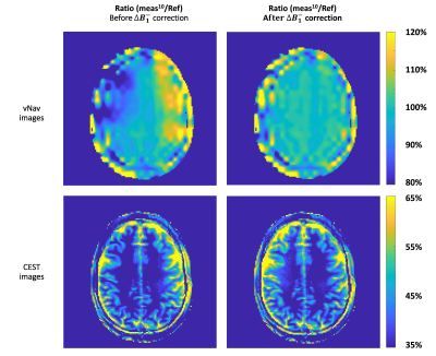 |
Real-time motion and retrospective receiver sensitivity correction for gluCEST using volumetric navigators (vNavs) at 7T
Esau Poblador Rodriguez1, Philipp Moser1, Sami Auno2, Korvinian Eckstein1, Stephan Gruber1, Benjamin Schmitt3, Andre van de Kouwe4, Siegfried Trattnig 1, and Wolfgang Bogner1
1High Field MR Centre, Biomedical Imaging and Image-guided Therapy, Medical University of Vienna, Vienna, Austria, 2Department of Physics, University of Helsinky, Helsinki, Finland, 3Siemens Healthineers, Sydney, Australia, 4Harvard Medical School, Boston, MA, United States
CEST is susceptible to subject motion not only due to misregistration of MR images acquired over time and subsequent alterations of B0 field, but also due to the related receiver sensitivity (B1-) inhomogeneities. We propose to correct for ∆B1- from the images generated by an interleaved EPI navigator used for prospective motion correction. We show the benefit of the proposed method, in combination with B1+ and dynamic B0 corrections, to restore a glutamate weighted CEST map in an active healthy volunteer.
|
|
3090. |
On the feasibility of a single scan B1+ correction scheme in combination with Multiple Interleaved Mode Saturation for quantitative CEST at 7 Tesla
Andrzej Liebert1, Katharina Tkotz1, Juergen Herrler2, Patrick Liebig3, Rene Gumbrecht3, Arnd Doerfler2, Frederik B. Laun1, Michael Uder1, Moritz Zaiss2,4, and Armin M. Nagel1,5
1Institute of Radiology, University Hospital Erlangen, Friedrich-Alexander-Universität Erlangen-Nürnberg, Erlangen, Germany, 2Department of Neuroradiology, University Hospital Erlangen, Friedrich-Alexander-Universität Erlangen-Nürnberg, Erlangen, Germany, 3Siemens Healthcare GmbH, Erlangen, Germany, 4Max Planck Institute for Biological Cybernetics, Tuebingen, Germany, 5Institute of Medical Physics, University Hospital Erlangen, Friedrich-Alexander-Universität Erlangen-Nürnberg, Erlangen, Germany
In Chemical Exchange Saturation Transfer MR both B1+-inhomogeneity correction and mitigation have their limitations, in particular if large field-of views shall be covered. To overcome these limitations a Multiple Interleaved Mode Saturation scheme was applied together with a linear B1+ inhomogeneity correction method. Repeatability and reproducibility of the MTRRex metric for rNOE and APT were investigated. A combination of MIMOSA and a simple single point correction allows achieving repeatable and reproducible CEST contrast in whole brain with an acquisition time of 4min 54s.
|
|
3091. |
Ultrafast 3D Steady-State Chemical Exchange Saturation Transfer (CEST) MRI with Incoherent Sampling at 7T
Sugil Kim1, HoonJae Lee2, and Seong-Gi Kim2,3
1Siemens healthineers Ltd, Seoul, Republic of Korea, 2Center for Neuroscience Imaging Research (CNIR), Institute for Basic Science (IBS), Suwon, Republic of Korea, 3. Department of Biomedical Engineering, Sungkyunkwan University, Suwon, Republic of Korea
CEST MRI exploits saturation transfer-induced proton exchange and its corresponding, indirect loss of water signal, has been shown to provide a novel contrast mechanism in MRI. However, CEST MRI is prone to B0 inhomogeneities. To resolve this problem, CEST MRI requires multiple-acquisition. To acquire z-spectrum of whole brain at ultrahigh field of 7T, there are multiple issues to be tackled; a prohibitively long acquisition time, potential high power deposition, and B0 drift during a long scanning time. In this work, we proposed the rapid steady-state CEST MRI pulse sequence incorporating with incoherent sampling in 3D segmented EPI at 7T
|
|
3092. |
Investigation of a parallel transmission (pTx) GRE-readout with customized pulses for CEST MRI at 7 Tesla
Katharina Tkotz1, Andrzej Liebert1, Jürgen Herrler2, Patrick Liebig3, Arnd Dörfler2, Michael Uder1, Moritz Zaiss2,4, and Armin M. Nagel1,5
1Institute of Radiology, University Hospital Erlangen, Friedrich-Alexander-Universität Erlangen-Nürnberg, Erlangen, Germany, 2Department of Neuroradiology, University Hospital Erlangen, Friedrich-Alexander-Universität Erlangen-Nürnberg, Erlangen, Germany, 3Siemens Healthcare GmbH, Erlangen, Germany, 4Max Planck Institute for Biological Cybernetics, Tuebingen, Germany, 5Institute of Medical Physics, Friedrich-Alexander-Universität Erlangen-Nürnberg, Erlangen, Germany
In Chemical Exchange Saturation Transfer MRI B1+-inhomogeneity influences both saturation and acquisition of the signal. Typically only a correction of the inhomogeneity of the CEST saturation is performed while the inhomogeneity of the readout is neglected. The influence of the readout was investigated in measurements using standard inhomogeneous 1Tx readout and a homogenized readout with customized pTx pulses. Compared to the 1Tx readout the readout with pTx pulses shows an increase of the homogeneity of the CEST contrast for CEST agents with low SNR like the amides and in regions with low 1Tx flip angle like the cerebellum.
|
|
3093. |
Protein-sensitive CEST-MRI of Alzheimer’s patients at 3 T
Steffen Goerke1, Johannes Breitling1, Katharina M. Kubera2, Moritz Zaiss3, Dusan Hirjak4, Robert C. Wolf2, Anoshirwan A. Tavakoli5, Heinz-Peter Schlemmer5, Daniel Paech5, Mark E. Ladd1, and Peter Bachert1
1Divsion of Medical Physics in Radiology, German Cancer Research Center (DKFZ), Heidelberg, Germany, 2Center for Psychosocial Medicine, Department of General Psychiatry, Heidelberg University, Heidelberg, Germany, 3Neuroradiology, University of Erlangen-Nürnberg, Erlangen, Germany, 4Departement of Psychiatry and Psychotherapy, Central Institute of Mental Health, Medical Faculty Mannheim, Heidelberg University, Mannheim, Germany, 5Departement of Radiology, German Cancer Research Center (DKFZ), Heidelberg, Germany
In this study, the potential of CEST-MRI as a tool for diagnostic imaging of Alzheimer’s patients was evaluated. Although the study size was yet too small to draw final conclusions, a significant decrease of the rNOE-CEST signal in Alzheimer’s patients was observed. This finding was in line with previous in vitro studies on the monitoring of protein aggregation using CEST-MRI. A diagnostic tool for Alzheimer’s based on CEST-MRI would allow avoiding invasive examinations and thus more frequent follow-ups allowing an improved level of patient care.
|
|
3094. |
Investigation of Optimizing Chemical Exchange Saturation Transfer Imaging for Tuberous Sclerosis Epilepsy at 3 Tesla
Qingqing Wen1, Kang Wang2, Yi-Cheng Hsu3, Yi Sun3, Dan Wu1,2, and Yi Zhang1,2
1Key Laboratory for Biomedical Engineering of Ministry of Education, Department of Biomedical Engineering, College of Biomedical Engineering & Instrument Science, Zhejiang University, Hangzhou, China, 2Department of Neurology, First Affiliated Hospital, College of Medicine, Zhejiang University, Hangzhou, China, 3Siemens Healthcare Ltd., Shanghai, China
Epilepsy is a prevalent neurological manifestation of Tuberous Sclerosis Complex (TSC). Here, the feasibility of applying Chemical Exchange Saturation Transfer (CEST) imaging to TSC epilepsy is investigated, with the radio-frequency saturation power, duration and frequency offsets optimized. The maximum CEST contrast between cortical tubers and normal white matter was achieved for an optimal saturation duration of 1000ms, while the optimal saturation frequency offset was related to the saturation power. Importantly, distinct contrast between tubers and normal white matter was demonstrated for TSC epilepsy for the first time, indicating CEST may serve as a potentially useful tool for diagnosing TSC.
|
|
3095. |
Frequency-stabilized chemical exchange saturation transfer imaging with free induction decay readout
Ruibin Liu1, Hongxi Zhang2, Yi-Cheng Hsu3, Caixia Fu4, Yi Sun3, Dan Wu1, and Yi Zhang1
1Key Laboratory for Biomedical Engineering of Ministry of Education, Department of Biomedical Engineering, College of Biomedical Engineering & Instrument Science, Zhejiang University, Hangzhou, Zhejiang, China, 2Department of Radiology, Children's Hospital, Zhejiang University School of Medicine, Hangzhou, Zhejiang, China, 3MR Collaboration, Siemens Healthcare Ltd., Shanghai, China, 4Siemens Shenzhen Magnetic Resonance Ltd., Shenzhen, China
CEST imaging is highly sensitive to temporal B0 drift, for which a frequency-stabilized CEST (FS-CEST) sequence was recently proposed by inserting a frequency stabilization module in front of the conventional non-frequency-stabilized CEST (NFS-CEST) sequence. Here, the frequency stabilization module in the FS-CEST sequence was further simplified by replacing the original gradient-echo readout with free induction decay (FID) readout. The proposed FS-CEST sequence with FID readout in the frequency stabilization module was validated in phantoms on a 3T Siemens Prisma scanner and in 15 volunteers on a 3T Philips Achieva scanner, both leading to improved CEST maps.
|
|
3096. |
Snapshot whole brain CEST MRI at 3T with 3D-EPI
Sebastian Mueller1, Rüdiger Stirnberg2, Suzan Akbey2, Philipp Ehses2, Klaus Scheffler1,3, Tony Stöcker2,4, and Moritz Zaiss1,5
1High-field Magnetic Resonance Center, Max Planck Institute for Biological Cybernetics, Tuebingen, Germany, 2German Center for Neurodegenerative Diseases (DZNE), Bonn, Germany, 3Department of Biomedical Magnetic Resonance, Eberhard Karls University Tuebingen, Tuebingen, Germany, 4Department of Physics and Astronomy, University of Bonn, Bonn, Germany, 5Department of Neuroradiology, University Hospital Erlangen, Erlangen, Germany
CEST MRI provides metabolite-based contrasts but often suffers from poor volume coverage or spatial resolution. We optimized and included a snapshot 3D-EPI readout and propose a suitable post-processing pipeline to generate CEST contrast in the whole brain at clinical B0=3T. It is shown that CEST MRI with 1.8mm isotropic nominal resolution at a field of view of 256x224x156mm³ is feasible within 4.3s per presaturation frequency offset. The approach is adaptable for any presaturation scheme. Exemplarily low power saturation was performed and fitted Lorentzian amplitudes gave a coefficient of variation <8.5% across three healthy subjects.
|
|
3097.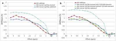 |
Glioma staging with CEST asymmetry curves and amides/amines ratio.
Laura Mancini1,2, Stefano Casagranda3, Francisco Torrealdea4, Marilena Rega4, Enrico De Vita1,5, Bruno Lopez3, Sebastian Brandner6,7, Benjamin Schmitt8, Patrick Liebig8, Eser Sanverdi1, Xavier Golay1,2, and Sotirios Bisdas1,2
1Lysholm Dept of Neuroradiology, National Hospital for Neurology & Neurosurgery, UCL Hospitals NHS Foundation Trust, London, United Kingdom, 2UCL Institute of Neurology, London, United Kingdom, 3Olea Medical, La Ciotat, France, 4University College of London Hospitals NHS Foundation Trust, London, United Kingdom, 5Biomedical Engineering Department, School of Biomedical Engineering and Imaging Sciences, King's College London, London, United Kingdom, 6Department of Neurodegenerative Disease, Institute of Neurology UCL, London, United Kingdom, 7National Hospital for Neurology & Neurosurgery, UCL Hospitals NHS Foundation Trust, London, United Kingdom, 8Siemens Healthcare Limited, Erlangen, Germany
Gliomas are the most common primary brain tumour, whose staging depends on the IDH and 1p/19q status and is reflected in different prognoses and clincal management. CEST is a highly sensitive MRI technique detecting amide- and amine-containing mobile proteins. The CEST amides/amines ratio has been proposed as a measure of pH in stroke. We show that CEST amides/amines ratio is much more sensitive than separate amides and amines CEST in differentiating gliomas with the best prognosis (IDH-mutant_1p/19q-retained) from those with the worst prognosis (IDH-wildtype). The different shapes in CEST asymmetry spectra could also potentially help in glioma staging.
|
|
3098.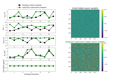 |
AutoCEST: a Machine-Learning Approach for Optimal CEST-MRI Experiment Design and Quantitative Mapping
Or Perlman1, Bo Zhu1,2, Moritz Zaiss3,4, Matthew S. Rosen1,2, and Christian T. Farrar1
1Athinoula A. Martinos Center for Biomedical Imaging, Massachusetts General Hospital and Harvard Medical School, Charlestown, MA, United States, 2Department of Physics, Harvard University, Cambridge, MA, United States, 3Magnetic Resonance Center, Max Planck Institute for Biological Cybernetics, Tübingen, Germany, 4Department of Neuroradiology, University Clinic Erlangen, Erlangen, Germany
The most common metric for CEST analysis is the magnetization-transfer-ratio asymmetry. Although qualitatively useful, it is affected by a mixed contribution from several exchange properties and requires experiment-specific protocol optimization. Herein, we propose a machine-learning framework for simultaneously tackling two challenging tasks: (1) automatic design of the optimal CEST acquisition schedule; (2) automatic extraction of fully quantitative CEST maps from the acquired data. The method was evaluated in simulations and phantoms at 4.7T. The resulting data acquisition and reconstruction times were 52 s and 36 ms respectively, providing quantitative exchange-rate and volume fraction maps with good agreement to ground-truth.
|
|
3099.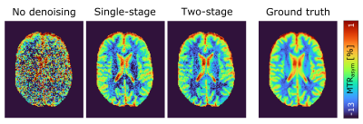 |
Two-stage denoising of CEST MRI data by principal component analysis of spectral groups
Johannes Breitling1, Steffen Goerke1, Mark E. Ladd1, Peter Bachert1, and Andreas Korzowski1
1German Cancer Research Center (DKFZ), Heidelberg, Germany
In this study a novel method for the denoising of CEST MRI data is presented, combining the formation of subsets of similar spectra and the subsequent application of a principal component analysis. Exploiting only the subtle spectral differences of these reduced datasets – as opposed to using all spectra for the analysis – allows for a better identification and isolation of the obscured underlying spectral features. The proposed denoising resulted in an SNR gain by approximately a factor of four compared to the noisy initial data and an additional 14% compared to the conventional principal component analysis denoising.
|
|
3100.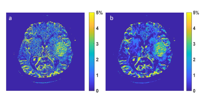 |
Deep Learning Reconstruction improves CEST MRI
Shu Zhang1, Xinzeng Wang2, F. William Schuler1, R. Marc Lebel3, Mitsuharu Miyoshi4, Ersin Bayram2, Elena Vinogradov5, Jason M. Johnson6, Jingfei Ma7, and Mark D. Pagel1
1Cancer Systems Imaging, The University of Texas MD Anderson Cancer Center, Houston, TX, United States, 2Global MR Applications & Workflow, GE Healthcare, Houston, TX, United States, 3Global MR Applications & Workflow, GE Healthcare, Calgary, AB, Canada, 4Global MR Applications & Workflow, GE Healthcare Japan, Tokyo, Japan, 5Radiology, UT Southwestern Medical Center, Dallas, TX, United States, 6Neuroradiology, The University of Texas MD Anderson Cancer Center, Houston, TX, United States, 7Imaging Physics, The University of Texas MD Anderson Cancer Center, Houston, TX, United States
Image reconstruction using deep learning (DL Recon) is capable of enhancing image signal-to-noise ratio (SNR) without losing image resolution or altering the image contrast. Our study demonstrates that CEST imaging and quantification, which are often limited by SNR and long scan time, can be improved with DL Recon. Our results clearly indicated that DL Recon can be used for CEST imaging with higher spatial resolution without or with only a mild increase in scan time or for CEST imaging in reduced scan time by using parallel imaging without the typical SNR penalty.
|
|
3101.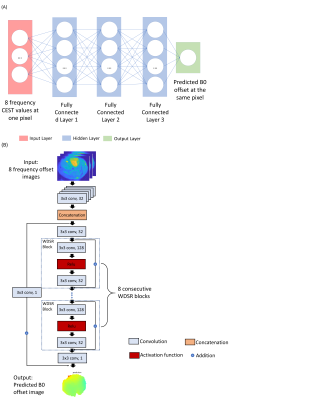 |
Deep learning-based B0 inhomogeneities mapping using sparse CEST spectral data
Yiran Li1, Danfeng Xie1, Hanlu Yang1, Li Bai1, Guanshu Liu2, and Ze Wang3
1Department of Electrical and Computer Engineering, Temple University, PHILADELPHIA, PA, United States, 2Russell H. Morgan Department of Radiology and Radiological Science, Johns Hopkins University School of Medicine, Baltimore, MD, United States, 3Department of Diagnostic Radiology and Nuclear Medicine, University of Maryland School of Medicine, Baltimore, MD, United States
Chemical Exchange Saturation Transfer (CEST) is an MR based imaging method that can image compounds containing protons exhibiting a suitable exchange rate with bulk water. One of the crucial technical hurdles in CEST MRI is, as CEST signal highly depends on the saturation frequency, how to accurately correct the B0 inhomogeneity in each voxel. We proposed two deep learning (DL) based methods for estimating B0 inhomogeneities to accelerate CEST imaging using spare samples. While only a small sample size was used, our study shows the potential of DL-based B0 mapping, which can greatly reduce the total CEST acquisition time.
|
 Watch the Video
 View the Poster
Session Topic: CEST, Magnetization Transfer and Relaxometry
Session Sub-Topic: CEST Friends & Family
Digital Poster
Contrast Mechanisms
3102. |
In vivo proton exchange rate MRI of stroke patients
Zhenxiong Wang1,2, Mehran Shaghaghi2, Yiran Zhou1, Wenzhen Zhu1, and Kejia Cai2
1Radiology, Tongji Hospital, Tongji Medical College, Huazhong University of Science and Technology, Wuhan, China, 2Departments of Radiology, Department of Bioengineering, and the Center for MR Research, University of Illinois at Chicago, Chicago, IL, USA, Chicago, IL, United States
In this study, we demonstrated that in vivo proton exchange rate MRI based on improved omega plot can serve as a novel and independent MRI contrast for assessing ischemic brain tissues of stroke patients.
|
|
3103.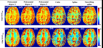 |
To Evaluate Effect of Interpolation Methods for B0 inhomogeneity correction on computation of CEST MRI Contrast in Human Brain at 3T and 7T
Anup Singh1,2, Ayan Debnath1,3, Rakesh Kumar Gupta4, and Ravinder Reddy3
1Centre for Biomedical Engineering, Indian Institute of Technology Delhi, New Delhi, India, 2Biomedical Engineering, AIIMS, New Delhi, India, 3Radiology, University of Pennsylvania, Philadelphia, PA, United States, 4Radiology, Fortis Memorial Research Institute, Gurugram, India
CEST MRI provides high resolution mapping of molecules such as Glutamate, Creatine, labile proteins/peptide, etc. CEST MRI contrast computation using asymmetry analysis require interpolation of data at different frequency offsets for B0 inhomogeneity corrections. In this study, different interpolation methods such as polynomial, cubic, spline and smoothingspline were compared for B0 inhomogeneity correction of various CEST contrasts(GluCEST-w, APT-w, CrCEST-w) at different field strength (3T, 7T). The 2nd and 3rd degree polynomial interpolations provided better B0 inhomogeneity correction for in vivo data from human brain. Polynomial interpolations for APT-w also improved differentiation of high-grade-glioma and low-grade-glioma tumors at 3T.
|
|
3104. |
Post-acquisition correction of the T1 relaxation effect for fast multi-slice CEST MRI
Tao Jin1
1University of Pittsburgh, Pittsburgh, PA, United States
Rapid multi-slice CEST MRI can be achieved by the immediate acquisition of multiple slices after a single irradiation pulse. However, the signals from the slices acquired after the 1st slice are contaminated by the T1-relaxation relaxation effect. In this work, we propose a simple post-acquisition correction method to compensate for the relaxation effect in the multi-slice CEST signals.
|
|
3105. |
Exchangeable Proton Spectroscopy using RACETE-FLEX
Fabian Tobias Gutjahr1,2, Simon Mayer2, and Peter M Jakob2
1Comprehensive Heart Failure Center, University Hospital Wuerzburg, Wuerzburg, Germany, 2Experimental Physics 5, University Wuerzburg, Wuerzburg, Germany
RACETE-FLEX is a combination of the novel RACETE-method and the spectroscopic FLEX method. RACETE is a method for imaging chemical exchange with positive contrast with concurrent water suppression. Combining the RACETE approach with the FLEX frequency labeling strategy leads to a high sensitivity exchangeable proton spectroscopy method.
|
|
3106.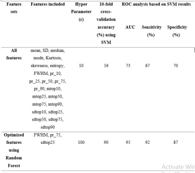 |
Differentiation between Intra-Cranial Mass Lesions using Machine Learning approach on Amide Proton Transfer-weighted (APT-w) CEST MRI
Ayan Debnath1, Manish Awasthi1, Neha Vats1, Rakesh Kumar Gupta2, and Anup Singh1,3
1Centre for Biomedical Engineering, Indian Institute of Technology Delhi, New Delhi, India, 2Department of Radiology, 2Fortis Memorial Research Institute, Gurgaon, Haryana, India, 3Department of Biomedical Engineering, All India Institute of Medical Science, New Delhi, India
It is challenging to differentiate between intra-cranial mass lesions (ICMLs) due to similar appearance using conventional MRIs. Amide-proton-transfer-weighted(APT-w) MRI provides differentiation among ICMLs with lower sensitivity ad specificity. The accuracy of classification between neo-plastic mass lesions and infective mass lesions as well as differentiation between low-grade-glioma and high-grade-glioma improves by implementing machine learning classifier based on features from APT-w CEST MRI. An optimized support-vector-machine with 10-fold cross-validation and optimal set of features extracted using Random forest based feature selection provided high accuracy of around 90%.
|
|
3107. |
Significant changes of APT signal in the Substantia Nigra and striatum of subacute PD mouse induced by MPTP
Quan Tao1, Peiwei Yi1, Zimeng Cai1, Yingjie Mei2, Zhifeng Chen1, Ruiyuan Liu1, Wufan Chen1, and Yanqiu Feng1
1School of Biomedical Engineering, Southern Medical University, Guangzhou, China, 2Philips healthcare, Guangzhou, China
Chemical exchange saturation transfer (CEST) MRI has been widely investigated for the early diagnosis of neurological diseases, such as brain tumor and neurodegenerative disorders. The goal of this study was to develop an imaging biomarker for the future intervention and treatment of PD in clinic. For such purpose, a novel radial-sampling steady-state CEST sequence based ultrashort echo time (UTE) readout was used to acquire the Z-spectrum in the mouse brain of subacute PD model induced by MPTP.
|
|
3108. |
Amide Proton Transfer imaging for differentiation of Tuberculomas from High-Grade Gliomas
Karthik Kulanthaivelu1, Sanita Raju2, Jitender Saini1, Atchayaram Nalini2, Nishanth Sadashiva3, Shashank Hegde4, Narayana Krishna Rolla5, and Indrajit Saha5
1Department of Neuroimaging and Interventional Radiology, National Institute of Mental Health and Neurosciences, Bengaluru, India, 2Department of Neurology, National Institute of Mental Health and Neurosciences, Bengaluru, India, 3Department of Neurosurgery, National Institute of Mental Health and Neurosciences, Bengaluru, India, 4Philips Healthcare, Bengaluru, India, 5Philips Health Systems, Philips India Ltd, Bengaluru, India
Amide proton transfer imaging was investigated for its potential to discriminate tuberculomas from high-grade gliomas. The diagnosis was confirmed by histopathology, CSF examination or response to anti-tubercular therapy. The MTRasym value of the Tuberculomas (mean 2.32± 0.50 s.d.) was significantly lower than high-grade gliomas (mean 4.32±0.84s.d.). Lower MTRasym values in tuberculomas are suggestive of relatively reduced mobile amide protons compared to the tumoral microenvironment. Perilesional elevated APT values in tuberculomas are a unique observation and may reflect a milieu of inflammation.
|
|
3109. |
Investigating the dependence of APT-CEST imaging in the human breast at 7 Tesla on the menstrual cycle
Lisa Loi1, Ferdinand Zimmermann2, Andreas Korzowski2, Jan-Eric Meissner2, Johannes Breitling2, Peter Bachert2, Mark Edward Ladd2, Heinz-Peter Schlemmer1, Sarah Schott3, Sebastian Bickelhaupt4, Steffen Goerke2, and Daniel Paech1
1Radiology, German Cancer Research Center, Heidelberg, Germany, 2Medical Physics in Radiology, German Cancer Research Center, Heidelberg, Germany, 3Gynecology, University Hospital Heidelberg, Heidelberg, Germany, 4German Cancer Research Center, Heidelberg, Germany
Compared to conventional dynamic contrast-enhanced MR-mammography, which is known to be significantly affected by the phase of the menstrual cycle, the menstrual cycle-related effect on APT CEST MRI in the human breast is still unknown. This is the first study investigating the influence of the menstrual cycle on APT CEST MRI in fibroglandular breast tissue of seven healthy premenopausal women. No significant signal intensity differences in fibroglandular breast tissue between the follicular and the luteal phase of the menstrual cycle were observed, which suggests that APT contrasts are comparable regardless of the phase of the menstrual cycle in premenopausal women.
|
|
3110. |
Relaxation-compensated APT CEST imaging in breast cancer diagnostics at 7T
Lisa Loi1, Ferdinand Zimmermann2, Andreas Korzowski2, Jan-Eric Meissner2, Peter Bachert2, Mark Edward Ladd2, Heinz-Peter Schlemmer1, Sebastian Bickelhaupt3, Steffen Goerke2, Sarah Schott4, and Daniel Paech1
1Radiology, German Cancer Research Center, Heidelberg, Germany, 2Medical Physics in Radiology, German Cancer Research Center, Heidelberg, Germany, 3German Cancer Research Center, Heidelberg, Germany, 4Gynecology, University Hospital Heidelberg, Heidelberg, Germany
Relaxation-compensated, fat-corrected and B1-corrected APT CEST MRI is a novel MR imaging technique that generates complementary information to conventional MR-mammography. In this study, the ability of this improved APT CEST metric was investigated in nine patients with newly diagnosed breast cancer. Compared to normal appearing fibroglandular breast tissue, significantly increased APT CEST signal intensities in breast cancer tissue were observed. Consequently, this MRI approach represents a contrast agent-free method that may enable a non-invasive differentiation of breast cancer and normal appearing breast tissue and, therefore, help to increase diagnostic accuracy in breast cancer imaging.
|
|
3111.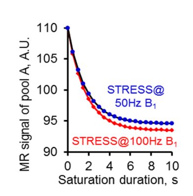 |
Induced Saturation Transfer Recovery Steady State (iSTRESS) for proton exchange rate imaging with MRI
Mehran Shaghaghi1 and Kejia Cai1,2,3
1Radiology, University of Illinois - Chicago, Chicago, IL, United States, 2Bioengineering, University of Illinois - Chicago, Chicago, IL, United States, 3Center for MR Research, University of Illinois - Chicago, Chicago, IL, United States
We present a novel method to induced saturation-transfer-recovery steady-states for determining proton exchange (kex) with low saturation power, short duration, and hence negligible SAR deposition. Our previous study has demonstrated that omega plotting with direct saturation (DS) signal removed can be used to map kex of healthy brains in vivo. However, omega plot, based on steady-state saturation assumption, requires long scanning time and potential high SAR deposition, which makes it non-applicable for clinical studies. Our suggested method, validated by simulations and phantom imaging, shows great promise for in vivo proton exchange rate imaging and clinical applications.
|
|
3112.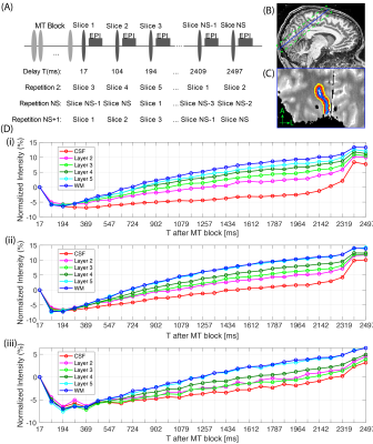 |
Layer-dependent Transient Saturation Transfer MRI at 7T
Catarina Rua1, Sanne Kaalund2, James B Rowe2,3, Christopher T Rodgers1, and Guy B Williams1
1Wolfson Brain Imaging Centre, Department of Clinical Neurosciences, University of Cambridge, Cambridge, United Kingdom, 2Department of Clinical Neurosciences, University of Cambridge, Cambridge, United Kingdom, 3Medical Research Council Cognition and Brain Sciences Unit, University of Cambridge, Cambridge, United Kingdom
The human cortical mantle has distinct non-uniform layers of gray matter. In this study we used ultra high-resolution MT-weighted imaging at 7T with varying mixing times to measure the transient effects of the magnetization suppression through the primary visual cortex (V1). The MT effect was monitored across different cortical depths to evaluate the effect of the macromolecular pool (MP) and myelination.
|
|
3113.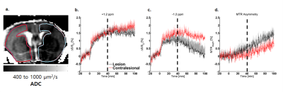 |
Disparity between Glucose Exchange and Osmolality in Pathological Tissue using Dynamic Glucose Enhanced Magnetic Resonance Imaging (DGE-MRI)
Julius Juhyun Chung1, Geun Ho Im2, Jung Hee Lee2,3, Tao Jin1, and Seong-Gi Kim2
1Radiology, University of Pittsburgh, Pittsburgh, PA, United States, 2Center for Neuroscience Imaging Research, Suwon, Republic of Korea, 3Radiology, Samsung Medical Center, Seoul, Republic of Korea
CEST has been used as a way to monitor glucose transport as a way of studying uptake as well as permeability. However, signal is not solely affected by exchange but also osmolality shifts with significant intravenous dosage. Using MCAO, we study disparity between these two effects and how they may lead to obfuscate signal sources. With the injection of glucose after the onset of ischemia, +1.2 ppm , -1.2 ppm, and MTR asymmetry curves behave quite differently. Both sides of the spectrum must be scrutinized to have a better picture of what is going on during glucose dosage.
|
|
3114.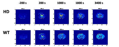 |
Glucose Metabolism by Dynamical Glucose Enhanced imaging
Yu-Wen Chen1 and Dennis Wenhan Hwang2
1, IBMS N123, Academia Sinica, Taipei, Taiwan, 2IBMS, Academia Sinica, Taipei, Taiwan
Dynamic glucose-enhanced (DGE) images, detected by CEST, was used to provide direct information on glucose metabolism. It also provides the spatial distribution of the metabolic processes. The time-dependent glucose concentration variation in the striatum of HD shows that the glucose uptake in the HD mice striatum decreases with age. The groups of older than 9-11 weeks HD mice were easy to find the symptoms of HD, and their glucose uptaking also show a significant decrement. By contrast, WT mice show a mild decrease in the glucose uptake for the mice with similar age.
|
|
3115. |
Dynamic glucose-enhanced (DGE) MRI for brain tumors: a small-scale clinical observational study
Jianhua Mo1, Xiang Xu2,3, Xianglong Wang1, Linda Knutsson2,4, Akansha A Sehgal2,3, Peter C. M. van Zijl2,3, and Zhibo Wen1
1Zhujiang Hospital, Southern Medical University, Guangzhou, China, 2Russell H. Morgan Department of Radiology and Radiological Science, Johns Hopkins University School of Medicine, Baltimore, MD, United States, 3F.M. Kirby Research Center for Functional Brain Imaging, Kennedy Krieger Research Institute, Baltimore, MD, United States, 4Department of Medical Radiation Physics, Lund University, Lund, Sweden
Dynamic glucose-enhanced (DGE) MRI has shown potential for imaging glucose delivery and blood brain barrier permeability. Previous reports focused on the technical development of DGE and only a few clinical brain tumor cases have been reported. Here we incorporated the current DGE protocol into our routine MRI examination at 3T and performed a small-scale clinical observational study on patients with different types of brain tumors. Despite the technical challenges of DGE at clinical scanners, we observed different (non)-enhancement patterns with various brain tumor types.
|
Session Topic: CEST, Magnetization Transfer and Relaxometry
Session Sub-Topic: Diet CEST (No Glucose)
Digital Poster
Contrast Mechanisms
3116. |
Repeatability of Creatine Recovery Constants in Exercise Muscle Measured using 3D Creatine Chemical Exchange Saturation Transfer Imaging at 7T
Dushyant Kumar1, Ravi Prakash Reddy Nanga1, Deepa Thakuri1, Abigail Cember1, Neil Wilson2, Hari Hariharan1, and Ravinder Reddy1
1Radiology, University of Pennsylvania, Philadelphia, PA, United States, 2Siemens Medical Solutions USA Inc, Malvern, PA, United States
Creatine CEST is a relatively new imaging technique, with the proven potential to assess the systemic energy deficiency in form of delayed creatine recovery in exercised skeletal muscles. However, this 2D method still suffers from limited volume coverage in slice encoding direction. Since the distribution of disease in many musculoskeletal disorders may vary across the muscle, there is a need to increase coverage in slice encoding direction. Towards this goal, we demonstrate the feasibility of 3D CrCEST, while still maintaining 30s time resolution necessary to capture underlying dynamics and also demonstrate its repeatability using data from five healthy volunteers.
|
|
3117. |
Accelerated 3D chemical exchange saturation transfer imaging using compressed SENSE for full Z-spectrum acquisition
Tatsuhiro Wada1, Chiaki Tokunaga1, Osamu Togao2, Masami Yoneyama3, Yasuo Yamashita1, Kouji Kobayashi1, Toyoyuki Kato1, and Hidetake Yabuuchi4
1Division of Radiology, Department of Medical Technology, Kyushu University Hospital, Fukuoka, Japan, 2Department of Clinical Radiology, Graduate School of Medical Sciences, Kyushu University, Fukuoka, Japan, 3Philips Japan, Fukuoka, Japan, 4Department of Health Sciences, Faculty of Medical Sciences, Kyushu University, Fukuoka, Japan Poster Permission Withheld
Multi-slice chemical exchange saturation transfer (CEST) imaging is difficult to use for clinical studies because data acquisition of the full z-spectrum is time-consuming. To accelerate the scan time for obtaining multi-slice CEST imaging, we applied the compressed sensing (CS) and sensitivity encoding (SENSE) technique (CS-SENSE) to 3D CEST imaging. The 3D CEST imaging combined with CS-SENSE was obtained without reducing the image contrast of the 2D CEST imaging. Moreover, 10 slice CEST images could be acquired in approximately 7 minutes including B0 map. 3D CEST imaging combined with CS-SENSE was shown to be useful for clinical study.
|
|
3118. |
A Steady State Approach to Positive Contrast Chemical Exchange Imaging using RACETE
Fabian Tobias Gutjahr1,2, Simon Mayer2, and Peter M Jakob2
1Comprehensive Heart Failure Center, University Hospital Wuerzburg, Wuerzburg, Germany, 2Experimental Physics 5, University Wuerzburg, Wuerzburg, Germany
RACETE is a novel method for positive contrast imaging of chemical exchange. In this work the RACETE sequence is extended to allow steady state imaging by interleaving the preparation with small flip angle RACETE read-outs. It can be shown that the efficiency of the steady state RACETE is improved over the equilibrium RACETE. This is an important step towards speeding up the acquisition process to make RACETE a viable option for future in vivo experiments.
|
|
3119.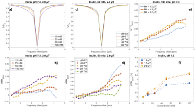 |
Inulin as a biodegradable contrast agent for CEST MRI
Anina Seidemo1, Malte Knutsson2, Patrick M. Lehmann1, Pia C. Sundgren3,4, Anthony Aletras5,6, Peter C.M. van Zijl7,8, and Linda Knutsson1,7
1Department of Medical Radiation Physics, Lund University, Lund, Sweden, 2Procivitas, Malmö, Sweden, 3Department of Diagnostic Radiology, Lund University, Lund, Sweden, 4Lund University Bioimaging Center, Lund University, Lund, Sweden, 5Department of Clinical Physiology, Clinical Sciences, Lund University and Lund University Hospital, Lund, Sweden, 6Laboratory of Computing and Medical Informatics, School of Medicine, Aristotle University of Thessaloniki, Thessaloniki, Greece, 7Russell H. Morgan Department of Radiology and Radiological Science, Johns Hopkins University School of Medicine, Baltimore, MD, United States, 8F.M. Kirby Research Center for Functional Brain Imaging, Kennedy Krieger Institute, Baltimore, MD, United States
GlucoCEST (chemical exchange saturation transfer imaging using glucose as a contrast agent) has shown potential in tumor imaging. However, glucose enters cells and is rapidly metabolized, leading to disappearance of the glucoCEST signal over time. Inulin, a polysaccharide, is non-toxic and acts like an intravascular tracer when injected intravenously. A phantom including both glucose and inulin at different pH and concentrations was scanned at 3T to investigate the potential of inulin as a CEST agent. This study indicates that inulin shows CEST contrast comparable to glucose on a per-OH-unit basis and has potential as a biodegradable CEST agent.
|
|
3120.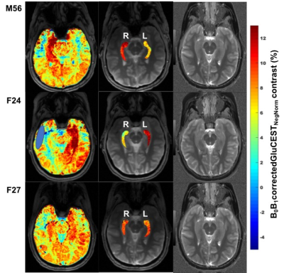 |
Application of GluCEST in monitoring abnormal glutamate dehydrogenase activity in Hyperinsulinism/Hyperammonemia (HI/HA) syndrome at 7.0T
Ravi Prakash Reddy Nanga1, Elizabeth A Rosenfeld2, Deepa Thakuri1, Mark Elliott1, Ravinder Reddy1, and Diva D De Leon2
1Radiology, Perelman School of Medicine at The University of Pennsylvania, Philadelphia, PA, United States, 2Division of Endocrinology and Diabetes, Children's Hospital of Philadelphia, Philadelphia, PA, United States
Hyperinsulinism/Hyperammonemia (HI/HA) syndrome is an orphan disease characterized by fasting and protein-induced hypoglycemia, hyperammonemia, and has high prevalence of epilepsy, developmental delays, and learning disabilities. Understanding the mechanism involved in brain phenotype remains limited. Glutamate weighted chemical exchange saturation transfer (GluCEST) imaging was used to spatially map the glutamate levels of hippocampus. We observed a higher GluCEST contrast in the hippocampus of some of these subjects following a unilateral pattern. This preliminary study demonstrates for the first time the application of GluCEST MRI for studying the abnormal function of glutamate dehydrogenase (GDH) enzyme activity in human subjects with HI/HA syndrome.
|
|
3121. |
Optimized CEST acquisition and analysis for treatment assessment of response to neoadjuvant chemotherapy in triple negative breast cancer
Shu Zhang1, Abeer H. Abdelhafez2, Jong Bum Son3, Benjamin C. Musall3, Mitsuharu Miyoshi4, Xinzeng Wang5, Ken-Pin Hwang3, Gaiane M. Rauch2, Jingfei Ma3, and Mark D. Pagel1
1Cancer Systems Imaging, The University of Texas MD Anderson Cancer Center, Houston, TX, United States, 2Abdominal Imaging, The University of Texas MD Anderson Cancer Center, Houston, TX, United States, 3Imaging Physics, The University of Texas MD Anderson Cancer Center, Houston, TX, United States, 4Global MR Applications & Workflow, GE Healthcare Japan, Tokyo, Japan, 5Global MR Applications & Workflow, GE Healthcare, Houston, TX, United States
In our ongoing study of 13 completed patients, we compared two saturation power levels (2.0 μT vs. 0.9 μT) and two analysis methods (MTRasym vs. Lorentzian line fitting) of CEST for assessing treatment response to neoadjuvant chemotherapy of triple-negative breast cancer (TNBC). A consistently decreasing trend of the CEST signals was observed with the longitudinal treatment when a higher saturation power of 2.0 μT was used with the amide MTRasym analysis method. In contrast, the same trend was observed when a lower saturation power of 0.9 μT was used for the Lorentzian line fitting analysis method.
|
|
3122.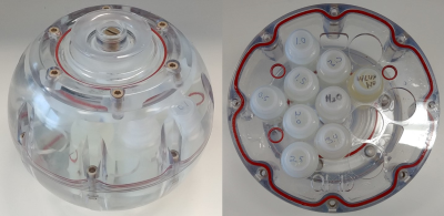 |
Development of a standard phantom for Chemical Exchange Saturation Transfer MRI
Shu Zhang1, F. William Schuler1, Tianzhe Li1, Ken-Pin Hwang2, Mitsuharu Miyoshi3, Xinzeng Wang4, and Mark D. Pagel1
1Cancer Systems Imaging, The University of Texas MD Anderson Cancer Center, Houston, TX, United States, 2Imaging Physics, The University of Texas MD Anderson Cancer Center, Houston, TX, United States, 3Global MR Applications & Workflow, GE Healthcare Japan, Tokyo, Japan, 4Global MR Applications & Workflow, GE Healthcare, Houston, TX, United States
Many clinical CEST MRI methods have been developed and disseminated during the last few years, and the expansion of clinical CEST MRI will likely continue. A common phantom is needed to standardize the development and implementation of CEST methods. In this study, we have developed a clinical CEST MRI phantom with high-quality gelatin to test a range of concentrations, pH values, and T1 relaxation times. The experimental conditions of the CEST saturation and various post-saturation acquisition methods can be tested. This standard phantom can be used to support many applications within the CEST MRI research community.
|
|
3123.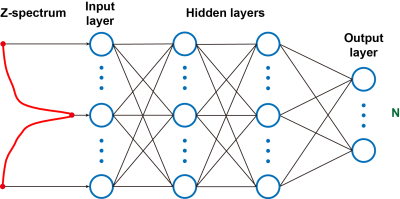 |
Alzheimer’s Disease identification by an artificial neutral network based on chemical exchange saturation transfer (ANNCEST) MRI
Joseph H. C. Lai1, Jianpan Huang1, Xiongqi Han1, Jiadi Xu2,3, and Kannie W. Y. Chan1,2
1Department of Biomedical Engineering, City University of Hong Kong, Hong Kong, Hong Kong, 2Russell H. Morgan Department of Radiology and Radiological Science, Johns Hopkins University School of Medicine, Baltimore, MD, United States, 3F.M. Kirby Research Center for Functional Brain Imaging, Kennedy Krieger Research Institute, Baltimore, MD, United States
There is an urgent need to develop an efficient and noninvasive methods to diagnose AD at an early stage. Artificial neural network (ANN) is a powerful model for prediction and classification of diseases, thus, it has been applied to facilitate prognosis and diagnosis. We propose to apply ANN based on chemical exchange saturation transfer (CEST) MRI at 3T to detect AD. Our phantom and AD mouse results showed that the trained ANN was able to identify AD from age-matched wild-type (WT) mice with high accuracy, which could provide valuable information for AD diagnosis.
|
|
3124. |
The perfusion contributing significantly to the CEST signal acquired by a UTE-CEST sequence
Quan Tao1, Peiwei Yi1, Zimeng Cai1, Yingjie Mei2, Zhifeng Chen1, Ruiyuan Liu1, Wufan Chen1, and Yanqiu Feng1
1School of Biomedical Engineering, Southern Medical University, Guangzhou, China, 2Philips healthcare, Guangzhou, China
Chemical exchange saturation transfer (CEST) as a novel molecule MRI technique, was approved to detect some diseases, like tumor grading, kidney injure, osteoarthritis (OA) and etc. However, it is crucial to purify the CEST signal. In this study, we explored the perfusion contribution to the CEST signal acquired by a UTE-CEST sequence.
|
|
3125.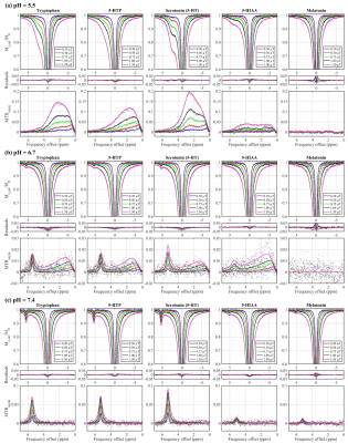 |
In Vitro Characterization of Serotonin Biosynthesis Pathway by CEST MRI
Ryan T Oglesby1,2, Wilfred W Lam2, and Greg J Stanisz1,2
1Medical Biophysics, University of Toronto, Toronto, ON, Canada, 2Physical Sciences, Sunnybrook Research Institute, Toronto, ON, Canada
This study demonstrates the in vitro characterization of tryptophan, 5-HTP, serotonin (5-HT), 5-HIAA (all members of the serotonin biosynthesis pathway), and melatonin at precise pH, temperature, and concentration. At pH 5.5, CEST contrast between 0.6 and 1.9 ppm, originating from the NH3+ side chain, is exhibited by tryptophan, 5-HTP, and 5-HT. All five molecules at pH 7.4 exhibit CEST contrast between 5.11 and 5.47 ppm, originating from the NH proton on the indole ring. If sensitive enough in vivo, these measurements could improve the objectivity of clinically diagnosed psychiatric disorders and could provide a new biological understanding of serotonergic dysfunction.
|
|
3126.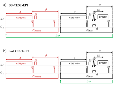 |
Fast 2D EPI multislice imaging for full APT-CEST brain coverage
Jan-Rüdiger Schüre1, Manoj Shrestha2, Eike Steidl3, Ralf Deichmann2, Elke Hattingen3, Marlies Wagner3, and Ulrich Pilatus3
1Neuroradiology, Goethe University Hospital, Frankfurt am Main, Germany, 2Brain Imaging Center (BIC), Frankfurt am Main, Germany, 3Goethe University Hospital, Frankfurt am Main, Germany
We present a fast 2D EPI multislice sequence that allows to acquire the APT-CEST contrast in 16 slices within 8 seconds. The fast CEST-EPI sequence was compared in vitro and in vivo with a steady-state CEST sequence, where the saturation is applied for 4 s for each frequency offset. The reduced acquisition time in the fast sequence can be used for measurement repetitions or additional MR examinations (e.g. MR spectroscopic imaging).
|
|
3127.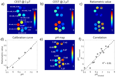 |
Investigation of the reliability in pH quantification with MR chemical exchange saturation transfer (CEST) power ratiometric imaging at 3 Tesla
Jie Liu1, Zongwei Xu1, Qi Liu2, Hui Liu2, Jian Xu2, Xin Liu1, Hairong Zheng1, and Yin Wu1
1Paul C. Lauterbur Research Center for Biomedical Imaging, Shenzhen Institutes of Advanced Technology, Chinese Academy of Sciences, Shenzhen, China, 2United Imaging Healthcare America, Houston, TX, United States
Chemical exchange saturation transfer (CEST) using iodinated agents has been tested for extracellular pH imaging on high-field animal scanners. However, the reliability in the pH quantification on low magnetic strengths remains elucidated. In this study, CEST imaging was performed on iobitridol phantom twice with a three-day interval at 3T. pH was quantified by ratioing iobitridol CEST effects at 5.6 ppm under B1 of 1.0 and 2.0 μT. Results show accurate pH quantification in both scans. Good consistency of paired measures was observed between the two scans, suggesting the reliability of the method and its potential clinical applicability at 3T.
|
|
3128.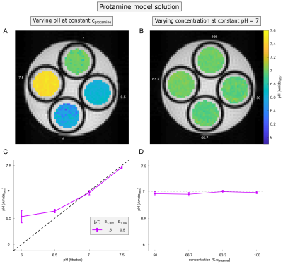 |
Concentration-independent absolute pH mapping using amide CEST-MRI at 9.4 T
Philip S. Boyd1, Johannes Breitling1, Mark E. Ladd1, Peter Bachert1, and Steffen Goerke1
1Division of Medical Physics in Radiology, German Cancer Research Center (DKFZ), Heidelberg, Germany
In this study, we developed an absolute pH mapping method based on endogenous amide CEST-MRI which simultaneously compensates for concentration changes, the semi-solid magnetization transfer, and spillover dilution. This was realized by a ratiometric approach of two different B1 in combination with the inverse metric and polynomial Lorentzian-fitting of the amide signal. Compensation for concomitant effects was theoretically demonstrated in simulations and verified experimentally in protein model solutions and porcine brain lysates. Consequently, amide signal-based absolute pH mapping is now in principle also reliably applicable for tumor imaging which was previously prevented by the concomitant effects.
|
|
3129. |
Turnkey qMT via Selective Inversion Recovery
Richard Dortch1
1Division of Neuroimaging Research, Barrow Neurological Institute, Phoenix, AZ, United States
MTR offers a measure of myelin content, but is also sensitive to non-physiological parameters. Quantitative MT methods remove these confounding effects; however, they require long scan times and complicated acquisition/analysis strategies. Selective inversion recovery is a simple method that may address many of these limitations, but requires long scan times and specialized inversion recovery sequences. The issue of long scan times was previously addressed through optimized sampling schemes. The need for specialized sequences is addressed herein by using a standard MP-RAGE sequence and modifying the SIR signal model. The resulting “turnkey” approach was tested numerically and applied in the brain.
|
|
3130.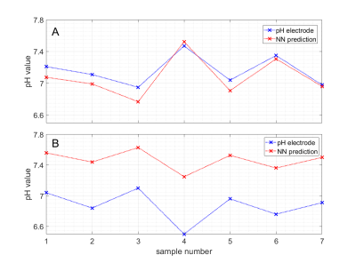 |
pH mapping of brain tissue by a deep neural network trained on 9.4T CEST MRI data – pH-deepCEST
Sebastian Mueller1, Felix Glang1, Klaus Scheffler1,2, and Moritz Zaiss1,3
1High-field Magnetic Resonance Center, Max Planck Institute for Biological Cybernetics, Tuebingen, Germany, 2Department of Biomedical Magnetic Resonance, Eberhard Karls University Tuebingen, Tuebingen, Germany, 3Department of Neuroradiology, University Hospital Erlangen, Erlangen, Germany
The pH value is of major importance for most physiological processes and may change due to altered metabolism in pathologies. In the present work, we exploit the inherent dependency of CEST MR data on pH with a new approach: train neural networks to map voxel-by-voxel from multi-B1+ CEST spectra to pH value. Measurements were performed in homogenate of pig brain tissue at 9.4T ultra high field. Prediction of absolute pH values was possible and predictions were stable against inhomogeneity in B1+. We hope this proof of concept might be a first small step towards high-resolution 3D pH maps in vivo.
|
|
3131.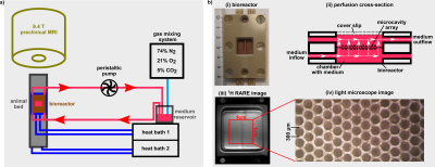 |
The cellular heat shock response monitored by chemical exchange saturation transfer MRI
Dennis Kleimaier1, Steffen Goerke2, Cordula Nies3, Moritz Zaiss4, Patrick Kunz5, Eric Gottwald3, and Lothar R. Schad1
1Computer Assisted Clinical Medicine, Heidelberg University, Mannheim, Germany, 2Division of Medical Physics in Radiology, German Cancer Research Center (DKFZ), Heidelberg, Germany, 3Institute of Functional Interfaces, Karlsruhe Institut of Technology, Karlsruhe, Germany, 4Neuroradiology, University of Erlangen-Nürnberg, Erlangen, Germany, 5Division of Functional Genome Analysis, German Cancer Research Center (DKFZ), Heidelberg, Germany
Chemical exchange saturation transfer of the relayed nuclear Overhauser effect(rNOE) enables detection of mobile protein protons via their exchange with the water signal. Recent studies have shown a close relationship between the rNOE-signal and the protein conformation. This study used the -3.5ppm rNOE-signal to monitor the heat shock response of HepG2 cells in a microcavity array-based bioreactor system. A significant drop of the rNOE-signal to 92.4±1.3% and a slow, steady increase of the rNOE-signal for 136.1±13.6min after heat shock were observed. Therefore, the rNOE-signal in CEST spectroscopic imaging is a sensitive readout for the cellular heat shock response.
|
Session Topic: CEST, Magnetization Transfer and Relaxometry
Session Sub-Topic: Magnetization Transfer Imaging
Digital Poster
Contrast Mechanisms
3132. |
qMTNet: Accelerated Quantitative Magnetization Transfer Imaging with Neural Networks
Huan Minh Luu1, Dong-Hyun Kim1, Jae-Woong Kim1, Seung-Hong Choi2, and Sung-Hong Park1
1Bio and Brain Engineering, Korea Advanced Institute of Science and Technology, Daejeon, Republic of Korea, 2Department of Radiology, Seoul National University Hospital, Seoul, Republic of Korea
Quantitative magnetization transfer (qMT) imaging overcomes the drawbacks of traditional MT imaging by producing more quantitative parameters. However, data acquisition and processing can be time-consuming, which limits its usage. In this study, an artificial neural network, qMTNet, is proposed to accelerate both the acquisition and fitting of qMT data. For data acquired from both conventional and inter-slice acquisition strategies, our approach demonstrated consistent fitting results with those from a previous dictionary-driven fitting method. The network reduces the time for both data acquisition and qMT fitting by a factor of 3 and 5000 times, respectively, compared to the conventional methods.
|
|
3133. |
One Minute Whole-Brain Magnetization Transfer Ratio Imaging with Intrinsic B1-Correction
Roya Afshari1,2, Francesco Santini1,2, Rahel Heule3, Craig H. Meyer4, Josef Pfeuffer5, and Oliver Bieri1,2
1Division of Radiological Physics,Department of Radiology, University Hospital Basel, University of Basel, Basel, Switzerland, 2Department of Biomedical Engineering, University of Basel, Basel, Switzerland, 3High Field Magnetic Resonance, Max Planck Institute for Biological Cybernetics, Tübingen, Germany, 4Department of Biomedical Engineering, University of Virginia, Charlottesville, VA, United States, 5Siemens Healthcare, Application Development, Erlangen, Germany Poster Permission Withheld
Magnetization transfer (MT), reflecting the exchange of magnetization between mobile and bound protons, has shown good potential for the diagnosis and prognosis of various neurological disorders, such as multiple sclerosis. Frequently, MT effects are assessed by measuring the contrast between two scans performed with and without saturation of the bound pool protons. Evidently, saturation is affected by B1 inhomogeneity and should be accounted for. In this work, we report on a very rapid one-minute whole-brain magnetization transfer ratio (MTR) imaging method offering intrinsic B1-correction.
|
|
3134. |
Enhancing Magnetization Transfer Contrast (MTC): A proof-of-concept study of MTC-STAGE Imaging
Jun Chen Li1,2, Yu Liu1, Yongsheng Chen3, Zhijia Jin1, Naying He1, Weibo Chen4, Fuhua Yan1, and Ewart Mark Haacke1,5,6
1Radiology, Ruijin Hospital, Shanghai, China, 2Radiology, Changshu Hospital Affiliated to Nanjing University of Chinese Medicine, Changshu, China, 3Neurology, Wayne State University, Detroit, MI, United States, 4Philips Healthcare, Shanghai, China, 5Radiology, Wayne State University, Detroit, MI, United States, 6Biomedical Engineering, Wayne State University, Detroit, MI, United States
Magnetization transfer contrast (MTC) imaging has been used to study neuromelanin (NM) in Parkinson’s disease. By suppressing the background tissue using an MTC pulse in a T1W sequence, the NM becomes visible, supposedly because of its reduced T1. However, we show using STAGE (strategically acquired gradient echo) imaging with/without an MTC pulse that this is not the reason for its visibility. Rather, it is the increased water content relative to surrounding tissue that keeps the signal high. Using the appropriate choice of flip angles and resolution, the NM contrast on MTC images can be significantly increased.
|
|
3135. |
Quantitative magnetization transfer (MT) of tissue from subjects with multiple sclerosis using inhomogeneous MT (ihMT) data
Gopal Varma1, Hemant Varma2, Cody Callahan1, Olivier M Girard3, Guillaume Duhamel3, Aaron K Grant1, and David C Alsop1
1Division of MR Research, Radiology, Beth Israel Deaconess Medical Center, Harvard Medical School, Boston, MA, United States, 2Department of Pathology, Beth Israel Deaconess Medical Center, Harvard Medical School, Boston, MA, United States, 3CNRS, CRMBM, Aix-Marseille Univ, Marseille, France
Quantitative magnetization transfer (MT) was carried out using inhomogeneous MT (ihMT) data, following optimization of an acquisition protocol. The optimized protocol was applied in ex-vivo brain tissue from a donor with multiple sclerosis (MS), as well as tissue containing an MS plaque. Parameter maps output from quantitative MT showed white and grey matter contrast, as well as a lower restricted or bound pool fraction in the area of the MS plaque from histology. Results from quantitative MT using ihMT were compared with those using selective inversion recovery, as well as quantitative outputs from diffusion tensor MRI.
|
|
3136.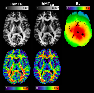 |
Inhomogeneous magnetization transfer saturation (ihMTsat): efficient centric-encoded GRE implementation with B1 inhomogeneity correction
Christopher D Rowley1,2, Zhe Wu2,3, Ilana R. Leppert2, Jennifer S.W. Campbell 2, David A. Rudko2,4,5, G. Bruce Pike6, and Christine L. Tardif1,2,4
1Neurology and Neurosurgery, McGill University, Montreal, QC, Canada, 2McConnell Brain Imaging Center, McGill Unversity, Montreal, QC, Canada, 3Techna Institute, University Health Network, Toronto, ON, Canada, 4Department of Biomedical Engineering, McGill Unversity, Montreal, QC, Canada, 5Department of Neurology and Neurosurgery, McGill Unversity, Montreal, QC, Canada, 6Hotchkiss Brain Institute and Departments of Radiology and Clinical Neuroscience, University of Calgary, Calgary, AB, Canada
Inhomogeneous magnetization transfer (ihMT) contrast in the brain has been reported to be a myelin-specific biomarker, but can be impacted by B1 inhomogeneities, reducing its accuracy. ihMT equations incorporating B1 correction assume a single excitation and readout, either a k-space line or plane, per saturation module. Here we use an arbitrary number of readout segments collected after an MT saturation preparation module. T1 and B1 variations are included in the signal equations to increase specificity of the contrast to the microstructure. The resulting implementation yields a balance between acquisition efficiency and contrast resolution for different brain imaging applications.
|
|
3137.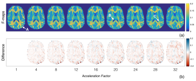 |
Accelerated quantitative magnetization transfer (qMT) imaging using compressed sensing and parallel imaging
Melany McLean1, R. Marc Lebel1,2, M. Ethan MacDonald1, Mathieu Boudreau3,4, and G. Bruce Pike1
1Departments of Radiology and Clinical Neuroscience, Hotchkiss Brain Institute, University of Calgary, Calgary, AB, Canada, 2GE Healthcare, Calgary, AB, Canada, 3Montreal Heart Institute, Université de Montreal, Montreal, QC, Canada, 4NeuroPoly Lab, Institute of Biomedical Engineering, Polytechnique Montreal, Montreal, QC, Canada
Quantitative magnetization transfer (qMT) is a Z-spectrum based imaging technique used to study white matter. The need to acquire many images with unique RF saturation pulses leads to long acquisition times. We aim to shorten qMT imaging times using a sparseSENSE technique that combines parallel imaging and compressed sensing to reduce the amount of acquired data. Retrospectively undersampled data was reconstructed for a range of acceleration factors using wavelet and total variation sparsifying domains. Pool size ratio (F) maps were accelerated by a factor of 4×, and acceleration factors of 8-12× may be possible in future work.
|
|
3138.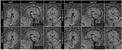 |
High-resolution magnetization transfer ratio maps using spiral-phyllotaxis Cartesian FLASH and compressed sensing in under five minutes
Gabriele Bonanno1,2,3, Tom Hilbert4,5,6, Arun Joseph1,2,3, Emilie Mussard4,5,6, Christoph Forman7, Gian Franco Piredda4,5,6, and Tobias Kober4,5,6
1Advanced Clinical Imaging Technology, Siemens Healthcare AG, Bern, Switzerland, 2Translational Imaging Center, sitem-insel AG, Bern, Switzerland, 3Departments of Radiology and Biomedical Research, University of Berne, Bern, Switzerland, 4Advanced Clinical Imaging Technology, Siemens Healthcare AG, Lausanne, Switzerland, 5Department of Radiology, University Hospital (CHUV) and University of Lausanne (UNIL), Lausanne, Switzerland, 6LTS5, École Polytechnique Fédérale de Lausanne, Lausanne, Switzerland, 7Magnetic Resonance, Siemens Healthcare GmbH, Erlangen, Germany
Magnetization Transfer Ratio (MTR) imaging may be a valuable tool for the diagnosis and follow-up of demyelinating diseases. However, MTR maps require long scan times for whole-brain coverage and high isotropic resolution. We present a novel MTR imaging method based on a spiral-phyllotaxis Cartesian FLASH sequence and compressed sensing, called MTRSparse, and compare it to fully sampled and parallel-imaging-accelerated acquisitions in healthy volunteers. MTRSparse showed good contrast and similar MTR values in comparison to the reference method with up to 87% reduction in acquisition time. This can help facilitate implementation of MTR imaging in the clinical practice.
|
|
3139.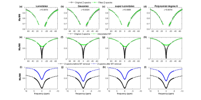 |
Estimation of Glutamate-weighted CEST Contrast after Removal of Magnetization Transfer Effect in Human Brain and Rat Brain with Tumor
Ayan Debnath1,2, Hari Hariharan2, Ravi Prakash Reddy Nanga2, Ravinder Reddy2, and Anup Singh1,3
1Centre for Biomedical Engineering, Indian Institute of Technology Delhi, New Delhi, India, 2Center for Magnetic Resonance & Optical Imaging, University of Pennsylvania, Philadelphia, PA, United States, 3Department of Biomedical Engineering, All India Institute of Medical Science, New Delhi, India
Asymmetry in MT causes erroneous GluCEST computation. The study focuses on removal of MT effects from Z-spectra for better quantification of GluCEST contrast using in-vivo human volunteer at 7T and rat brain with tumor at 9.4T. Different lineshapes for modelling and fitting broad MT spectrum has been compared. Lorentzian lineshape provided significant difference between GluCEST contrast before and after MT removal and also preserves the gray-matter to white-matter glutamate concentration ratio validated with MR spectroscopic based estimation. After removal of MT effect using Lorentzian lineshape, the specificity of GluCEST to glutamate concentration increases which can helps better diagnosis of diseases.
|
|
3140. |
Variable-density Fast-Spin-Echo (FSE) for volumetric inhomogeneous Magnetization Transfer (ihMT) imaging
Manuel Taso1, Fanny Munsch1, Arnaud Guidon2, Olivier M. Girard3, Guillaume Duhamel3, David C. Alsop1, and Gopal Varma1
1Division of MRI research, Department of Radiology, Beth Israel Deaconess Medical Center, Harvard Medical School, Boston, MA, United States, 2Global MR Applications and Workflow, GE Healthcare, Boston, MA, United States, 3CRMBM, Aix-Marseille Univ, CNRS, Marseille, France
While the original inhomogeneous magnetization transfer (ihMT) implementations for myelin imaging relied heavily on single-slice imaging, recent developments have enabled volumetric acquisitions using rapid gradient-echo sequences. But in vivo volumetric spin-echo acquisitions have been unexplored so far although they provide a theoretical advantage over GRE. We report here the implementation of a variable-density FSE with Compressed-Sensing for time-efficient volumetric ihMT imaging with high SNR. Provisional experiments show promising results even at high acceleration rates while also identifying areas for potential improvement, paving the way for future use of ihMT FSE for whole brain and potentially spinal cord imaging.
|
|
3141.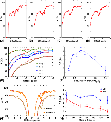 |
Magnetization transfer contrast (MTC) suppressed relayed nuclear Overhauser enhancement (rNOE) imaging at 3 T
Jianpan Huang1, Xiongqi Han1, Lin Chen2,3, Xiang Xu2,3, Peter C. M. van Zijl2,3, Jiadi Xu2,3, and Kannie W. Y. Chan1,2
1Department of Biomedical Engineering, City University of Hong Kong, Hong Kong, China, 2Russell H. Morgan Department of Radiology and Radiological Science, The Johns Hopkins University School of Medicine, Baltimore, MD, United States, 3F.M. Kirby Research Center for Functional Brain Imaging, Kennedy Krieger Research Institute, Baltimore, MD, United States
Relayed nuclear Overhauser enhancement (rNOE) imaging indirectly detects the aliphatic groups of biomolecules and can be used to diagnose protein or lipid signals and related pathology involving such signals. Current rNOE imaging has been studied mainly on high-field MRI scanners (≥7T). When implementing the technique on low clinical MRI fields (≤3T), rNOE contrast is more obscured by semisolid magnetization transfer contrast (MTC) and water direct saturation (DS). We developed a pulsed-CEST/MT method at 3T MRI that can suppress MTC and DS efficiently, not compromising rNOE contrast.
|
|
3142. |
Quantification of Magnetization Transfer and Inhomogeneous Magnetization Transfer with an MPRAGE sequence and an MTsat Approach
David C Alsop1, Fanny Munsch1, Gopal Varma1, Olivier Girard2, and Guillaume Duhamel2
1Radiology, Beth Israel Deaconess Medical Center and Harvard Medical School, Boston, MA, United States, 2CRMBM, Aix Marseille Univ, CNRS, Marseille, France
Methods for quantification of Inhomogeneous Magnetization Transfer (ihMT), are not yet well established. This is especially true for ihMT prepared sequences such as ihMT MPRAGE, where no steady state solution is available. Here we adapt the MTsat approach to quantification of MT and ihMT in an MPRAGE sequence.
|
|
3143.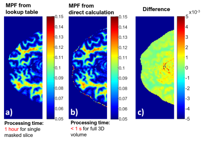 |
Analytic solution for macromolecular proton fraction (MPF) determined from single point magnetization transfer experiment
Kimberly L. Desmond1
1Research Imaging Centre, CAMH, Toronto, ON, Canada
Single point magnetization transfer (MT) is a quantitative method which reduces the number of unknowns in the MT signal equation by fixing tissue parameters which are effectively constant such that the macromolecular proton fraction (MPF) is the only remaining variable. In this work, the nonlinear signal equation describing single point MT was inverted to obtain an analytic expression for MPF as a function of observed normalized signal (MT∆/MT0). This enables rapid computation of MPF maps from matrix operations performed on the 3D images of the MT-weighted image (MT∆), the MT reference image, (MT0), and separately acquired T1 and B1 maps.
|
|
3144.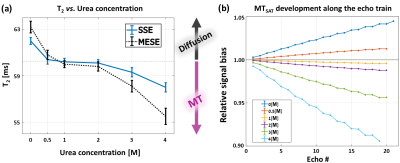 |
Distinguishing between macromolecular-driven magnetization transfer (MTSAT) and direct water saturation (MTDIR)
Dvir Radunsky1, Tamar Blumenfeld-Katzir1, and Noam Ben-Eliezer1,2,3
1Bio-medical Engineering, Tel Aviv University, Tel Aviv, Israel, 2Sagol School of Neuroscience, Tel Aviv University, Tel Aviv, Israel, 3Center for Advanced Imaging Innovation and Research (CAI2R), New-York University Langone Medical Center, New York, NY, United States
Mapping of T2 values is highly valuable for a wide range of applications. Still, accurate mapping is challenging due to the inherent bias of rapid Multi-Echo Spin-Echo (MESE) protocols by stimulated echoes, and also by magnetization transfer (MT). In this work, we investigate the different effects of macromolecular-driven MT (MTSAT) and direct water saturation (MTDIR) mechanisms on MESE signals, and their respective influence on T2 values. The investigation includes quantitative MT measurements between protocols with different scan settings and protocol schemes, aiming to isolate the individual roles of MTSAT and MTDIR.
|
|
3145.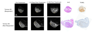 |
An Analysis of Post-Radiation Response in DU145 Prostate Tumour Xenografts Using Magnetization Transfer MRI
Leedan Murray1, Wendy Oakden1, Wilfred W. Lam1, and Greg J. Stanisz1
1Physical Sciences, Sunnybrook Research Institute, Toronto, ON, Canada
The response to radiation treatment of DU145 prostate tumour xenografts was studied by comparing magnetization transfer-weighted Z-spectra before and after treatment. ROIs were drawn in homogenous sections of the tumour and analyzed based on corresponding TUNEL histology images. MTR decreased more than 1 week post-treatment in tumours that responded to treatment but was unchanged for non-responding tumours. The technique introduced in this study could potentially introduce a non-invasive way of determining treatment efficacy based on tumour necrosis/apoptosis.
|
|
3146.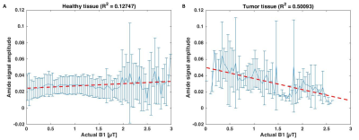 |
Towards understanding of CEST signal in breast cancer; relation between APT- MT- and B1 effects.
Elles Elschot1, Lieke van den Wildenberg1, Vitaly Khlebnikov1, Dennis Klomp1, and Jannie Wijnen1
1Radiology Department, UMC Utrecht, Utrecht, Netherlands
This study investigates the relation between B1+ and signal amplitude of the APT and MT exchange pools in CEST MRI. We examined 19 breast cancer patients that underwent NAC treatment with CEST MRI at 7T. The data indicates evidence for an extra exchanging pool with strong B1 dependence, that is more abundant in tumor tissue compared to healthy tissue. By identifying the exchanging components in this pool, a new biomarker for tumor tissue could be found and used to understand changes in response to NAC early during treatment.
|
Session Topic: CEST, Magnetization Transfer and Relaxometry
Session Sub-Topic: Relaxation
Digital Poster
Contrast Mechanisms
3147.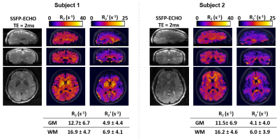 |
Alternating 3D Unbalanced SSFP With Z-shimming for Background Field Corrected R2’ Measurement in the Human Brain
Hyunyeol Lee1, Dongyeop Han2, Cheng-Chieh Cheng1, and Felix W Wehrli1
1Radiology, University of Pennsylvania, Philadelphia, PA, United States, 2Electrical and Electronic Engineering, Yonsei University, Seoul, Korea, Republic of
The relaxation parameter R2’ characterizes susceptibility-induced voxel signal modulations, for example, in the presence of deoxygenated hemoglobin in brain microvasculature or iron deposits in deep gray matter structures. Current R2’ measurement methods build on a spin-echo sequence configuration, and hence require impractically long scan time for volumetric 3D R2’ mapping. Furthermore, large susceptibility gradients around air/tissue interfaces result in signal distortions with increasing echo times, thus making it challenging to achieve accurate R2’ estimation in deep GM regions. Here, we propose an alternating, 3D z-shimmed, unbalanced steady-state-free-precession (SSFP) technique for rapid and B0-corrected R2’ mapping in the human brain.
|
|
3148. |
Assessment of R2* dependency on fiber orientation and myelin concentration in normal-appearing white matter in multiple sclerosis
Renat Sibgatulin1, Daniel Güllmar1, Andreas Deistung2, Stefan Ropele3, and Jürgen Rainer Reichenbach1,4,5,6
1Medical Physics Group, Institute of Diagnostic and Interventional Radiology, Jena University Hospital - Friedrich Schiller University Jena, Jena, Germany, 2Department of Radiology, University Hospital Halle (Saale), Halle, Germany, 3Department of Neurology, Medical University of Graz, Graz, Austria, 4Michael Stifel Center Jena for Data-Driven and Simulation Science, Friedrich Schiller University Jena, Jena, Germany, 5Abbe School of Photonics, Friedrich Schiller University Jena, Jena, Germany, 6Center of Medical Optics and Photonics, Friedrich Schiller University Jena, Jena, Germany
The effective transverse relaxation rate (R2*) is increasingly used in quantitative MRI, due to its sensitivity to iron and myelin content of the tissue. Separating the contributions from both sources is of particular interest in relation to multiple sclerosis. This work attempts to factor out contribution from myelin using orientation information derived from dMRI, and magnetization transfer saturation (MTS). Application of such correction to a group of multiple sclerosis patients and a control group, suggests that sensitivity of R2* to iron might be improved by accounting for fiber orientation in WM, while incorporation of MTS might not show clear benefit.
|
|
3149. |
Fat DESPOT for MR-oximetry validated by STEAM MRS
Véronique Fortier1,2 and Ives R. Levesque1,2,3
1Medical Physics Unit, McGill University, Montreal, QC, Canada, 2Biomedical Engineering, McGill University, Montreal, QC, Canada, 3Research Institute of the McGill University Health Centre, Montreal, QC, Canada
The R1 relaxation rate of fat is a promising biomarker for mapping tissue oxygenation. Existing techniques to map fat R1 are limited to single-voxel or 2D imaging with long scan times. To address these limitations, this work presents a 3D technique to map fat R1 using a fat-water-separated variable flip angle (VFA) approach. The sensitivity of this technique to oxygenation variations was evaluated in a phantom. The results showed that fat R1 can be measured using a technique based on 3D VFA at 3 T and is feasible for MR-oximetry.
|
|
3150. |
T1 relaxation in fat and its dependence on fat content
Véronique Fortier1,2 and Ives R. Levesque1,2,3
1Medical Physics Unit, McGill University, Montreal, QC, Canada, 2Biomedical Engineering, McGill University, Montreal, QC, Canada, 3Research Institute of the McGill University Health Centre, Montreal, QC, Canada
The T1 relaxation of triglyceride molecules is of interest for fat-water separation and fat quantification. A better understanding of the T1 of fat could benefit modeling techniques for applications in MR-oximetry and fatty liver disease. In this work, the T1 relaxation and its dependence on fat content was evaluated for five spectral peaks present in triglyceride molecules, over a range of fat fractions in a homogeneous fat-water mixture. The T1 of water in mixture was also studied. A model is proposed to describe the two-pool relaxation in a fat-water mixture as a function of the fraction of each pool.
|
|
3151. |
Multi-seed myelin water imaging using gradient echo: beyond the initial guess in exponential-sum fit
Hyeong-Geol Shin1, Se-Hong Oh2, Sooyeon Ji1, Jieun Lee1, Woojin Jung1, Eun-Jung Choi1, and Jongho Lee1
1Department of Electrical and Computer Engineering, Seoul National University, Seoul, Korea, Republic of, 2Biomedical Engineering, Hankuk University of Foreign Studies, Yongin, Korea, Republic of
In this work, we investigated the effects of initial parameter guess on non-linear least square (NLLS) method for myelin water imaging (MWI). We demonstrated that an inappropriate initial guess induces error in MWI and proposed a multi-seed algorithm to reduce the initial guess-dependent error. To do so, we applied the multi-seed MWI to synthetic and in-vivo data and compared the outcomes with the conventional algorithm (i.e., single-seed MWI). MWI results estimated by the multi-seed algorithm showed better agreement with the model than the single-seed algorithm, suggesting a potential solution to mitigate the ill-posed condition of MWI.
|
|
3152. |
Adaptive and slice-specific z-shimming approach for signal rephasing in 2D multi gradient echo imaging
Martin Soellradl1, Johannes Strasser1, Stefan Ropele1, and Christian Langkammer1
1Department of Neurology, Medical University of Graz, Graz, Austria
Intravoxel dephasing due to macroscopic field variations along the slice-selective direction z can be compensated by application of compensation gradients in z-direction (“z-shimming”). Compensation gradients applied between echo acquisition allow to estimate R2* also in areas with strong field gradients. However, if equally strong compensation gradients are applied in each slice the signal dephases in homogenous areas. We therefore propose an adaptive method where slice-specific compensation gradients are estimated for each slice from a fast pre-scan. With the proposed approach improved R2*-maps, compared to constant compensation gradient strategies, with higher SNR and accuracy can be achieved.
|
|
3153. |
Relaxometry Differences Between Magno- and Parvocellular Human LGN Subdivisions Revealed by In- and Ex-vivo Quantitative MRI
Christa Müller-Axt1,2, Cornelius Eichner1, Louise Kauffmann1,3, Pierre-Louis Bazin1,4, Henriette Rusch5, Markus Morawski5, Alfred Anwander1, and Katharina von Kriegstein2
1Max Planck Institute for Human Cognitive and Brain Sciences, Leipzig, Germany, 2Faculty of Psychology, Technical University of Dresden, Dresden, Germany, 3LPNC, Grenoble Alpes University, Grenoble, France, 4Integrative Model-based Cognitive Neuroscience Research Unit, Department of Psychology, University of Amsterdam, Amsterdam, Netherlands, 5Paul Flechsig Institute of Brain Research, University of Leipzig, Leipzig, Germany
The human lateral geniculate nucleus (LGN) is the central station for visual processing before information reaches the cerebral cortex. It is characterized by subdivisions with distinct cyto- and myeloarchitecture. Due to its small size, imaging of the LGN and especially its subdivisions is challenging. Here, we show that the LGN and its subdivisions can be identified using in-vivo and ex-vivo high-field quantitative MRI with ultra-high resolution. We present the to-date first atlas of the LGN and its estimated subdivisions. This work will serve as a highly valuable tool both for neuroscientists and clinicians investigating the visual system and its disorders.
|
|
3154.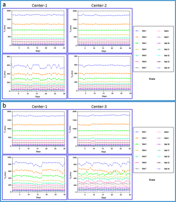 |
Quantitative Imaging Metrics Derived from MR Fingerprinting using ISMRM/NIST System Phantom: An International Multi-Center Test-Retest Study
Amaresha Konar Shridhar1, Enlin Qian2, Sairam Geethanath2, Guido Buonincontri3, Maggie M Fung4, Nancy A Obuchowski5, Pedro Gomez6, Rolf Schulte7, Lawrence H Schwartz8, and Amita Shukla-Dave1,9
1Department of Medical Physics, Memorial Sloan Kettering Cancer Center, New York, NY, United States, 2Columbia Magnetic Resonance Research Center, Columbia University, New York, NY, United States, 3Imago7 Foundation, Pisa, Italy, 4MR Apps & Workflow, GE Healthcare, New York, NY, United States, 5Department of Quantitative Health Sciences, Cleveland Clinic Foundation, Cleveland, OH, United States, 6Global Research, GE Healthcare, Munich, Germany, 7MR Workflow & Application Team, GE Healthcare, New York, NY, United States, 8Department of Radiology, Columbia University Medical Center, New York, NY, United States, 9Department of Radiology, Memorial Sloan Kettering Cancer Center, New York, NY, United States
The present study performed the repeatability and reproducibility test for T1, and T2 values estimated using MR fingerprinting (MRF) method. MRF data have been acquired over 30 days at three different centers (center-1 (USA): 1.5T and 3T, center-2 (USA): 3T and center-3 (Italy): 1.5T) using ISMRM/NIST MRI system phantom. MRF based estimated T1 and T2 values are compared with the vendor provided values and standard inversion recovery spin echo and a multiple single-echo spin-echo method as scanner Ground Truth (GT) values. The results show that the T1 and T2 values estimated using the MRF method are highly repeatable and reproducible.
|
|
3155.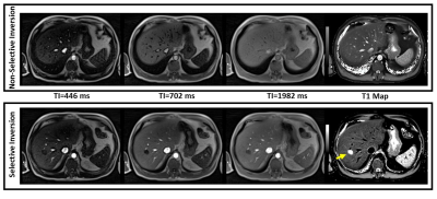 |
An Efficient, Robust, and Reproducible Quantitative Radial T1 Mapping Technique
Mahesh Bharath Keerthivasan1, Marcel Dominik Nickel2, Fei Han3, Xiaodong Zhong3, Maria Altbach4, Berthold Kiefer2, and Vibhas Deshpande5
1Siemens Healthcare USA, Tucson, AZ, United States, 2Siemens Healthcare Gmbh, Erlangen, Germany, 3Siemens Healthcare USA, Los Angeles, CA, United States, 4University of Arizona, Tucson, AZ, United States, 5Siemens Healthcare USA, Austin, TX, United States
There has been renewed interest in the estimation of T1 relaxation times as a quantitative method for characterization of pathologies in the abdomen. While various techniques have been presented for T1 mapping they have not been systematically evaluated. In this work, we present a multi-slice radial Look-Locker FLASH technique for robust and reproducible abdominal T1 mapping. We investigate the effect of various pulse sequence and reconstruction parameters on the T1 estimation performance.
|
|
3156.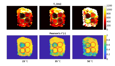 |
Low Field MR Thermometry with a Fast, Interleaved Look-Locker based T1 Mapping Approach.
Marco Fiorito1, Maksym Yushchenko1, Mathieu Sarracanie1,2, and Najat Salameh1
1Laboratory for Adapatable MRI Technology (AMT lab), Dpt of Biomedical Engineering, University of Basel, Basel, Switzerland, 2Medical Image Analysis Centre (Miac AG), Basel, Switzerland
Low field scanners present several advantages for interventional applications, however no standard method has yet arisen from temperature mapping. In this study we investigated the relationship between T1 and temperature at 0.1 T. MR images of four water phantoms, characterised by different T1, were acquired at different temperatures using a multi-slice FLASH sequence based on Look-Locker. Pixel-wise estimation of T1 relaxation through data fitting showed a growing trend with temperature. In light of the large number of time points, compromising SNR by a factor of 2 produced similar results for an almost 4 times faster acquisition.
|
|
3157.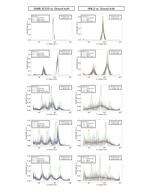 |
A novel T2 relaxation analysis method SAME-ECOS: Spectrum Analysis for Multiple Exponentials via Experimental Condition Oriented Simulation
Hanwen Liu1,2, Qing-San Xiang1,3, Roger Tam3,4, Piotr Kozlowski3, David K.B. Li3, Alex L. MacKay1,3, John K. Kramer2,5, and Cornelia Laule1,2,3,6
1Physics & Astronomy, University of British Columbia, Vancouver, BC, Canada, 2International Collaboration on Repair Discoveries, Vancouver, BC, Canada, 3Radiology, University of British Columbia, Vancouver, BC, Canada, 4Biomedical Engineering, University of British Columbia, Vancouver, BC, Canada, 5Kinesiology, University of British Columbia, Vancouver, BC, Canada, 6Pathology & Laboratory Medicine, University of British Columbia, Vancouver, BC, Canada
We propose a novel T2 relaxation data analysis method called spectrum analysis for multiple exponentials via experimental condition oriented simulation (SAME-ECOS), which was developed based on a combination of information theory and deep learning neural network algorithms. SAME-ECOS is tailored for different MR experimental conditions to decompose the multi-exponential decay data into a T2 spectrum, which has been considered an ill-posed problem using conventional fitting algorithms including the commonly used non-negative least squares (NNLS). Our results demonstrated that, compared with NNLS, SAME-ECOS can yield much more reliable T2 spectra in a dramatically shorter time.
|
|
3158.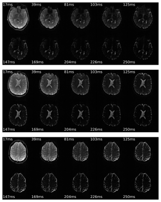 |
A Novel 10-echo GESE multi-parametric sequence based on the EPIK readout for simultaneous quantification of T2 and T2*
Fabian Küppers1, Seong Dae Yun1, and Nadim Jon Shah1,2,3,4
1Institute of Neuroscience and Medicine 4, Forschungszentrum Jülich, Jülich, Germany, 2Department of Neurology, RWTH Aachen, Aachen, Germany, 3Institute of Neuroscience and Medicine 11, Forschungszentrum Jülich, Jülich, Germany, 4JARA - BRAIN - Translational Medicine, Aachen, Germany
MRI sequences with a mixed GE/SE signal have been shown to be suitable for a wide range of applications due to the wide variety of accessible parameters. This work presents in vivo results of a 10-echo GESE sequence based on an EPIK readout to demonstrate the feasibility of whole-brain acquisitions below 5min, including T2 and T2* maps; comparison is made to reference methods. The generated images are artefact free and the T2*-maps correspond with conventional multi-echo GE results. The T2 values are higher than those from conventional SE, but are correctable using a reduced TE and improved fitting.
|
|
3159.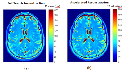 |
Accelerated reconstruction of T2 maps based on the echo modulation curve signal model, and using PCA and gradient-descent search algorithms
Guy Shpringer1 and Noam Ben-Eliezer1,2,3
1Department of Biomedical Engineering, Tel Aviv University, Tel Aviv, Israel, 2Sagol School of Neuroscience, Tel Aviv University, Tel Aviv, Israel, 3Center for Advanced Imaging Innovation and Research (CAI2R), New-York University Langone Medical Center, New York, NY, United States
Quantification of T2 values is important for a wide range of research and clinical applications. The Echo modulation Curve Algorithm allows accurate mapping of T2 values at clinical scan-times based on multi-echo spin echo data. Reconstruction, however, is still done offline and requires 10’s of minutes of processing times, thereby hampering the integration of this technique into real-time applications. We present in this work two approaches for accelerating maps reconstruction, based on PCA and gradient descent search algorithm. These offer up to x20 acceleration in reconstruction time, and can be potentially generalized to other reconstruction procedures involving dictionary search.
|
|
3160.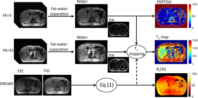 |
Joint estimation of PDFF and T1 in abdomen with simultaneous B1+ correction at 3T
Hao Peng1,2, Liwen Wan1, Qian Wan1, Jianxun Lv1, Chuanli Cheng1, Xin Liu1, Hairong Zheng1, and Chao Zou1
1Shenzhen Institutes of Advanced Technology,Chinese Academy of Sciences, Shenzhen, China, 2Huazhong University of Science & Technology, Wuhan, China
T1 quantification is a valuable biomarker for liver function estimation and fibrosis staging. The presence of fat would confound the measurement of tissue T1. Chemical shift encoded fat-water separation combined variable flip angle (CSE-VFA) T1 mapping could remove the bias from fat signals, but B1 inhomogeneity would be a serious problem in abdominal applications. In this work, we proposed to combine the DREAM sequence with CSE-VFA, so that PDFF/T1/B1/R*2 could be simultaneously estimated. The deviation of the T1 values between the results with and without B1 correction verified the
necessity of B1 measurements in the abdominal application.
|
|
3161.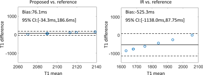 |
Multi-compartment T1 relaxometry with inversion recovery sequence: a fat-water phantom study
Hao Peng1, Liwen Wan1, Qian Wan1, Chuanli Cheng1, Xin Liu1, Hairong Zheng1, and Chao Zou1
1Shenzhen Institutes of Advanced Technology,Chinese Academy of Sciences, Shenzhen, China
T1 relaxation time has been a valuable biomarker with emerging applications in cardiac diseases and liver function. Traditional inversion-recovery T1 quantification method is frequently applied as the reference for various T1 quantification methods. When applied in targets where fat and water signals co-exists, the single component fitting model would be in a dilemma. Combining the inversion-recovery T1 quantification with existing fat-water quantification methods, this work presented a solution for multi-component T1 quantification. Proposed method has been tested on fat-water phantoms and showed good consistency over a wide range of fat fractions.
|
|
3162.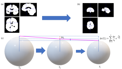 |
Quantification of tissue shrinkage due to formalin fixation of entire post-mortem human brain
Tobias Streubel1, Francisco Javier Fritz1, Herbert Mushumba2, Klaus Püschel2, and Siawoosh Mohammadi1
1Institute for Systems Neuroscience, University Medical Center Hamburg-Eppendorf, Hamburg, Germany, 2Institute of Legal Medicine, University Medical Center Hamburg-Eppendorf, Hamburg, Germany
This work investigates tissue shrinkage during fixation and how it is related to associated changes in three quantitative MRI parameters (longitudinal relaxation $$$R_1$$$ and effective transverse relaxation $$$R_2^*$$$ rates, and magnetization transfer saturation rate MT). We proposed a new model to estimate tissue shrinkage from brain volume changes and found that shrinkage was $$$7.7\%$$$. No apparent relation between changes in MT and tissue shrinkage were found, whereas it was remarkable for $$$R_1$$$ and $$$R_2^*$$$, indicating that mostly the extra-axonal space is reduced during fixation.
|
Session Topic: CEST, Magnetization Transfer and Relaxometry
Session Sub-Topic: Contrast Mechanisms: Quantitation & Validation
Digital Poster
Contrast Mechanisms
3163.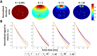 |
Simulating the effect of axonal dispersion and noise on anisotropic R2* relaxometry in white matter
Francisco Javier Fritz1, Luke J. Edwards2, Tobias Streubel1,2, Kerrin J. Pine2, Nikolaus Weiskopf2, and Siawoosh Mohammadi1,2
1Institute of System Neuroscience, Universitätklinikum Hamburg-Eppendorf, Hamburg, Germany, 2Department of Neurophysics, Max Planck Institute for Human Cognitive and Brain Sciences, Leipzig, Germany
We introduced a forward model to simulate the effect of fibre dispersion on the angle-dependent GRE signal decay in different SNR conditions. We compared the classical signal model against three variations of the signal model derived from a second-order Taylor expansion in time of Wharton and Bowtell’s HCFM model, including a new model that accounts for fibre dispersion. We found a noise enhancement in the model parameters when using the second-order models. Moreover, we found that the orientation dependency of the GRE signal diminished for high dispersions.
|
|
3164. |
The effect of noise on myelin assessment by multi-exponential fitting for varying white matter fibre geometries
Tonima S Ali1, Vaibhavi S Itkyal1,2, Kiran Thapaliya1,3, and Markus Barth1,2,3
1University of Queensland, Brisbane, Australia, 2Indian Institute of Technology, Madras, India, 3Griffith University, Gold Coast, Australia
The use of multi-echo gradient echo (mGRE) with signal compartment modelling is an increasingly popular choice for myelin assessment by measuring myelin T2* (T2*My), myelin water fraction (MWF) and g-ratio. In this simulation study, we show the effects of varying white matter microstructure and noise levels on GRE and the efficiency of commonly used fitting algorithms for predicting the true T2*My, MWF and g-ratio.
|
|
3165.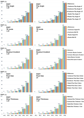 |
A stability study of breath-hold and free-breathing liver fat and R2* quantification at 1.5T and 3T
Chang Gao1,2,3, Peng Hu1,2, Brian Dale4, Marcel D. Nickel5, Stephan A.R. Kannengiesser5, Berthold Kiefer5, Vibhas Deshpande6, and Xiaodong Zhong3
1Department of Radiological Sciences, David Geffen School of Medicine, University of California, Los Angeles, CA, United States, 2Physics and Biology in Medicine Inter-departmental Graduate Program, David Geffen School of Medicine, University of California, Los Angeles, CA, United States, 3MR R&D Collaborations, Siemens Healthcare, Los Angeles, CA, United States, 4MR Training, Siemens Healthcare, Cary, NC, United States, 5MR Application Development, Siemens Healthcare GmbH, Erlangen, Germany, 6MR R&D Collaborations, Siemens Healthcare, Austin, TX, United States
Accurate quantification of proton-density fat fraction (PDFF) and R2* requires appropriate compensation of various confounding factors. A stability study was performed to investigate the impacts of different protocols and imaging parameters using both breath-hold Cartesian and free breathing radial sequences on a standardized phantom and a group of 5 same normal subjects at both 1.5T and 3T. This study revealed that flip angle, TE mode and coil usage were important protocol parameters to investigate further in larger patient population.
|
|
3166.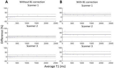 |
Quality assurance of multiparametric mri protocol for multi-centre prostate cancer sequential imaging clinical trial
Yu-Feng Wang1,2, Gary Liney2,3, Robba Rai2,3, Sirisha Tadimalla1, Jonathan Goodwin4, Lois Holloway2,3, and Annette Haworth1
1Institute of Medical Physics, The University of Sydney, Camperdown, Australia, 2Ingham Institute for Applied Medical Research, Liverpool, Australia, 3Liverpool and Macarthur Cancer Therapy Centre, Liverpool Hospital, Liverpool, Australia, 4Radiation Oncology Department, Calvary Mater Newcastle, Newcastle, Australia
Inter-/intra-scanner differences contribute to uncertainties in results from multi-centre longitudinal imaging studies. The Sequential Imaging Biofocussed Radiotherapy (SI-BiRT) trial is a multi-centre longitudinal imaging clinical trial that aims to develop imaging biomarkers using mpMRI to predict radiation therapy response in prostate cancer. Here we quantify the inter-/intra-scanner variabilities in the mpMRI protocol and scanners used in the SI-BiRT trial using commercial phantoms. ADC measurements were highly accurate while VFA T1 mapping overestimated reference values. ADC and T1 showed high intra-scanner reproducibility while R2* showed higher variability. The quantified uncertainties will be considered when interpreting results from the SI-BiRT trial.
|
|
3167.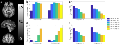 |
Assessment of the sensitivity of inhomogeneous Magnetization Transfer (ihMT) brain imaging with B1+ variations at 3T
Lucas Soustelle1,2, Samira Mchinda1,2, Thomas Troalen3, Maxime Guye1,2, Jean-Philippe Ranjeva1,2, Gopal Varma4, David C Alsop4, Guillaume Duhamel1,2, and Olivier M Girard1,2
1Aix-Marseille Univ, CNRS, CRMBM, Marseille, France, 2APHM, Hôpital Universitaire Timone, CEMEREM, Marseille, France, 3Siemens Healthcare SAS, Saint-Denis, France, 4Division of MR Research, Radiology, Beth Israel Deaconess Medical Center, Harvard Medical School, Boston, MA, United States
Inhomogeneous Magnetization Transfer (ihMT) is a novel myelin imaging technique. As for many other MRI techniques special attention must be paid to local B1+ variations in applications at high field. In this work, a detailed analysis of the ihMT ratio sensitivity to B1+ inhomogeneities is performed in the context of human brain imaging at 3T. It is shown that concentrating RF power is an efficient way to reduce B1+ induced bias and that the overall performance of the technique lies on a tradeoff between high ihMT ratios (sensitivity) and reduced sensitivity to B1+ inhomogeneities.
|
|
3168.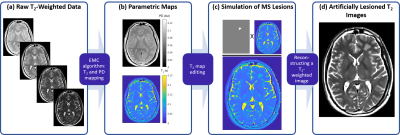 |
Quantitative Estimation of Visual Sensitivity to Early Pathological Changes in T2-Weighted Images
Chen Solomon1, Tamar Blumenfeld-Katzir1, Moti Salti2,3, Dvir Radunsky1, Noam Omer1, Neta Stern1, and Noam Ben-Eliezer1,4,5
1Department of Biomedical Engineering, Tel Aviv University, Tel Aviv, Israel, 2Brain Imaging Research Center (BIRC), Soroka Medical Center, Beer Sheva, Israel, 3Zlotowski Center for Neuroscience, Ben-Gurion University of the Negev, Beer Sheva, Israel, 4Center for Advanced Imaging Innovation and Research (CAI2R), New-York University Langone Medical Center, New York, New York, NY, United States, 5Sagol School of Neuroscience, Tel-Aviv University, Tel Aviv, Israel
Quantitative MRI (qMRI) may provide higher sensitivity to early pathological changes than standard qualitative assessment. This advantage, however, is yet to be rigorously compared with conventional diagnostic methods. This study aims to quantify the human ability to detect predefined changes in T2-weighted images as part of the diagnostic process of multiple sclerosis (MS). A visual diagnostic test was performed on neurosciences students, suggesting that the human vision has lower sensitivity to subtle MS lesions in T2-weighted images, in comparison to quantitative assessment of the change in T2 values.
|
|
3169.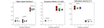 |
Exploring the anatomical variability in the precision of frequency difference mapping in the corpus callosum at 7T
Chiara Casella1, Elena Kleban1, and Derek K Jones1
1Department of Psychology, Cardiff University Brain Research Imaging Centre, Cardiff, United Kingdom
We explored the anatomical variability in reliability of complex mGRE signal, and of parameters estimated by modelling this signal as a superposition of complex myelin, intra- and extra-axonal water signals, in the corpus callosum at 7T. Our data showed to be repeatable in the posterior portion of the corpus callosum, while reliability was lower in more anterior portions. These findings have important implications and need to be considered when investigating callosal microstructure with mGRE in health and disease.
|
|
3170.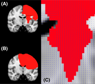 |
A method to remove the influence of fixative concentration on post-mortem T2 maps using a kinetic-tensor model
Feng Qi1, Karla Miller1, Sean Foxley2, Menuka Pallebage-Gamarallage3, Ricarda AL Menke1, Olaf Ansorge3, Samuel A Hurley4, and Benjamin C Tendler1
1Wellcome Centre for Integrative Neuroimaging, Nuffield Department of Clinical Neurosciences, University of Oxford, Oxford, United Kingdom, 2Department of Radiology, University of Chicago, Chicago, IL, United States, 3Nuffield Department of Clinical Neurosciences, University of Oxford, Oxford, United Kingdom, 4School of Medicine and Public Health, University of Wisconsin-Madison, Madison, WI, United States
Postmortem imaging allows for validation of the origins of image contrast through comparisons with histology. However, the inclusion of formalin fixative substantially reduces the T2. This reduction is (approximately) linear with concentration. Prior to scanning, samples are often placed in a fluid that has more favourable properties for imaging (e.g. perfluorocarbon fluids). This may lead to an outflow of fixative and an increase in T2 at the tissue surface. Here we propose to correct for T2 inhomogeneity by modelling the outflow of fixative within whole, human brains using a novel kinetic tensor model, which incorporates the effects of diffusion anisotropy.
|
|
3171.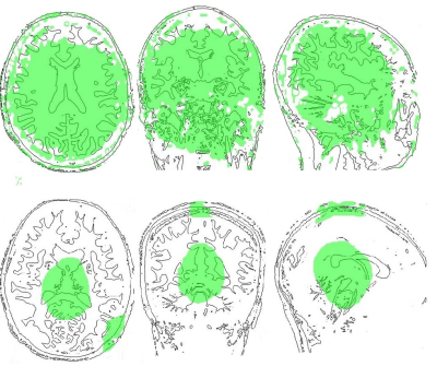 |
Simulation of B1+ inhomogeneities errors induced on T1w/T2w contrast at 3T and 7T
Mathilde Ripart1,2, Vincent Gras1, Rachida Elamrani1, Lucie Hertz-Pannier1, and Alexandre Vignaud1
1CEA, Neurospin, Université Paris-Saclay, Gif-sur-Yvette, France, 2Centrale Lyon, Ecully, France
The T1w/T2w ratio is commonly used at 3T as a biomarker of pathological changes in brain's structure. However, this method faces strong B1+ inhomogeneities that induce bias in the contrast. Simulations of this bias at 3T and 7T were done using an EPG model, and reported on the anatomical contours of human brains. At 3T, only a central part of the brain provides acceptable bias in the contrast (≤5%). At 7T, more than half of the brain gives unreliable contrast values. Therefore, T1w/T2w needs to be employed with cautiousness at 3T, and might not be a reliable method at 7T.
|
|
3172.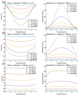 |
Investigation on the estimation performance of phase-based T2 mapping in the presence of motion
Daiki Tamada1, Xiaoke Wang1, Timothy J. Colgan1, Diego Hernando1,2, and Scott B. Reeder1,2,3,4,5
1Radiology, University of Wisconsin-Madison, Madison, WI, United States, 2Medical Physics, University of Wisconsin-Madison, Madison, WI, United States, 3Biomedical Engineering, University of Wisconsin-Madison, Madison, WI, United States, 4Medicine, University of Wisconsin-Madison, Madison, WI, United States, 5Emergency Medicine, University of Wisconsin-Madison, Madison, WI, United States
The estimation performance of phase-based T2 mapping in the presence of motion was investigated using Bloch equation simulations. The steady-state signal of RF-phase modulated gradient echo imaging in the presence of motion was calculated. Linear, sinusoidal, and random motion were considered for the simulation. The results showed that phase errors induced by motion may lead to underestimation of T2 when using a gradient echo phase-based T2 mapping method.
|
|
3173.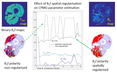 |
Reducing parameter estimate bias due to B1 field error ambiguity in CPMG MRI T2 relaxometry
Nick Zafeiropoulos1, Stephen Wastling1, Mary M Reilly2, Enrico De Vita3, Tarek Yousry1, and John Thornton1
1UCL Queen Square Institute of Neurology, London, United Kingdom, 2MRC Centre for Neuromuscular Diseases, London, United Kingdom, 3King's College London, London, United Kingdom
B1 field deviations above and below 100% of the nominal prescribed values have been noted by previous authors as a potential source of error in MRI CPMG T2 relaxometry. We show in simulations of clinically realistic acquisition protocols that this effect causes fit instability and loss of precision, and corroborate this with in vivo lower limb muscle image data. We then introduce a B1 error polarity spatial regularization scheme, and show that this improves parameter estimation accuracy.
|
|
3174.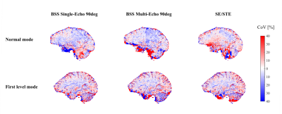 |
R1 reproducibility at 3T and 7T using Bloch-Siegert based corrections for transmit field inhomogeneity
Nadège Corbin1 and Martina F Callaghan1
1Wellcome Centre for Human Neuroimaging, UCL Institute of Neurology, London, United Kingdom
High reproducibility of longitudinal relaxation rate (R1) estimation using the variable flip angle approach requires unbiased and precise estimation of the transmit field inhomogeneity. In this context, the Bloch-Siegert (BSS)-based B1+ mapping approaches were evaluated at 3T and 7T and compared to the SE/STE approach. R1 values in grey and white matter were slightly higher with the BSS-based techniques. At 3T, the highest reproducibility was observed with the single-echo BSS approach, using an RF spoiling increment of 90°. At 7T, the reproducibility of the techniques were equivalent in well B0-shimmed regions but diverged in regions of high B0 inhomogenity.
|
|
3175.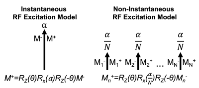 |
Effect of RF Pulse Duration on Chemical Shift Encoded MRI
Nathan Tibbitts Roberts1,2, Timothy Colgan1, and Scott B Reeder1,3,4,5,6
1Radiology, University of Wisconsin - Madison, Madison, WI, United States, 2Electrical and Computer Engineering, University of Wisconsin - Madison, Madison, WI, United States, 3Medical Physics, University of Wisconsin - Madison, Madison, WI, United States, 4Biomedical Engineering, University of Wisconsin - Madison, Madison, WI, United States, 5Medicine, University of Wisconsin - Madison, Madison, WI, United States, 6Emergency Medicine, University of Wisconsin - Madison, Madison, WI, United States
The effects of RF pulse duration on chemical shift encoded MRI (CSE-MRI) methods is a previously unexplored potential confounder for accurate and precise estimation of proton density fat fraction (PDFF) and R2*. Most signal models of CSE-MRI model RF excitation as instantaneous rotation of bulk magnetization; however, instantaneous excitation is not physically realizable. In this work we use Bloch equation simulations to characterize the effect of pulse duration on PDFF and R2* bias. We found that R2* bias was more sensitive than PDFF bias to RF pulse duration and note that clinically meaningful bias can be observed.
|
|
3176.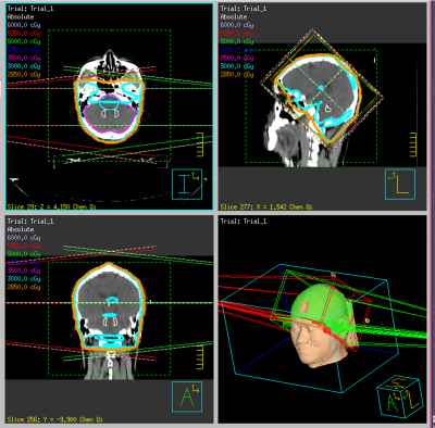 |
Signal Intensity in Dentate Nucleus Does Not Always Correlate with Accumulative Gadolinium doses after Whole-Brain Radiotherapy
Rongbiao Tang1, E. Mark Haacke2, Yibin Zhang2, Qingrou Wang2, Naying He2, Weibo Chen3, Ke-min Chen2, and Fuhua Yan2
1Ruijin Hospital, Shanghai, China, 2Ruijin Hosptial, Shanghai, China, 3Philips Healthcare, Shanghai, China
Whole-brain radiotherapy (WBRT) is often performed asynchronously with head contrast-enhanced MRI examination (CEME) in patients with brain metastasis (BM). Data regarding T1 signal intensity (SI) changes in the dentate nucleus (DN) in BM patients after WBRT and repeated exposure to gadolinium (Gd)-based contrast agents (GBCAs) are lacking. In this study, we explored the correlation between T1 SI in the DN and cumulative doses of linear GBCAs in BM patients treated with WBRT.
|
|
3177.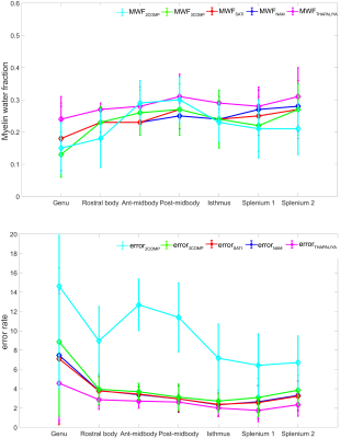 |
MR signal modelling comparison and its influence on tissue parameters using gradient-echo MRI at 7 Telsa
KIRAN THAPALIYA1,2, Viktor Vegh2, Steffen Bollmann2, and Markus Barth2,3
1Menzies Health Institute Queensland, Griffith University, Gold coast, Australia, 2Centre for Advanced Imaging, The University of Queensland, Brisbane, Australia, 3School of Information Technology and Electrical Engineering, The University of Queensland, Brisbane, Australia
Quantitative assessment of model parameters (water fraction and frequency shift) estimated using a multi-compartment model can be useful to study tissue properties in white matter. In this work, we compared common existing complex signal models for multi-echo gradient echo data acquired at 3T and 7T. We investigate the variation of model parameters that could potentially affect different models (number of compartments and parameters) on the estimation of tissue parameters. We show that the tissue parameters vary across the sub-regions of the corpus callosum and are influenced by different modelling choices.
|
|
3178.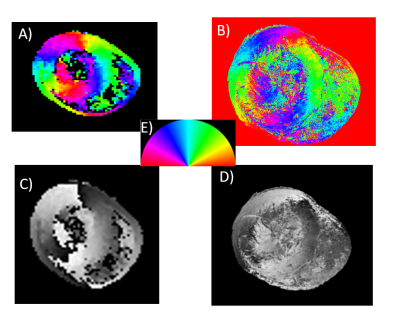 |
Local Structure Orientation (LSO) for histology-based validation of MRI: evaluation in a physical phantom study
Wadha Alyami1,2, Andre Kyme3, and Roger Bourne1
1Medical Imaging Sciences, Faculty of Health Sciences, University of Sydney, Sydney, Australia, 2Medical imaging sciences, King Saud University, Riyadh, Saudi Arabia, 3Biomedical Engineering, School of Aerospace Mechanical & Mechatronic Engineering, Faculty of Engineering and IT, University of Sydney, Sydney, Australia
Histology-based validation of magnetic resonance imaging (MRI) is essential to confirm imaging technique specificity and accuracy. One-to-one correspondence is complicated by histological preparation and differences in MRI and histology contrast mechanisms. A new approach, based on local structure orientation (LSO) and using microstructural features, is proposed. LSO facilitates registration based on direct correspondence between tissue fibre structure to emulate a common contrast mechanism. Using a physical phantom derived from a chicken heart with embedded fibrous structures, residual displacement was 14.1 mm for advanced mean squares (AMS), 2.2 mm for modified AMS (AMSR), and 3.3 mm for mutual information (MI).
|