
Digital Poster Session
Contrast Mechanisms: Quantitative Tissue Properties and Novel Contrast Mechanisms
Contrast Mechanisms
3179 -3194 Quantitative Tissue Properties and Novel Contrast Mechanisms - Electromagnetic Properties Mapping
3195 -3209 Quantitative Tissue Properties and Novel Contrast Mechanisms - QSM & Its Methods
3210 -3225 Quantitative Tissue Properties and Novel Contrast Mechanisms - QSM in Applications
3226 -3241 Quantitative Tissue Properties and Novel Contrast Mechanisms - Novel Contrast & Water/Fat Separation
3242 -3257 Quantitative Tissue Properties and Novel Contrast Mechanisms - Potpourri of Contrast Mechanisms
3179.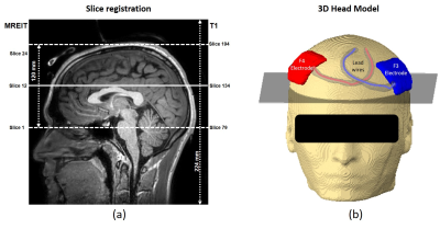 |
Current Density Measurements in the Human Brain in-vivo during TES treatment, using Multi-Band methods
Munish Chauhan1, Sulagna Sahu1, Saurav Zaman Khan Sajib1, Enock Boakye1, Michael Schär2, and Rosalind J Sadleir1
1School of Biological and Health System Engineering, Arizona State University, Tempe, AZ, United States, 2Department of Radiology, Johns Hopkins University, Baltimore, MD, United States
Current density distribution measured in the brain can guide and verify electrical stimulation therapies. Recent studies have demonstrated current density images of human heads during TES using Magnetic Resonance Electrical Impedance Tomography (MREIT). Earlier MREIT approaches permitted imaging of three 5-mm-thick slices in a sequence lasting 6 minutes, a typical therapeutic TES administration time. MREIT sequences must be accelerated to obtain whole brain coverage. In this study, we demonstrate in-vivo use of multiband-accelerated multi-echo-gradient-echo MREIT acquisition methods to acquire 24 5-mm-thick slices over 6 minutes. Computed current density maps measured in the brain using both methods are compared.
|
|
3180.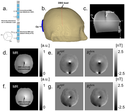 |
Imaging of Current Density Distribution in Deep Brain Stimulation (DBS)
Munish Chauhan1, Saurav Zaman Khan Sajib1, Sulagna Sahu1, Willard S Kasoff2, and Rosalind J Sadleir1
1School of Biological and Health System Engineering, Arizona State University, Tempe, AZ, United States, 2Department of Surgery, University of Arizona, Tucson, AZ, United States
Computational methods have been widely used to estimate field distributions produced by conventional or recently introduced directional DBS electrodes. To date there have been no experimental measurements of fields produced by DBS. We implanted either conventional or directional DBS leads into a head-shaped homogeneous phantom filled with agar gel and compared the current densities produced by each DBS electrode type using magnetic resonance current density imaging (MRCDI). This first experimental demonstration demonstrates the expected reduced spread for directional electrodes. This approach will be used in future studies to produce detailed images of DBS fields relative to target structures.
|
|
3181.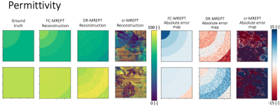 |
Machine learning methods applied to MR-based Electrical Properties Tomography to improve noise robustness and boundary accuracy.
Adan Jafet Garcia Inda1, Shao Ying Huang2,3, Stefano Mandija4,5, and Wenwei Yu1,6
1Department of Medical Engineering, Chiba University, Chiba, Japan, 2Department of Surgery, National University of Singapore, Singapore, Singapore, 3Engineering Product Development, Singapore University of Technology and Design, Singapore, Singapore, 4Department of Radiotherapy, University Medical Center Utrecht, Utrecht, Netherlands, 5Computational Imaging Group for MR diagnostic & therapy, University Medical Center Utrecht, Utrecht, Netherlands, 6Center for Frontier Medical Engineering, Chiba University, Chiba, Japan MREPT is a technique used to non-invasively estimate the electrical properties (EPs) of tissues based on Maxwell equations from MRI measurements. However, most reconstruction techniques are susceptible to noise and have severe boundary artifacts. In this work, we designed problem-oriented machine learning methods to improve the MREPT reconstructions. Through numerical experiments with 2-D cylindrical phantoms and comparison with cr-EPT, we demonstrate the feasibility of ML approaches to provide more noise robust EPT reconstructions with lower boundary artifacts. |
|
3182. |
Assessment of the sensitivity of Phase-Only Helmholtz-EPT to spatially-dependent conductivity changes using a glioma-brain-phantom
Lucia Bossoni1, Andrew Webb1, Loes Huijnen2, Remco Overdevest2, and Wyger Brink1
1Leiden University Medical Center, Leiden, Netherlands, 2Leiden University, Leiden, Netherlands
Phase-only Helmholtz-Electrical Property Tomography (PO-HEPT) has recently shown promise for the detection and grading of diffuse glioma. However, a brain glioma is usually located away from the centre of the brain where the transceive phase and phase-only approximations used in reconstruction may not be valid. Here we assessed the sensitivity of PO-HEPT in an anatomically realistic brain phantom where an off-centre glioma-compartment was incorporated, and using a clinically applicable MR sequence. Our results show that while the accuracy of the PO-HEPT deteriorates, its sensitivity is mostly unaffected, thus allowing correlative studies of tumour grading.
|
|
3183.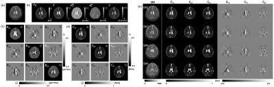 |
In Vivo Conductivity Tensor Imaging of Human Brain
Nitish Katoch1, Bup Kyung Choi1, In Ok Ko2, Ji Ae Park2, Yong Soo Cho3, Jin Woong Kim3, Hyung Joong Kim1, and Eung Je Woo1
1Kyung Hee University, Seoul, Korea, Republic of, 2Korea Institute of Radiological and Medical Sciences, Seoul, Korea, Republic of, 3Chosun University Hospital and Chosun University College of Medicine, Gwangju, Korea, Republic of
Human brain mapping of the electrical conductivity can facilitate the understanding of brain function. The low-frequency conductivity distribution of biological tissue exhibit the anisotropic tissue property and can be expressed as tensor. Considering the most physiological events occurs in frequency below 1 kHz, we developed a new conductivity tensor imaging method which can be implemented in conventional clinical MRI scanner without using any current injections for anisotropic conductivity measurement.
|
|
3184.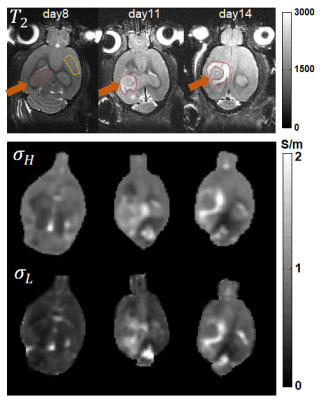 |
Conductivity Tensor Imaging (CTI): a novel contrast mechanism based on ionic conductivity for characterization of brain tumors
Nitish Katoch1, Clémentine Lesbats2, Atul Singh Minhas3, Hyung Joong Kim1, Eung Je Woo1, and Harish Poptani2
1Department of Biomedical Engineering, Kyung Hee University, Seoul, Republic of Korea, 2Physiology, University of Liverpool, Liverpool, United Kingdom, 3School of Engineering, Macquarie University, Sydney, Australia
Unlike the previously reported method of diffusion tensor magnetic resonance electrical impedance tomography (DT-MREIT), conductivity tensor imaging (CTI) does not require external electrodes for current injection. The low frequency conductivity (σL) value can be estimated from the high frequency conductivity (σH) measured using B1 map. The σL is influenced by cell size and density, which makes it an effective technique to characterize the cellular changes in brain tumors. Rat brain tumors were studied using 9.4 T MRI scanner using CTI protocol. Changes in ionic concentration cellular states depending on tumor growth were reflected in both high and low-frequency CTI images.
|
|
3185. |
In vivo pelvis conductivity mapping with a 3D patch-based convolutional neural network trained on in silico MR data
Soraya Gavazzi1, Cornelis AT van den Berg1,2, Mark HF Savenije1,2, H Petra Kok3, Lukas JA Stalpers3, Jan JW Lagendijk1, Hans Crezee3, and Astrid LHMW van Lier1
1Radiotherapy Department, University Medical Center Utrecht, Utrecht, Netherlands, 2Computational Imaging Group for MR diagnostic & therapy, University Medical Center Utrecht, Utrecht, Netherlands, 3Radiation Oncology Department, Amsterdam University Medical Center, Amsterdam, Netherlands
Pelvis conductivity is typically reconstructed with Helmholtz-based EPT. To overcome typical limitations of Helmholtz-based EPT in this challenging body site we explored reconstructing pelvis conductivity with deep learning. A 3D patch-based convolutional neural network was trained on in silica MR data (either a full complex B1+ field or transceive phase only) with realistic noise levels. These data were related to realistic pelvic anatomies and electrical properties. Preliminary results indicate that the network retrieved anatomically-detailed conductivity maps, without a priori anatomical knowledge given in input. Quantitatively, conductivity estimates on in vivo volunteer MR data were in line with literature.
|
|
3186.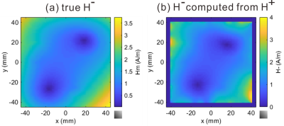 |
Computation of H- from H+ and its application to regularization for Magnetic Resonance Electrical Properties Tomography (MREPT)
Takaaki Nara1 and Motofumi Fushimi1
1Graduate School of Information Science and Technology, The University of Tokyo, Tokyo, Japan We derive an explicit formula to compute the negative circularly polarized component of the magnetic field, $$$H^-$$$, from the measured positively polarized component, $$$H^+$$$, for magnetic resonance electrical properties tomography (MREPT). Then, using the measured $$$H^+$$$ and the computed $$$H^-$$$, a novel regularization term for MREPT is introduced based on the $$$x$$$- and $$$y$$$-components of Ampere’s law and the $$$z$$$-component of Faraday’s law that have not been used so far for the reconstruction of EPs. This physically reasonable constraint well regularizes EPs at low convection field region. |
|
3187. |
Experimental Validation of Conductivity Tensor Imaging using Giant Vesicle Suspension
In Ok Ko1, Bup Kyung Choi2, Nitish Katoch2, Ji Ae Park1, Yong Soo Cho3, Jin Woong Kim3, Hyung Joong Kim2, and Eung Je Woo2
1Korea Institute of Radiological and Medical Sciences, Seoul, Korea, Republic of, 2Kyung Hee University, Seoul, Korea, Republic of, 3Chosun University Hospital and Chosun University College of Medicine, Gwangju, Korea, Republic of
Conductivity tensor can realize volume conductor model of brain for neuroimaging and electrical stimulation. We report validation of electrodeless conductivity tensor imaging (CTI) method [1]. From CTI imaging using giant vesicle suspension at 9.4T MRI, relative error in conductivity tensor image was found to be less than 1.7% compared with the measured values using an impedance analyzer. High- and low-frequency conductivity can quantify total and extracellular water contents, respectively, at every pixel. Their difference can quantify intracellular water content at every pixel. Current CTI method can separately quantify the contributions of ion concentrations and mobility to the conductivity tensor.
|
|
3188. |
Feasibility of Phase-based EPT using clinical routine sequences.
Jun-Hyeong Kim1, Kyu-Jin Jung1, Mina Park2, and Dong-Hyun Kim1
1electrical electronic engineering, yonsei University, seoul, Korea, Republic of, 2Department of Radiology, Gangnam Severance Hospital, seoul, Korea, Republic of
In this study, we test whether the preparation pulse and compensation gradient affect B1-phase and suggested a method for merging B1-phase obtained from different pulse sequences. We also try to reconstruct conductivity with the routine of guide images for surgery which have high resolution and low SNR.
|
|
3189.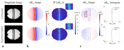 |
Acquisition-weighted MR Current Density Imaging (AW-MRCDI) improves sensitivity and spatial resolution
Cihan Göksu1,2, Lars G. Hanson1,3, Fróði Gregersen1,3,4, Klaus Scheffler2,5, and Axel Thielscher1,3
1Danish Research Centre for Magnetic Resonance, Centre for Functional and Diagnostic Imaging and Research, Copenhagen University Hospital, Hvidovre, Denmark, 2High-Field Magnetic Resonance Center, Max-Planck-Institute for Biological Cybernetics, Tübingen, Germany, 3Center for Magnetic Resonance, DTU Health Tech, Technical University of Denmark, Kgs. Lyngby, Denmark, 4Sino-Danish Center for Education and Research, Aarhus, Denmark, 5Department of Biomedical Magnetic Resonance, University of Tübingen, Tübingen, Germany
Exact knowledge of currents flowing inside the human brain is important for several neuroscientific applications. MRCDI combines MR with externally injected weak currents, and uses measurements of the current-induced magnetic field to estimate spatial current distribution. The method’s accuracy highly depends on the sensitivity and spatial resolution of the field measurements. Here, we improve the currently most sensitive MRCDI method based on steady-state free precession free induction decay (SSFP-FID) by using an acquisition-weighted scheme (AW-MRCDI). We compared weighted and conventional schemes by phantom experiments. AW-MRCDI demonstrated 59% increase in sensitivity and significantly improved the spatial resolution.
|
|
3190. |
Properties and implementation issues of phase based cr-MRECT for conductivity imaging
Yusuf Ziya Ider1 and Merve Nur Akyer1
1Dept of EEE, Bilkent University, Ankara, Turkey
Phase based cr-MRECT aims at reconstructing tissue conductivity without boundary artefacts. In this work some properties and implementation issues of the Phase based cr-MRECT are studied, namely, discretization methods, ill conditioning, bias due to neglecting the gradient of abs(B1+), and regularization. It is shown that by properly weighing the regularising artificial diffusion, bias-correction, and the convection term, artefact free and accurate conductivity images can be obtained. The system matrices are shown to be relatively well-conditioned and that there is no need to specify Dirichlet boundary conditions.
|
|
3191.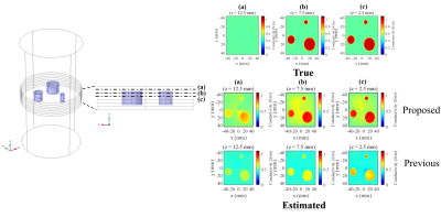 |
Noniterative Electrical Properties Tomography Reconstruction Method Based on Three-Dimensional Integral Equation for the Electric Field
Naohiro Eda1, Motofumi Fushimi1, Keisuke Hasegawa1, and Takaaki Nara1
1The University of Tokyo, Tokyo, Japan
This paper proposes a novel, noniterative reconstruction method for magnetic resonance electrical properties tomography (MREPT). For this method, we first estimate the electric field using a novel, three-dimensional integral equation, derived from Maxwell's equations and vector calculus identities. The EPs are then reconstructed from the estimated electric field, using Ampere’s law. Our method can estimate an arbitrary three-dimensional distribution of EPs, including those varying in the z-axis. The efficacy of our proposed method was validated through numerical simulations and a phantom experiment.
|
|
3192.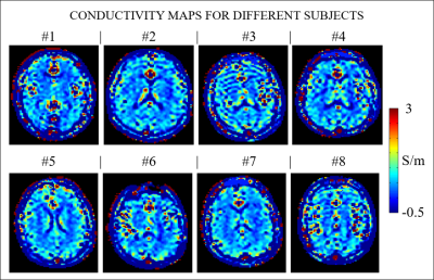 |
An in-vivo study to provide reference conductivity values of the human brain at 3T
Stefano Mandija1,2, Petar Petrov3, Jord Vink3, Sebastian Neggers3, and Cornelis A.T. van den Berg1,2
1Computational Imaging Group for MR diagnostic and therapy, Center for Image Sciences, University Medical Center Utrecht, Utrecht, Netherlands, 2Department of Radiotherapy, Division of Imaging and Oncology, University Medical Center Utrecht, Utrecht, Netherlands, 3Rudolf Magnus Institute of Neuroscience, University Medical Center Utrecht, Utrecht, Netherlands
First in-vivo brain conductivity reconstructions have been recently published. However, a large variation in the reconstructed conductivity values is reported and these results substantially differ from ex-vivo measurements. Given this lack of agreement, we performed an in-vivo study on eight healthy subjects to provide reference brain conductivity values. The measured in-vivo mean WM and GM conductivity values verify for the first time the literature values measured ex-vivo, while the reconstruction accuracy was verified in simulation settings. The presented values can therefore be used as a verified in-vivo reference for future studies, where new reconstruction algorithms are tested in-vivo.
|
|
3193.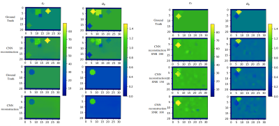 |
On the usage of deep neural networks as a tensor-to-tensor translation between MR measurements and electrical properties
Ilias Giannakopoulos1,2, José Serallés2, Georgy Guryev1,2, Luca Daniel2, Elfar Adalsteinsson2,3, Lawrence Wald4,5,6, Daniel Sodickson7,8,9, Jacob White2, and Riccardo Lattanzi7,8,9
1Skoltech Center for Computational and Data-Intensive Science and Engineering, Skolkovo Institute of Science and Technology, Moscow, Russian Federation, 2Department of Electrical Engineering & Computer Science, Massachusetts Institute of Technology, Cambridge, MA, United States, 3Institute for Medical Engineering and Science, Cambridge, MA, United States, 4Department of Radiology, Massachusetts General Hospital, Charlestown, MA, United States, 5Department of Radiology, Harvard Medical School, Boston, MA, United States, 6Harvard-MIT Health Sciences and Technology, Cambridge, MA, United States, 7Center for Advanced Imaging Innovation and Research (CAI2R), Department of Radiology, New York University School of Medicine, New York, NY, United States, 8The Bernard and Irene Schwartz Center for Biomedical Imaging (CBI), Department of Radiology, New York University School of Medicine, New York, NY, United States, 9Sackler Institute of Graduate Biomedical Sciences, New York University School of Medicine, New York, NY, United States Electrical properties (EP) can be retrieved from magnetic resonance measurements. We employed numerical simulations to investigate the use of convolutional neural networks (CNN) as a tensor-to-tensor translation between transmit magnetic field pattern ($$$b_1^+$$$) and EP distribution for simple tissue-mimicking phantoms. Given the volumetric nature of the problem, we chose a 3D UNET and trained the network on $$$10000$$$ data. We investigated on the usage of regularization to account for overfitting and observed that multiple dropouts through the layers of the network yield optimal EP reconstructions for $$$1000$$$ testing data. |
|
3194.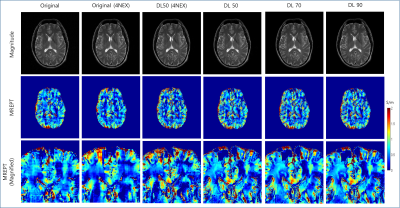 |
Feasibility of magnetic resonance based electrical properties tomography with deep learned reconstruction based denoising
Ho-Joon Lee1, Yeonah Kang1, Marc Lebel2, Joon-Hyeong Kim3, Dong-Hyun Kim3, and Sung-Min Gho4
Video Permission Withheld
1Department of Radiology, Haeundae Paik Hospital, Busan, Republic of Korea, 2MR Collaboration and Development, GE Healthcare, Calagary, AB, Canada, 3Department of Electrical and Electronic Engineering, Yonsei University, Seoul, Republic of Korea, 4MR Collaboration and Development, GE Healthcare, Seoul, Republic of Korea With advances in deep learning, feasibility has been investigated for MREPT reconstruction showing interesting results. However whether images denoised with deep learned reconstruction will improve EPT map quality has not been investigated. After denoising of complex data acquired with a DL algorithm, EPT maps were generated with phase based 2D-weighted polynomial fitting. Use of DL, shows better results as compared to conventionally generated maps (i.e. decreased NRMSE, increased PSNR and SSIM, with increasing denoising levels), and results in sharper appearing maps. Spreading of boundary artifacts are not observed with increasing denoising factors. |
 View the Poster
 Watch the Video
3195.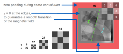 |
How to train a Deep Convolutional Neural Network for Quantitative Susceptibility Mapping (QSM)
Thomas Jochmann1, Jens Haueisen1, and Ferdinand Schweser2,3
1Department of Computer Science and Automation, Technische Universität Ilmenau, Ilmenau, Germany, 2Buffalo Neuroimaging Analysis Center, Dept. of Neurology, Jacobs School of Medicine and Biomedical Sciences, University at Buffalo, The State University of New York, Buffalo, NY, United States, 3Center for Biomedical Imaging, Clinical and Translational Science Institute, University at Buffalo, The State University of New York, Buffalo, NY, United States
Deep convolutional neural networks have recently gained popularity for solving the ill-posed dipole inversion problem in Quantitative Susceptibility Mapping (QSM). The training of the neural networks is performed with examples of χ and f that can either be obtained from physical simulations on synthetic source distributions, or through “classical” QSM methods on real data. For both choices, there is a plethora of decisions to make and parameters to set. Here we seek to present best practices regarding the modelling of synthetic source distributions and data augmentation.
|
|
3196.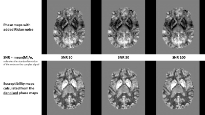 |
SOJU-Net—Denoising MR phase images with physics-informed deep learning using artificial Rician noise augmentation
Thomas Jochmann1, Nora Kuechler1, Jens Haueisen1, and Ferdinand Schweser2,3
1Department of Computer Science and Automation, Technische Universität Ilmenau, Ilmenau, Germany, 2Buffalo Neuroimaging Analysis Center, Dept. of Neurology, Jacobs School of Medicine and Biomedical Sciences, University at Buffalo, The State University of New York, Buffalo, NY, United States, 3Center for Biomedical Imaging, Clinical and Translational Science Institute, University at Buffalo, The State University of New York, Buffalo, NY, United States
Phase noise follows the Rician distribution, with a non-linear dependency of the phase noise on the local magnitude signal intensity. In this work, we present SOJU-Net, a deep-learning based denoising for MR phase images. SOJU-Net reduces Rician noise while preserving boundary contrast.
|
|
3197.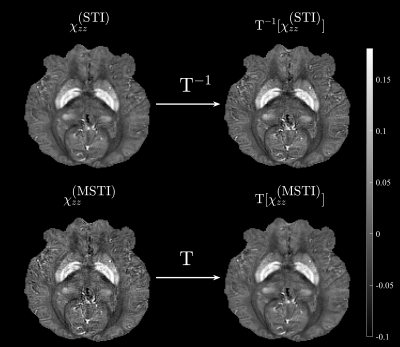 |
Sharper Images in Quantitative Susceptibility Mapping with Magnetometric Dipole Kernels
Anders Dyhr Sandgaard1 and Sune Nørhøj Jespersen1,2
1Center for Funcionally Integrative Neuroscience, Institute of Clinical Medicine, Aarhus University, Aarhus, Denmark, 2Department of Physics and Astronomy, Aarhus University, Aarhus, Denmark
The purpose of this study is to consider the GRE signal phase in a voxel for short echo times, from an MRI-visible fluid induced by MRI-invisible magnetic inclusions. We find that the signal phase relates to the magnetometric demagnetization field. In Fourier space, this defines a magnetometric dipole kernel which can be used to perform QSM. Using the data from the 2016 QSM challenge, we show that applying the magnetometric dipole kernel increases the sharpness, and that it is possible to increase the sharpness of already made STI susceptibility maps.
|
|
3198.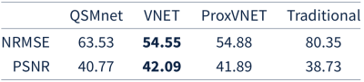 |
ProxVNET: A proximal gradient descent-based deep learning model for dipole inversion in susceptibility mapping
Christian Kames1,2, Jonathan Doucette1,2, and Alexander Rauscher1,2,3
1Department of Physics and Astronomy, University of British Columbia, Vancouver, BC, Canada, 2UBC MRI Research Centre, University of British Columbia, Vancouver, BC, Canada, 3Department of Pediatrics, University of British Columbia, Vancouver, BC, Canada
A deep learning model, ProxVNET, is proposed to solve the ill-posed dipole inversion in susceptibily mapping. ProxVNET is derived from unrolled proximal gradient descent iterations wherein the proximal operator is implemented as a V-Net and is itself learned. ProxVNET is shown to outperform the U-Net-based dipole inversion deep learning model QSMnet when compared to COSMOS reconstructed susceptibility maps.
|
|
3199.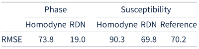 |
Recovering phase from homodyne filtered phase data using a residual dense network
Christian Kames1,2, Jonathan Doucette1,2, and Alexander Rauscher1,2,3
1Department of Physics and Astronomy, University of British Columbia, Vancouver, BC, Canada, 2UBC MRI Research Centre, University of British Columbia, Vancouver, BC, Canada, 3Department of Pediatrics, University of British Columbia, Vancouver, BC, Canada
In this work we train a residual dense network to recover homodyne filtered phase data. The proposed approach is able to accurately recover phase information. Susceptibility maps computed from the recovered phase yield the same accuracy as susceptibility maps computed from the original phase when compared to COSMOS.
|
|
3200.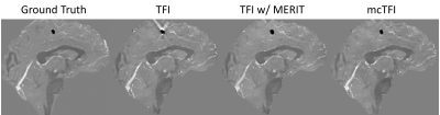 |
Improved signal modeling in Quantitative Susceptibility Mapping using multi-echo complex Total Field Inversion (mcTFI)
Yan Wen1,2, Thanh Nguyen2, Junghun Cho2,3, Pascal Spincemaille2, and Yi Wang3,4
1Meinig School of Biomedical Engineering, Cornell University, new york, NY, United States, 2Radiology, Weill Cornell Medicine, New York, NY, United States, 3Meinig School of Biomedical Engineering, Cornell University, New York, NY, United States, 4Radiology, Weill Cornell Medicine, new york, NY, United States
Multi-echo Complex Total Field Inversion (mcTFI) is a Total Field Inversion (TFI) method that computes QSM directly from the complex gradient echo data, for which the assumption of Gaussian noise is well justified. mcTFI demonstrated improvement over TFI in simulation and hemorrhage brain.
|
|
3201.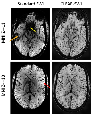 |
Improved Susceptibility Weighted Imaging using bipolar multi-echo acquisition and optimized processing of phase and magnitude data: CLEAR-SWI
Simon Daniel Robinson1,2,3, Gilbert Hangel2, Beata Bachrata2, Andreas Ehrmann2, Siegfried Trattnig2, Christian Enzinger3, Markus Barth1, and Korbinian Eckstein2
1Centre for Advanced Imaging, University of Queensland, Brisbane, Australia, 2Medical University of Vienna, Vienna, Austria, 3Medical University of Graz, Graz, Austria
We describe a systematic optimization of gradient-echo-based SWI at 7T, encompassing SNR in single-echo vs multi-echo acquisitions (both monopolar and bipolar), coil combination, phase unwrapping, filtering, echo weighting and correction of image inhomogeneity. The improvement achieved with the resulting Contrast-weighted, Laplace-unwrapped, bipolar multi-Echo, ASPIRE-combined, homogeneous, improved Resolution SWI (or CLEAR-SWI) is compared to ‘Standard’ single-echo, Homodyne filtered SWI in healthy subjects and patients. CLEAR-SWI reduces signal dropout, eliminates wrap artefacts and provides multi-echo phase and magnitude data for R2* mapping and QSM. Applied clinically, it provides improved visibility of multiple sclerosis lesions and the composition of tumors.
|
|
3202.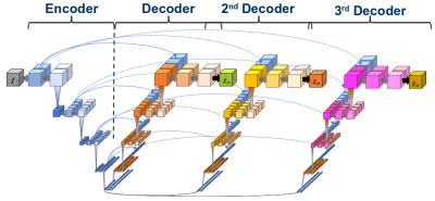 |
U3-Net for Deep Vector QSM – Solving the Susceptibility Tensor Phase Model in Single-Orientation MRI
Edith Franziska Baader1, Thomas Jochmann2, Jens Haueisen2, Robert Zivadinov1,3, and Ferdinand Schweser1,3
1Buffalo Neuroimaging Analysis Center, Department of Neurology at the Jacobs School of Medicine and Biomedical Sciences, University at Buffalo, The State University of New York, Buffalo, NY, United States, 2Department of Computer Science and Automation, Technische Universität Ilmenau, Ilmenau, Germany, 3Center for Biomedical Imaging, Clinical and Translational Science Institute, University at Buffalo, The State University of New York, Buffalo, NY, United States
Quantitative susceptibility mapping (QSM) is increasingly being used to study the brain iron homeostasis and white matter pathology. However, all QSM algorithms that are currently used in the clinical setting use a physical model that neglects the well-established anisotropic magnetic susceptibility of myelin. In this work, we demonstrate that an extended U-Net allows solving a Vector QSM model that accounts for field perturbations caused by off-diagonal tensor elements. The proposed Deep Vector QSM yielded improved estimates of χ33 compared to conventional QSM.
|
|
3203.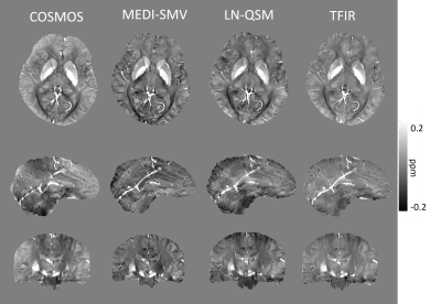 |
TFIR - A Spatial Frequency and R2* Informed Regularization for Total Field Inversion in Quantitative Susceptibility Mapping.
Priya S Balasubramanian1, Lingfei Guo2, Weiyuan Huang2, Pascal Spincemaille3, and Yi Wang4
Video Permission Withheld
1Electrical and Computer Engineering, Cornell University, Ithaca, NY, United States, 2Weill Cornell, Cornell University, New York, NY, United States, 3Weill Cornell, Cornell University, New York City, NY, United States, 4Biomedical Engineering, Cornell University, New York, NY, United States
TFIR is a novel regularization framework for total field quantitative susceptibility mapping. This method employs spatial frequency selection and R2* information within the L2 regularization method to map field to susceptibility source. It outperforms local field methods and existing regularization frameworks for total field susceptibility mapping, such as LN-QSM when computing error with respect to COSMOS and numerical and gadolinium phantoms. Hemorrhage cases and non-hemorrhage in vivo cases have reduced streaking and shadowing artifacts when reconstructed using TFIR compared to PDF-MEDI-SMV and LN-QSM. Future directions include spatial frequency selection automation in order to produce optimal signal to noise and accuracy.
|
|
3204.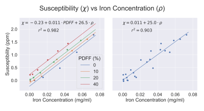 |
Phantom Validation of Simultaneous Mapping of PDFF, R2*, and Susceptibility
Collin J Buelo1,2, Ruiyang Zhao1,2, Ante Zhu2,3, Scott B. Reeder1,2,3,4,5, and Diego Hernando1,2,3
1Medical Physics, University of Wisconsin-Madison, Madison, WI, United States, 2Radiology, University of Wisconsin-Madison, Madison, WI, United States, 3Biomedical Engineering, University of Wisconsin-Madison, Madison, WI, United States, 4Medicine, University of Wisconsin-Madison, Madison, WI, United States, 5Emergency Medicine, University of Wisconsin-Madison, Madison, WI, United States
Chemical shift-encoded (CSE) MRI can be used to simultaneously obtain multi-parametric maps, including R2*, proton density fat fraction (PDFF), and tissue susceptibility, to aid in the diagnosis of diffuse liver disease. However, the presence of fat in tissue can confound both R2* and B0 field estimation and consequently quantitative susceptibility mapping (QSM). To validate this multi-parametric mapping, comparison of quantitative maps to phantoms with known iron concentrations were performed. This work demonstrated the feasibility of accurate PDFF mapping, and R2* and susceptibility measurements that are linearly dependent on both iron and fat concentration.
|
|
3205.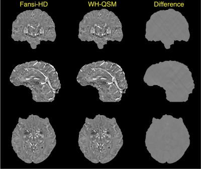 |
Hybrid Data fidelity term approach for Quantitative Susceptibility Mapping
Mathias Lambert1,2,3, Carlos Milovic1,2,3, and Cristián Tejos1,2,3
1Department of Electrical Engineering, Pontificia Universidad Católica de Chile, Santiago, Chile, 2Biomedical Imaging Center, Pontificia Universidad Católica de Chile, Santiago, Chile, 3Millennium Nucleus for Cardiovascular Magnetic Resonance, Santiago, Chile
Here we present the winner of the QSM challenge 2.0 in the RMS category, a novel hybrid data consistency method. We validated our method with in vivo data.
|
|
3206.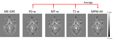 |
Can Multi-Parametric Mapping Sequences Be Used for Accurate Quantitative Susceptibility Mapping?
Russell Murdoch1, Jamie Kawadler2, David Carmichael2,3, Fenella Kirkham2, and Karin Shmueli1
1Department of Medical Physics and Biomedical Engineering, University College London, London, United Kingdom, 2Great Ormond Street Institute of Child Health, University College London, London, United Kingdom, 3Biomedical Engineering & Imaging Sciences, King's College London, London, United Kingdom
We compared the results of Quantitative Susceptibility Mapping (QSM) in each of the Proton Density (PD-), Magnetization Transfer (MT-) and T1-weighted Multi-Echo Gradient-Echo (ME-GRE) sequences in Multi-Parametric Mapping (MPM) with QSM from a conventional ME-GRE sequence. In deep grey matter (GM) regions, we found significant susceptibility (χ) correlations between each sequence pair. Correlation coefficients were lower in white matter (WM), particularly in T1-w and MT-w sequences. Averaging χ over MPM sequences increased correlations in WM and reduced noise in GM and WM. Whole-brain χ difference maps showed the largest χ differences in and around large veins and air spaces.
|
|
3207.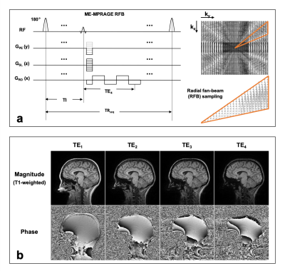 |
Simultaneous T1-weighted imaging, R2* mapping, and QSM from a multi-echo MPRAGE sequence using a radial fan-beam sampling scheme at 3 Tesla
Hongfu Sun1, M. Ethan MacDonald2, R. Marc Lebel3, and G. Bruce Pike2
1University of Queensland, Brisbane, Australia, 2University of Calgary, Calgary, AB, Canada, 3General Electric Healthcare, Calgary, AB, Canada
A multi-echo MPRAGE sequence with radial fan-beam segments is demonstrated at 3 T. The radial fan-beam sampling scheme permits any number of encoding steps by adjusting the fan size for each inversion segment, which allows longer echo time for magnetic susceptibility contrast. Simultaneous T1-weighted image, R2* map, and QSM were successfully extracted from the single acquisition and were compared with images reconstructed from standard single-echo MPRAGE and multi-echo GRE acquisitions.
|
|
3208. |
Use of high-resolution QSM to identify global patterns in the ex vivo human brain cortex at 7T
Leah Morgan1, Berkin Bilgic1, Andre van der Kouwe1,2, Jean Augustinack1,2, Jonathan Polimeni2,3, Anna Blazejewska1,2, Viviana Siless1,2, Allison Stevens1, Bram R Diamond1, Natalya Slepneva1, Bruce Fischl1, and Divya Varadarajan1
1A. A. Martinos Center for Biomedical Imaging, Massachusetts General Hospital, Boston, MA, United States, 2Harvard Medical School, Cambridge, MA, United States, 3Harvard-MIT Division of Health Sciences and Technology, Boston, MA, United States
Quantitative susceptibility mapping (QSM) can reflect the iron and myelin concentration within human brain tissue. Ex vivo MRI can allow us to study the cortical laminar structure at high-resolution. Here we conduct a study to combine the two, that is to analyze QSM maps of an ex vivo human brain cortex at 500um and 150um resolution. Our analysis shows matching global patterns of positive susceptibility at the gray-white boundary for both resolutions that were validated with iron-stain on a tissue-block. We find visual cortex, and localized regions of parietal and frontal lobe to contain the highest concentration of positive susceptibility.
|
|
3209.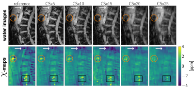 |
Highly Accelerated Compressed-Sensing-Based Field-mapping is Possible for Body QSM Applications
Christof Böhm1, Julio Oscanoa1,2, Sophia Kronthaler1, Maximilian N. Diefenbach1,3, Alexandra Gersing1, Jakob Meineke4, and Dimitrios C. Karampinos1
1TUM, Munich, Germany, 2Stanford University, Stanford, CA, United States, 3Division of Infectious Diseases and Tropical Medicine, University Hospital, LMU, Munich, Germany, 4Philips Research, Hamburg, Germany Body QSM relies on accurate magnetic field-mapping that particularly accounts for the presence of fat. Due to the need to account for T2* effects and perform water–fat separation, water–fat-imaging-based field-mapping typically acquires multiple echoes with appropriate echo time step, increasing the total required scan time especially for high-resolution body QSM. To reduce scan time, several acceleration schemes have been proposed, such as parallel imaging and compressed sensing (CS). This work demonstrates the feasibility of high CS acceleration factors to distinguish strong para- and diamagnetic susceptibility sources in body regions with CS acceleration factors of 10 and higher. |
3210.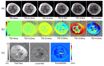 |
Ultrashort Echo Time Quantitative Susceptibility Mapping (UTE-QSM) as a Highly Sensitive Biomarker for Hemophilic Arthropathy
Hyungseok Jang1, Annette von Drygalski1, Xing Lu1, Yajun Ma1, Srila Gopal1, Jiang Du1, and Eric Chang1,2
1University of California, San Diego, San Diego, CA, United States, 2VA San Diego Healthcare System, San Diego, CA, United States
Hemophilia is a genetic bleeding disorder afflicting about 20,000 people in the US and over 400,000 people in the world. Severe hemophilia is characterized by frequent joint bleeding, resulting in debilitating arthropathy because of toxic iron depositions (e.g., hemosiderin) in synovium and cartilage. Development of a sensitive, non-invasive biomarker is of high importance to determine efficacy of costly treatment plans. In this study, we investigate the feasibility of ultrashort echo time-based QSM (UTE-QSM) to identify hemosiderin deposition and to provide a sensitive biomarker for joint disease in hemophilia.
|
|
3211.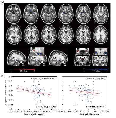 |
Quantitative susceptibility mapping and PET indicate iron-related cognitive decline during aging
Lin Chen1,2,3, Anja Soldan4, Kenichi Oishi1, Andreia Faria1, Marilyn Albert4, Peter C.M. Van zijl1,2, and Xu Li1,2
1Department of Radiology and Radiological Sciences, Johns Hopkins University, Baltimore, MD, United States, 2F.M. Kirby Research Center for Functional Brain Imaging, Kennedy Krieger Institute, Baltimore, MD, United States, 3Department of Electronic Science, Fujian Provincial Key Laboratory of Plasma and Magnetic Resonance, Xiamen University, XIAMEN, China, 4Department of Neurology, Johns Hopkins University, Baltimore, MD, United States
We investigated the associations between brain iron levels, as measured by QSM MRI, and amyloid-β plaque load as measured by 11C-PiB PET imaging, and their possible synergistic effect on both global composite and domain specific cognitive functions, including memory, visuospatial processing and language. Various association patterns were observed between iron load and amyloid-β deposition in voxel-based analysis. More importantly, in cognitively normal adults, iron levels in several brain regions were found negatively associated with cognition. This was independent of amyloid-β load, suggesting that the impact of iron on cognition may be related to other known molecular changes during early aging.
|
|
3212.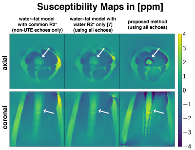 |
Sampling and Modeling UTE Signals is Important to Estimate Bone Marrow Susceptibility
Christof Böhm1, Sophia Kronthaler1, Maximilian N. Diefenbach1,2, Jakob Meineke3, and Dimitrios C. Karampinos1
1TUM, Munich, Germany, 2Division of Infectious Diseases and Tropical Medicine, University Hospital, LMU, Division, Munich, Germany, 3Philips Research, Hamburg, Germany
Body QSM relies on accurate magnetic field-mapping. A well-established water–fat voxel signal model with shared $$$R_2^*$$$ has successfully been used for body QSM based on multi-echo data at conventional echo times. However, when UTE echoes are recorded to obtain field-map information in previously unavailable MR signal voids, such as trabecular bone, the above water–fat model is no longer accurate. Therefore a joint 2-signal-model fitting using graph-cuts is proposed and applied. Further, this work demonstrates the importance of UTE echoes to obtain correct susceptibility values of cortical bone and more importantly, for bone marrow.
|
|
3213.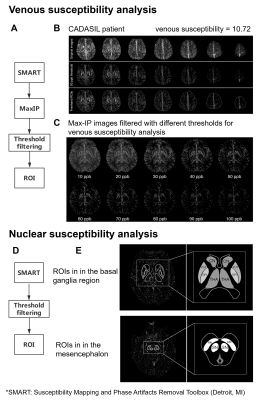 |
Increased susceptibility in cortical veins and nuclei in small vessel disease revealed by susceptibility weighted imaging and mapping at 7T
Yue Wu1,2,3, Qingle Kong1,2,3, Chen Ling4, Chengyue Sun4, Jing An5, Rong Xue1,2,3, Yan Zhuo1,2,3, Qi Yang6, Yun Yuan4, and Zihao Zhang1,2,3
1State Key Laboratory of Brain and Cognitive Science, Institute of Biophysics, Chinese Academy of Sciences, Beijing, China, 2University of Chinese Academy of Sciences, Beijing, China, 3CAS Center for Excellence in Brain Science and Intelligence Technology, Beijing, China, 4Department of Neurology, Peking University First Hospital, Beijing, China, 5Siemens Shenzhen Magnetic Resonance Ltd., Shenzhen, China, 6Department of Radiology, Xuanwu Hospital, Capital Medical University, Beijing, China
Cerebral autosomal dominant arteriopathy with subcortical infarcts and leukoencephalopathy (CADASIL) is the most common hereditary cerebral small vessel disease (SVD). This study aimed to use susceptibility weighted imaging and mapping (SWIM) at 7 Tesla to evaluate venous oxygen saturation (SvO2) and subcortical nuclear iron deposition and explore the correlation of these parameters with the clinical phenotypes of CADASIL patients. We found decreases in SvO2 and abnormal iron deposition in patients compared with healthy individuals. Also, we found associations between susceptibilities and clinical characteristics of CADASIL. These results suggest that SWIM can be used to assess clinical conditions of SVD patients.
|
|
3214.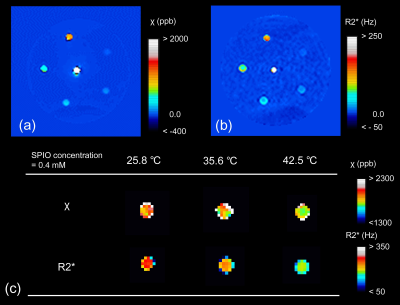 |
Sensitivity of Quantitative Susceptibility Mapping to Temperature: A Phantom Study
Hirohito Kan1,2, Yuto Uchida3,4, Masahiro Takizawa5, Tosiaki Miyati6, Hiroshi Kunitomo7, Nobuyuki Arai7, Harumasa Kasai7, and Yuta Shibamoto2
1Radiological and Medical Laboratory Sciences, Nagoya University Graduate School of Medicine, Nagoya City, Japan, 2Department of Radiology, Nagoya City University Graduate School of Medical Sciences, Nagoya City, Japan, 3Department of Neurology, Nagoya City University Graduate School of Medical Sciences, Nagoya City, Japan, 4Department of Neurology, Toyokawa City Hospital, Toyokawa, Japan, 5Healthcare Business Unit, Hitachi, Ltd., Tokyo, Japan, 6Faculty of Health Sciences, Institute of Medical, Pharmaceutical and Health Sciences, Kanazawa University, Kanazawa, Japan, 7Department of Radiology, Nagoya City University Hospital, Nagoya City, Japan Poster Permission Withheld
To date, we are unaware of any reports regarding quantitative susceptibility mapping (QSM) at different temperatures. To clarify the temperature dependence of susceptibility estimated by QSM analysis, we investigated the relationships between temperature and susceptibility using a simple cylinder phantom with varying temperatures. This study has demonstrated that a significant inverse correlation was found between the temperature and the susceptibility value estimated by QSM analysis. This dependence might be the confounding factor of QSM-based iron estimation.
|
|
3215.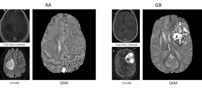 |
3D Texture Analysis of Quantitative Susceptibility Mapping distinguishes Anaplastic Astrocytoma from Glioblastoma
Jinwei Zhang1, Shun Zhang2, Hersh Patel3, Jacquelyn Knapp4, Gloria Chiang2, David Pisapia2, John Tsiouris2, Linda Heier2, Pascal Spincemaille2, Thanh Nguyen2, Yi Wang2, and Ilhami Kovanlikaya2
1Biomedical Engineering, Cornell University, Ithaca, NY, United States, 2Radiology, Weill Cornell Medical College, New York, NY, United States, 3New York Presbyterian Hospital, New York, NY, United States, 4Cornell University, New York, NY, United States
3D texture analysis-based feature extraction was deployed on QSM images of malignant astrocytoma (Anaplastic Astrocytoma (AA), grade III and Glioblastoma (GB), grade IV) and support vector classifier (SVC) (1) was trained and tested on multiple training-test dataset splits with different numbers of selected features using cross-validation (CV). Experiments indicate texture analysis on QSM is useful for differentiating AA and GB with high accuracy (94.7%).
|
|
3216.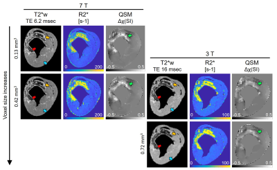 |
The effect of voxel size on magnetic susceptibility and R2* of intramyocardial hemorrhage at 3 and 7 T
Brianna F. Moon1, Srikant Kamesh Iyer2, Nicholas J. Josselyn2, James J. Pilla2, Joseph H. Gorman III3, Robert C. Gorman3, Cory Tschabrunn4, Giovanni Ferrari5, Yuchi Han4, Harold I. Litt2, and Walter R. Witschey2
1Bioengineering, University of Pennsylvania, Philadelphia, PA, United States, 2Radiology, Perelman School of Medicine, University of Pennsylvania, Philadelphia, PA, United States, 3Surgery, Perelman School of Medicine, University of Pennsylvania, Philadelphia, PA, United States, 4Medicine, Perelman School of Medicine, University of Pennsylvania, Philadelphia, PA, United States, 5Surgery, Columbia University Irving Medical Center, New York City, NY, United States
Intramyocardial hemorrhage is a complication of reperfused myocardial infarction. Quantitative susceptibility mapping (QSM), R2*-mapping and T2*-weighted images provide quantitative assessment and image contrast based on the presence of iron. We sought to determine the relationship between magnetic susceptibility and R2* with varying voxel size at 3 and 7 T, where tissue magnetic susceptibility showed independence of field strength but dependence on voxel size. At high resolution the detection of heterogeneous infarct magnetic susceptibility improved in a large animal model of myocardial infarction.
|
|
3217.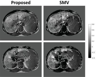 |
Comparison of Background Field Removal Strategies in Joint Quantitative Susceptibility Mapping in Abdomen
Julia Velikina1, Ante V Zhu2,3, Collin V Buelo1,4, Jing Zhou5, Timothy J Colgan2, Kritisha Rajlawot5, Bingjun He5, Jin Wang5, Scott B Reeder1,3,4,6,7, Alexey Samsonov4, and Diego Hernando1,2,3
1Medical Physics, University of Wisconsin, Madison, WI, United States, 2Radiology, University of Wisconsin - Madison, Madison, WI, United States, 3Biomedical Engineering, University of Wisconsin - Madison, Madison, WI, United States, 4Radiology, University of Wisconsin, Madison, WI, United States, 5Sun Yat-sen University, Guangzhou, China, 6Medicine, University of Wisconsin - Madison, Madison, WI, United States, 7Emergency Medicine, University of Wisconsin - Madison, Madison, WI, United States
Quantitative susceptibility mapping (QSM) is a promising technique for direct measure of iron concentration in vivo. In abdominal imaging, QSM faces challenges of strong background field, presence of fat, complex anatomy, low image resolution, and rapid signal decay with high iron concentration. Many methods for background field removal utilize spherical mean value property of harmonic functions, resulting in straightforward approach with known limitations in accuracy and edge preservation. Here, we propose an alternative background field removal method based on direct implementation of the Laplace operator and compare its performance with spherical mean value kernels within the joint QSM estimation framework.
|
|
3218.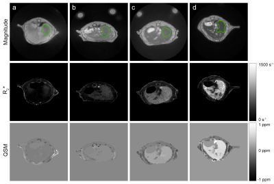 |
Liver iron content measurement in living mice with quantitative susceptibility mapping
Anton Abyzov1, Joao Piraquive1, Katell Peoc'h2, Philippe Garteiser1, and Bernard E. Van Beers1
1Laboratory of Imaging Biomarkers, Center of Research on Inflammation, UMR1149 Inserm-University Paris Diderot, Paris, France, 2Department of biochemistry, University Hospital Paris Nord - Beaujon, AP-HP, Clichy, France
To evaluate the feasibility of quantitative susceptibility mapping (QSM) in living mice liver and the robustness of QSM and transverse relaxation rate (R2*) based liver iron quantification. We show that QSM can be accurately performed in mice liver despite respiratory motion and that magnetic susceptibility measurements correlate more strongly with inductively coupled plasma mass spectrometry (ICP-MS) based iron quantification and have smaller standard deviations and narrower Bland-Altman agreement limits than R2* measurements.
|
|
3219.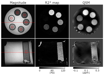 |
Using QSM to Quantify Microbubble Concentrations
Barbara Dymerska1, Bernard Siow2, and Karin Shmueli1
1Medical Physics and Biomedical Engineering, University College London, London, United Kingdom, 2Magnetic Resonance Imaging, The Francis Crick Institute, London, United Kingdom
Microbubbles are a well-established intravascular ultrasound contrast agent. There is increasing interest in MRI-guided microbubble-mediated focused ultrasound treatments such as thermal surgery. MRI magnitude has a non-linear and non-local dependence on microbubble size and volume fraction, making it unsuitable for estimating microbubble concentrations. Quantitative Susceptibility Mapping (QSM) is a strong candidate for tracking microbubble concentration, destruction and clearance because the susceptibility depends linearly on the volumetric bubble concentration. Here, we show the first QSM of microbubbles in a phantom and observe that the measured susceptibility has a high SNR and is directly proportional to the microbubble volumetric concentration.
|
|
3220.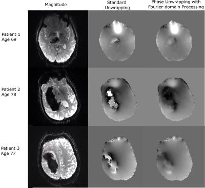 |
Application of Fourier-domain Analysis Based Unwrapping Technique in Quantitative Susceptibility Mapping (QSM) of Intracerebral Hemorrhage
Ashmita De1, Hongfu Sun1, Kenneth S. Butcher2, and Alan H. Wilman1
1Department of Biomedical Engineering, University of Alberta, Edmonton, AB, Canada, 2Division of Neurology, Department of Medicine, University of Alberta, Edmonton, AB, Canada
QSM offers a means to measure iron content changes in hemorrhage. However, the rapid T2* decay in hemorrhage causes a severe signal loss resulting in low SNR which cause failure of standard phase unwrapping in certain cases. Fourier-domain analysis based unwrapping technique may be useful to produce susceptibility maps of hemorrhages having low SNR. This method removes residual phase wraps resulting in artifact-free QSM with boundaries of the hemorrhage area more distinct to facilitate area measurement. Thus, this method can provide more precise susceptibility maps in cases where conventional unwrapping methods fail.
|
|
3221.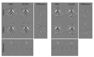 |
Clinical viable QSM using accelerated, segmented 3D EPI with inline, automated QSM post-processing
Monique Tourell1,2, Ashley Stewart1,2, Jin Jin2,3,4,5, Sunil Patil6, Kieran O'Brien1,2,3, Simon Robinson1,7,8, and Markus Barth1,2,5
1Centre for Advanced Imaging, University of Queensland, Brisbane, Australia, 2ARC Training Centre for Innovation in Biomedical Imaging Technology, The University of Queensland, Brisbane, Australia, 3Siemens Healthcare Pty Ltd, Brisbane, Australia, 4Mark and Mary Stevens Neuroimaging and Informatics Institute, University of Southern California, Los Angeles, CA, United States, 5School of Information Technology and Electrical Engineering, the University of Queensland, Brisbane, Australia, 6Siemens Healthcare Pty Ltd, Malvern, PA, United States, 7High Field Magnetic Resonance Centre, Department of Biomedical Imaging and Image-guided Therapy, Medical University of Vienna, Vienna, Austria, 8Department of Neurology, Medical University of Graz, Graz, Austria
Successful translation of QSM into the clinic requires the combination of fast imaging protocols with robust, automated on-scanner processing. In this work we present clinical viable QSM: acquisition of phase data using accelerated, segmented 3D EPI in 30 seconds, and automated QSM post-processing on the scanner in under 2 minutes. The resultant susceptibility maps were of comparable quality to those obtained using a GRE sequence. Analysis of the susceptibilities measured within the caudate, putamen and pallidum demonstrates that 3D EPI with on-scanner processing produces susceptibility values in good agreement with those obtained from a GRE sequence, and offline processing pipelines.
|
|
3222.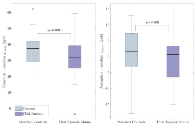 |
Reduced QSM values in the caudate and amygdala of first episode mania patients
Vanessa Wiggermann1,2, Enedino Hernández-Torres3, Christian Kames1,2, Leonardo E daSilveira4,5, Taj Dhanoa5, Alexander Rauscher1,2, and Lakshmi N Yatham5,6
1Physics and Astronomy, University of British Columbia, Vancouver, BC, Canada, 2Pediatrics, University of British Columbia, Vancouver, BC, Canada, 3Medicine (Neurology), University of British Columbia, Vancouver, BC, Canada, 4Laboratory of Molecular Psychiatry, Centro de Pesquisas Experimentais, Hospital de Clínicas de Porto Alegre, Porto Algre, Brazil, 5Mood Disorders Centre, University of British Columbia, Vancouver, BC, Canada, 6Psychiatry, University of British Columbia, Vancouver, BC, Canada
Bipolar I disorder is characterized by recurrent manic and depressive episodes. Both, dopamine and oxidative stress, resulting from mitochondrial dysfunction, have been implicated in BD and volumetric and metabolic changes have been reported. Nevertheless, it remains unknown whether changes result from treatment or ongoing disease, or if they are present at first episode mania. We used R2* and quantitative susceptibility mapping (QSM) to assess differences in brain iron and myelination in a cohort of first episode mania patients compared to controls. Patients exhibited lower QSM values than controls in the caudate and amygdala, possibly linked to iron loss or hypermyelination.
|
|
3223.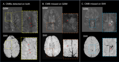 |
Increased sensitivity for cerebral microbleed detection on SWI compared to QSM
Sivakami Avadiappan1, Melanie A Morrison1, Yicheng Chen1, Angela Jakary1, Christopher P Hess1, and Janine M Lupo1
1University of California San Francisco, San Francisco, CA, United States
Although susceptibility weighted imaging (SWI) has been the standard method for quantifying and evaluating cerebral microbleeds, more recently total lesion susceptibility obtained from quantitative susceptibility mapping (QSM) has been found to correlate with measures of disease burden and can provide more accurate estimates of microbleed volume. This study found that 2.5 times as many CMBs can be detected on SWI compared to QSM images, with the number of CMBs missed by QSM increasing as a function of decreasing CMB size, highlighting the importance of including SWI for a more accurate assessment of disease burden in these patients.
|
|
3224.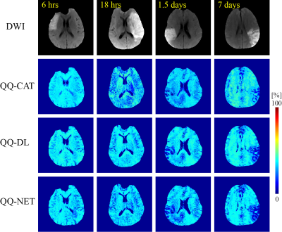 |
Sparsity based machine learning algorithms for oxygen extraction fraction mapping
Junghun Cho1, Hae-Yeoun Lee2, jinwei Zhang1, Pascal Spincemaille3, Hang Zhang4, Simon Hubertus5, Yan Wen1, Ramin Jafari1, Shun Zhang3, Thanh Nguyen3, Ajay Gupta3, and Yi Wang1,3
1Biomedical Engineering, Cornell University, New York, NY, United States, 2Computer Software Engineering, Kumoh National Institute of Technology, Gumi, Republic of Korea, 3Radiology, Weill Cornell Medical College, New York, NY, United States, 4Electrical and Computer Engineering, Cornell University, New York, NY, United States, 5Computer Assisted Clinical Medince, Heidelberg University, Mannheim, Germany
In this work, dictionary and deep learning based algorithms are developed that take advantage of sparse signal representations to improve the accuracy and speed of oxygen extraction fraction (OEF) mapping based on the QSM+qBOLD (QQ) modeling of multi-echo gradient echo data without vascular challenge. The developed dictionary learning (QQ-DL) and deep neural network (QQ-NET) algorithms are significantly faster and provide more accurate OEF maps in simulation than a current algorithm based on cluster analysis of time evolution (CAT). In ischemic stroke patients, QQ-DL and QQ-NET show OEF maps that are consistent with DWI-defined lesions.
|
|
3225.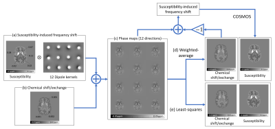 |
Generalized methods for separating susceptibility and chemical shift/exchange
Hwihun Jeong1, Hyunsung Eun1, and Jongho Lee1
1Department of Electrical and Computer Engineering, Seoul National University, Seoul, Republic of Korea
We proposed two generalized methods, weighted-average and least-squares methods, to separate susceptibility and chemical shift/exchange using multiple arbitrary B0 directional phase images. Compared to the previous method that requires three orthogonal directional data, the proposed methods can utilize data with smaller head rotation. A preliminary in-vivo result is presented.
|
3226.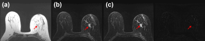 |
Semi-Supervised Image Domain Transfer for Dixon Water and Fat Separation
Jong Bum Son1, Ken-Pin Hwang1, Marion E. Scoggins2, Basak E. Dogan3, Gaiane M. Rauch2, Mark D. Pagel4, and Jingfei Ma1
1Imaging Physics Department, The University of Texas MD Anderson Cancer Center, Houston, TX, United States, 2Diagnostic Radiology Department, The University of Texas MD Anderson Cancer Center, Houston, TX, United States, 3Department of Diagnostic Radiology, The University of Texas Southwestern Medical Center, Dallas, TX, United States, 4Cancer Systems Imaging Department, The University of Texas MD Anderson Cancer Center, Houston, TX, United States
Deep learning neural-networks for Dixon imaging require a large number of “paired” input and output images for network training. Moreover, the previous methods require Dixon images as their network input, thus they could not be used to reconstruct water images from regular T1 or T2-weighted images. In this work, we propose an image domain transfer based deep-learning network which can reconstruct water images from either T1 or T2-weighted MR images. Using semi-supervised learning, two separate groups of “unpaired and unordered” input and output images were used to translate either T1 or T2-weighted images to their corresponding water-only images.
|
|
3227.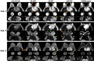 |
A Novel Chemical Shift-Selective Fat Saturation RF Pulse Design with Robustness to B0/B1 Inhomogeneity: A Demonstration on Breast MRI at 3T
Feng Xu1,2, Wenbo Li1,2, Dapeng Liu1,2, Dan Zhu3, Kelly Myers1, Michael Shär1, and Qin Qin1,2
1The Russell H. Morgan Department of Radiology and Radiological Science, Johns Hopkins University, School of Medicine, Baltimore, MD, United States, 2F.M. Kirby Research Center for Functional Brain Imaging, Kennedy Krieger Institute, Baltimore, MD, United States, 3Biomedical Engineering, Johns Hopkins University, School of Medicine, Baltimore, MD, United States
Chemical shift-selective fat saturation (CHESS) is the most commonly used fat suppression technique for clinical MRI. Conventional Gaussian-shaped pulses are sensitive to B1 inhomogeneity and their wide transitional band can be affected by B0 off-resonance. Uniform fat saturation across a large field of view (FOV) is especially challenging for body and breast MRI at 3T. Here, we designed a novel frequency-selective RF pulse based on the optimal control theory with robustness to a targeted wide range of B0/B1 conditions. Its superior performance than the regular ones was demonstrated using T1-weighted sequences with whole-breast coverage.
|
|
3228.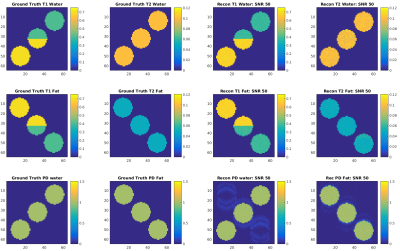 |
Multi-parametric quantification of multiple spectral components by an extended MR-STAT framework
Michael A. Eijbersen1, Hongyan Liu1, Oscar van der Heide1, Cornelis A.T. van den Berg1, and Alessandro Sbrizzi1
1Computational Imaging group for MR Diagnostics and Therapy, Center for Image Sciences, University Medical Center Utrecht, Utrecht, Netherlands
We present a novel method called Bicompartemental MR-STAT for simultaneous quantification T1, T2 and Proton density for two resonances based upon the MR-STAT framework. T1 and T2 may vary per compound in this framework. Additionally B0 is reconstructed by taking advantage of multple readouts within each TR which allows for separate but accurate B0 reconstruction.
|
|
3229.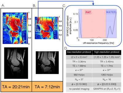 |
Optimized fast fat-fraction mapping in the knee using Signal Profile Asymmetries for Robust multi-Compartment Quantification (SPARCQ)
Adèle LC Mackowiak1,2,3, Tom Hilbert3,4, Giulia MC Rossi1,3, Tobias Kober3,4, and Jessica AM Bastiaansen1
1Department of Diagnostic and Interventional Radiology, Lausanne University Hospital (CHUV) and University of Lausanne (UNIL), Lausanne, Switzerland, 2Department of Physics, Ecole Polytechnique Fédérale de Lausanne (EPFL), Lausanne, Switzerland, 3Advanced Clinical Imaging Technology, Siemens Healthcare AG, Lausanne, Switzerland, 4LTS5, Ecole Polytechnique Fédérale de Lausanne (EPFL), Lausanne, Switzerland
The Signal Profile Asymmetries for Robust multi-Compartment Quantification (SPARCQ) framework was recently developed and used to quantify water and fat content in tissues. Applying SPARCQ on bSSFP signal profiles obtained from phase-cycled acquisitions provided reliable fat fractions with a total scan time of 20:21min at low resolution. In this study, SPARCQ was accelerated and optimized for water-fat separation using numerical simulations and in vivo experiments to obtain a clinically acceptable protocol. Images and fat-fraction maps of knees at an isotropic resolution of (1.25mm)3 were obtained with a scan time of 7:12min.
|
|
3230. |
Multiple dual-echo reconstructions to improve the robustness of water-fat MRI
Samir D. Sharma1
1Canon Medical Research USA, Mayfield Village, OH, United States
A challenge in water-fat imaging is robust separation of the water and fat components. In this work, we propose a method that performs a series of dual-echo reconstructions using different pairs of echoes. The estimates from the multiple reconstructions are then combined to generate a more robust initial estimate of the water and fat images. The final estimates are then calculated via voxel-wise minimization. The proposed multiple dual-echo method demonstrates an improvement to single dual-echo reconstruction for water-fat imaging.
|
|
3231.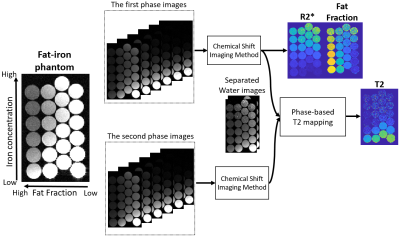 |
Multiparameter mapping of T2, R2*, fat fraction using a combination of chemical shift imaging and phase-based T2 mapping
Daiki Tamada1, Xiaoke Wang1, Diego Hernando1,2, and Scott B. Reeder1,2,3,4,5
1Radiology, University of Wisconsin-Madison, Madison, WI, United States, 2Medical Physics, University of Wisconsin-Madison, Madison, WI, United States, 3Biomedical Engineering, University of Wisconsin-Madison, Madison, WI, United States, 4Medicine, University of Wisconsin-Madison, Madison, WI, United States, 5Emergency Medicine, University of Wisconsin-Madison, Madison, WI, United States
A new multi-parameter method for quantitative mapping of T2, R2*, and fat fraction using CSE-MRI combined with phase-based T2 mapping was developed. This method can be performed using the RF phase-modulated gradient echo sequences that encode T2 information into the phase of the steady-state signal. A phantom study demonstrating the ability of this approach to obtain 3D high-resolution quantitative maps in reasonable acquisition time.
|
|
3232.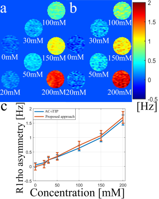 |
A pulse sequence for robust and efficient quantitative R1rho asymmetry imaging to probe chemical exchange effect
Baiyan Jiang1 and Weitian Chen1
1Department of Imaging and Interventional Radiology, The Chinese University of Hong Kong, Hong Kong, Hong Kong
The measurement of R1rho (1/T1rho) spectrum and its asymmetry have several advantages over Chemical Exchange Saturation Transfer (CEST) to probe chemical exchange effect. Previously reported AC-iTIP approach is able to obtain R1rho-spectrum and R1rho asymmetry robustly. However, it suffers from long scan time. In this work, we proposed a new AC-iTIP approach to reduce the scan time by approximately 30%~40%, without compromising the robustness.
|
|
3233.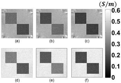 |
Single Current Diffusion Tensor Magnetic Resonance Electrical Impedance Tomography: A Simulation Study
Mehdi Sadighi1, Mert Şişman1, Berk Can Açıkgöz1, and B. Murat Eyüboğlu1
1Electrical and Electronics Engineering Dept., Middle East Technical University (METU), Ankara, Turkey
Diffusion Tensor Magnetic Resonance Electrical Impedance Tomography (DT-MREIT) is an imaging technique providing low-frequency conductivity tensor images. In all DT-MREIT applications in the literature two linearly independent current injection patterns are used to reconstruct the extra-cellular conductivity and diffusivity ratio (ECDR) which is the space-dependent scale factor between the conductivity and diffusion tensors in a porous medium. In this study, a new approach is proposed to reconstruct ECDR using only one current injection pattern. The proposed method is evaluated using simulated measurements with different SNR levels. The obtained results show similar performance compared to the conventional two current DT-MREIT.
|
|
3234.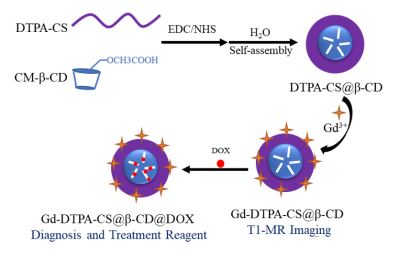 |
High-Efficient Metabolic Nano-Theranostics for MR Imaging and Cancer Therapy
Peng Wang1, Yaqiong Wang1, Jie Zhu2, Yong Zhang3, Ke-Xue Deng1, and Hui-Jun Dong1
1Radiology, The First Affiliated Hospital of University of Science and Technology of China, Hefei, China, 2Electronic information engineering, University of Aeronautics and Astronautics, Nanjing, China, 3GE Healthcare, Shanghai, China
In order to realize the nanotheranostics and reduce the toxicity of nanotheranostics agents to healthy tissues/organs, we constructed a novel nanotheranostics agents (Gd-DTPA-CS@β-CD@Dox) for MR imaging and Cancer treatment. Renal-clearable nanocomposites made it possible to overcome the long-term toxicity by accumulation in healthy living body. High uptake of the as-synthesized Gd-DTPA-CS@β-CD@Dox was achieved due to the longer blood circulation time. Moreover, the nanotheranostics agents possessed excellent performance in MRI. The nanotheranostics agents demonstrated a PH sensitive drug release. Such functional nanotheranostics agents applicable in highly integrated bio-modal imaging and multiple therapeutic functions may have great prospects in clinical practice.
|
|
3235.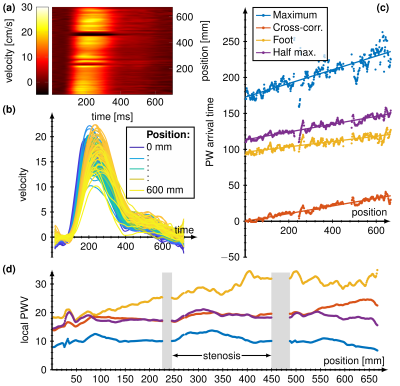 |
4D Flow for estimation of global and local pulse wave velocity in stenotic femoral arteries
Ingo Hermann1,2, Martin Sigl3, Stefan Schönberg4, Lothar R. Schad1, and Frank G. Zöllner1
1Computer Assisted Clinical Medicine, Medical Faculty Mannheim, University Heidelberg, Mannheim, Germany, 2Magnetic Resonance Systems Lab, Department of Imaging Physics, Delft University of Technology, Delft, Netherlands, 3First Department of Medicine, Medical Faculty Mannheim, Mannheim, Germany, 4Institute of Clinical Radiology and Nuclear Medicine, Medical Faculty Mannheim, Mannheim, Germany
Pulse wave velocity (PWV) is a promising biomarker for decreasing artery diameter and wall stiffness. Recent work showed the possibility to calculate the local PWV from the velocity profil time delay along the artery out of 4D Flow measurements. In this study we used 4 different models (half maximum, maximum, foot-to-foot, cross-correlation) to compare and calculate the PWV in the relatively small femoral arteries in five subjects with stenotic arteries. We found an increased PWV of up to 20 m/s compared to literature values of healthy volunteers. Cross-correlation and half maximum methods yielded consistent results.
|
|
3236.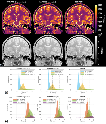 |
Parameter estimation with matrix-based signal models using VARPRO for transient- and steady-state imaging
Nam Gyun Lee1 and Krishna S. Nayak2
1Biomedical Engineering, University of Southern California, Los Angeles, CA, United States, 2Electrical and Computer Engineering, University of Southern California, Los Angeles, CA, United States VARPRO-based parameter estimation is extensively used in MR for its improved accuracy, precision, and convergence behavior compared to general nonlinear least-squares algorithms. This study investigates the feasibility of using matrix-based signal models with VARPRO instead of conventional analytic signal equations. Simulations and in-vivo study show that VARPRO with matrix-based signal models is identical to VARPRO with analytic signal equations, and both VARPRO approaches provide enhanced precision and accuracy in relaxometry maps compared to the conventional DESPOT1/2 methods from variable flip angle SPGR and bSSFP measurements. |
|
3237.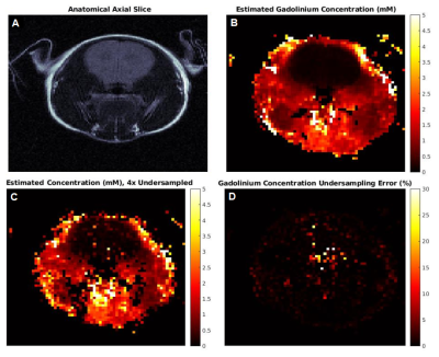 |
Simultaneous Quantification of SPIO and Gd contrast agents using MR Fingerprinting
Anna Marriott1,2, Chris Bowen1,2, James Rioux1,2, and Kimberley Brewer1,2
1Biomedical Translational Imaging Centre (BIOTIC), HALIFAX, NS, Canada, 2Department of Physics & Atmospheric Science, Dalhousie University, HALIFAX, NS, Canada
Superparamagnetic Iron Oxide (SPIO) contrast agents are used extensively in molecular imaging studies as a tool to evaluate various SPIO-labelled cells. These studies would be greatly improved if multiple contrast agents could be quantified simultaneously. We combined an SSFP MR fingerprinting sequence with TE variation, and an extension of a concentration-dependent linear model, to achieve dual quantification and show viability of SPIO labelled CD8+ T cell mapping. As a pilot study for in vivo application, we demonstrate concentration mapping of a mouse injected with gadolinium, and R2* quantification of an SPIO labelled immune therapy injection site.
|
|
 |
3238.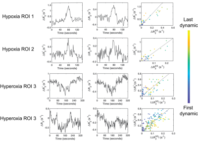 |
Vessel size imaging using hypoxia and hyperoxia gas challenges with spin- and gradient-echo (SAGE) MRI
Chau Vu1, Jian Shen1, and John C. Wood1,2
1Department of Biomedical Engineering, University of Southern California, Los Angeles, CA, United States, 2Departments of Pediatrics and Radiology, Children's Hospital Los Angeles, Los Angeles, CA, United States
This study demonstrated the feasibility of using hyperoxia and transient hypoxia gas challenges in conjunction with spin- and gradient-echo (SAGE) MRI to estimate vessel size index (VSI). Hypoxia yielded an average VSI of 14μm within acceptable range for healthy tissue, whereas hyperoxia severely underestimated vessel sizes. Test-retest in two patients demonstrated repeatability of hypoxia-VSI. Future work to optimize the acquisition and cross-validate against other VSI methodologies is required to assess the diagnostic value of this technique.
|
3239.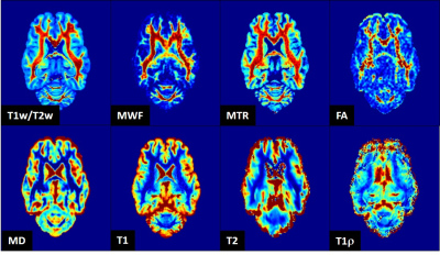 |
In Vivo Myelin Quantification by T1w/T2w: Its Relationship with Other MRI Quantifiers of Myelin and Specificity
Hiroyuki Hamaguchi1, Nina Patzke2, Yuta Urushibata3, Xinnan Li1, Isabel Fernandez2, and Khin Khin Tha4,5
1Department of Biomarker Imaging Science, Hokkaido University Graduate School of Biomedical Science and Engineering, Sapporo, Japan, 2Department of Biological Sciences, Faculty of Science, Hokkaido University, Sapporo, Japan, 3Siemens Healthineers, Tokyo, Japan, 4Department of Diagnostic and Interventional Radiology, Hokkaido University Hospital, Sapporo, Japan, 5Global Station for Quantum Medical Science and Engineering, Hokkaido University, Sapporo, Japan
The division of T1-weighted image by T2-weighted image (T1w/T2w) has recently been introduced as a myelin quantifier, which gained attention due to the ease of obtaining images. However, controversy exists about its performance. The aim of this study was to test the correlation of T1w/T2w with other MRI quantifiers of myelin, so as to determine its performance. Moderate to strong correlation of T1w/T2w with the other quantifiers was observed. T1w/T2w appears to be sensitive to detect an abnormality, but is not specific for myelin.
|
|
3240.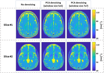 |
Accurate mapping of T2 relaxation times at low SNR, based on Bloch simulations and PCA image denoising
Neta Stern1, Dvir Radunsky1, Tamar Blumenfeld-Katzir1, and Noam Ben-Eliezer1,2,3
1Bio-medical Engineering, Tel Aviv University, Tel Aviv, Israel, 2Sagol School of Neuroscience, Tel Aviv University, Tel Aviv, Israel, 3Center for Advanced Imaging Innovation and Research (CAI2R), New-York University Langone Medical Center, New York, NY, United States Quantitative T2 mapping using Bloch simulations offers high mapping accuracy, albeit may suffer from reduced precision due to noisy data when operating at low SNR. In this work we tested the utility of PCA-based complex image denoising for increasing T2 mapping precision. Mapping was done using the echo modulation curve algorithm. Denoising was tested on phantom and in vivo scans. Comparing T2 maps before and after PCA complex image denoising showed an increase in T2 precision with no apparent loss of spatial resolution. |
|
3241.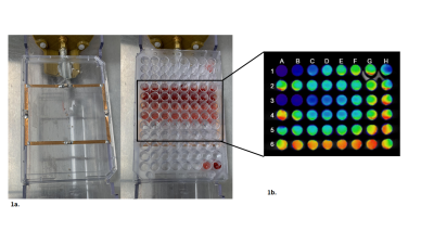 |
Rapid and Direct Quantification of Gadolinium from Blood using Time Resolved Fluorescence
James Tranos1, Ayesha Bharadwaj Das1, Zakia Ben Youss Gironda1, Nora Butler1, Jin Zhang1, Stewart Russell2, S. Gene Kim1, and Youssef Zaim Wadghiri1
1Bernard & Irene Schwartz Center for Biomedical Imaging (CBI), Department of Radiology and Center for Advanced Imaging Innovation and Research (CAI2R), New York University School of Medicine, New York, NY, United States, 2Institute for Ultrafast Spectroscopy and Lasers, The City College of New York, New York, NY, United States
There are increasing needs to quantify the concentration of gadolinium for use in pharmacokinetic modeling, and traditional MR techniques prove to be inadequate. As an alternative, Time Resolved Fluorescence (TRF) can be used to directly measure the concentration of gadolinium, using a GBCA conjugated to a fluorescent antenna moiety. Here we show that TRF serves as a sensitive and direct approach to quantify the concentration of a sensitized GBCA, as a bifunctional contrast agent.
|
3242.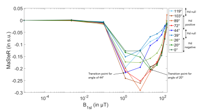 |
Magic Sandwich to Stimulated Echo Relative change to identify sign and intensity of dipolar interaction in anisotropic tissue
Eloïse MOUGEL1, Hélène RATINEY1, Eric VAN REETH1,2, Kevin TSE VE KOON1, Olivier BEUF1, and Denis GRENIER1
1Univ Lyon, INSA‐Lyon, Université Claude Bernard Lyon 1, UJM-Saint Etienne, CNRS, Inserm, CREATIS UMR 5220, U1206, F‐69616, LYON, France, 2CPE, VILLEURBANNE, France
Among the MR techniques playing on dipolar interaction (Hd), the magic sandwich echo sequence (MSE) is very seldom used in biological application. So far, the magic echo has been compared to the spin echo. However, MSE is closer to a stimulated echo thus a Magic Sandwich to Stimulated echo relative change (MaSteR) is of interest to study. The MaSteR evolution with spin-lock intensity increase was studied for thawed tendon for different orientations within B0 to modulate the dipolar interaction. We show that MaSteR is correlated to dipolar interaction, sample composition and its evolution with orientation is sensitive to Hd sign.
|
|
3243.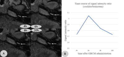 |
3D-FLAIR imaging with optimized scan parameters to visualize endolymphatic hydrops with intravenous gadolinium-based contrast media
Jinye Li1, Lixin Sun1, Long Li2, Hui Zhao1, Jing Tian1, Gesheng Song3, Na Hu1, Weiqiang Dou4, and Ruozhen Gong5
1Department of Radiology, Shandong Provincial ENT Hospital, Shandong Provincial ENT Hospital affiliated to Shandong University, Jinan, China, 2Department of Medical Service, Shandong Provincial ENT Hospital, Shandong Provincial ENT Hospital affiliated to Shandong University, Jinan, China, 3Shandong province Qianfoshan Hospital, Jinan, China, 4GE Healthcare, MR Research China, Beijing, China, 5Gong Ruozhen Innovation Studio, Shandong Medical Imaging Research Institute Affiliated to Shandong University, Jinan, China
We aimed to explore the optimal scan parameters of 3D-FLAIR technique in labyrinthine lymph imaging for patients with vertigo disease. With different scan times and scan baselines applied, the corresponding 3D-FLAIR images were systematically compared. We found that the optimal scan time was 6hours after intravenous administration of GBCM. Additionally, the optimal scan baseline, aiming to obtain the relative maximum areas of the saccule, utricle and lateral semicircular canal to be displayed at the same level, is about 10.74±2.24 degrees from the anterior skull base. Therefore, using these optimal parameters, an accurate determination of endolymphatic hydrops can be achieved.
|
|
3244.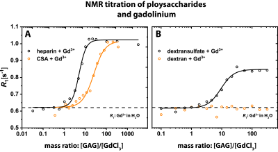 |
Biochemical Insights into the Molecular Mechanisms of Gadolinium Long-Term Deposition in the Human Body
Patrick Werner1,2, Patrick Schuenke1, Matthias Taupitz3, and Leif Schröder1
1Leibniz-Forschungsinstitut fuer Molekulare Pharmakologie, Berlin, Germany, 2BIOphysical Quantitative Imaging Towards Clinical Diagnosis (BIOQIC), Berlin, Germany, 3Department of Radiology, Charite Berlin, Berlin, Germany
The biochemical and molecular mechanisms of the gadolinium long-term deposition in biological tissues are widely unknown. In this study we prove that the observed hyperintensity of divined regions in the human body after clinical GBCA intervention can be caused by macromolecular species like glycosaminoglycans. We are able to show the importantance of sulfate groups and the influence of the sulfation state of these endogenous polysaccharides for this process by using MR relaxometry and isothermal titration calorimetry (ITC).
|
|
3245.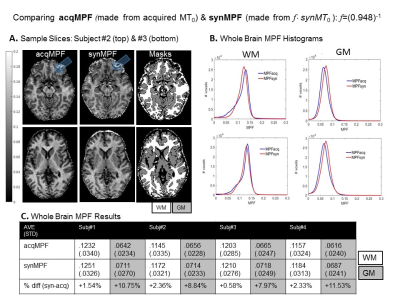 |
Macromolecular Pool Fraction (MPF) Maps in Minimal Scan Time Using a Modified Fast SPGR Sequence and a Calibrated Synthetic MT Reference Image
Kim Desmond1, Tobias C Wood2, and Sofia Chavez1
1CAMH, Toronto, ON, Canada, 2King's College, London, United Kingdom
MPF has been shown to correlate with myelin. Recent advances have reduced the required acquisitions to two: with (MTΔ) and without (MT0) the MT pulse. These, along with B1 and T1 maps, can be used to compute MPF. Scan time can be minimized by synthesizing MT0 from the T1 and B1 maps required for MPF mapping thus obviating the need to acquire MT0. Here we present advances that allow for MPF mapping in a minimal scan time (about 10 min) . These include optimizing B1 and T1 mapping implementations with calibration procedures to ensure accuracy and modifying an accelerated MT-prepared sequence.
|
|
3246.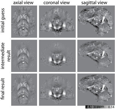 |
Shearlet-based susceptibility map reconstruction with iterative reweighting and nonlinear data fitting
Janis Stiegeler1,2 and Sina Straub1
1German Cancer Research Center (DKFZ), Heidelberg, Germany, 2Faculty of Physics and Astronomy, Heidelberg University, Heidelberg, Germany
The QSM algorithms tested in the 2016 Reconstruction Challenge showed oversmoothing effects and a notable loss of details. A multicale shearlet system was used to reconstruct the susceptibility map. Iterative reweighting of the coefficients led to a sparse image in the shearlet space. The result shows that shearlets are useful to obtain quantitative susceptibility maps which are rich in detail and simultaneously can achieve a top ten result in the ranking of the 2016 Reconstruction Challenge.
|
|
3247.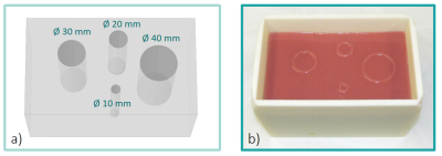 |
A low-cost, versatile phantom for elastography: validation via numerical simulations and MRE
Maksym Yushchenko1, Mathieu Sarracanie1,2, Jens Wuerfel2,3, and Najat Salameh1
1Laboratory for Adaptable MRI Technology (AMT lab), Dpt of Biomedical Engineering, University of Basel, Allschwil, Switzerland, 2Medical Image Analysis Center (MIAC AG), Basel, Switzerland, 3Quantitative Bioimaging Group (qbig), Dpt of Biomedical Engineering, University of Basel, Allschwil, Switzerland Poster Permission Withheld
We describe the fabrication of a low-cost, silicone-based phantom for elastography, which provides precise geometries and controlled stiffness. Simulations were carried out to produce synthetic MRE data and verify the performance of MRE reconstruction in ideal conditions. To our knowledge, this is the first time that simulations can be validated on exact same physical objects, making this approach very handy to optimize acquisition parameters and establish reconstruction validity criteria of elastography data. Such phantoms can be prepared with different mechanical properties, and could thus be broadly used as an accessible mean to assess the robustness of MRE tools.
|
|
3248.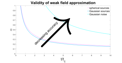 |
Signatures of microstructure in R2* decay: defining the limits of the weak field approximation
Pippa Storey1 and Dmitry S. Novikov1
1Department of Radiology, New York University School of Medicine, New York, NY, United States
R2* decay is nontrivial in the presence of magnetic microstructure; the logarithm of the signal is approximately quadratic over short times (the static dephasing regime) and asymptotically linear at long times (the diffusion narrowing regime). The transition can, in principle, provide an estimate of the characteristic length scales of the microstructure. Using Monte Carlo simulations of water molecules diffusing through a magnetic field perturbed by magnetic microstructure, we explore the regime of validity of the weak field approximation, in which the signal can be calculated analytically. We also illustrate how the signal behavior changes beyond the weak field limit.
|
|
3249.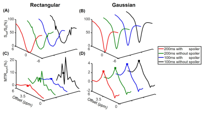 |
On the Saturation Scheme in Chemical Exchange Saturation Transfer Imaging with B0 Inhomogeneity
Ruibin Liu1, Qiuping Ding1, Yi-Cheng Hsu2, Yi Sun2, Dan Wu1, and Yi Zhang1
1Key Laboratory for Biomedical Engineering of Ministry of Education, Department of Biomedical Engineering, College of Biomedical Engineering & Instrument Science, Zhejiang University, Hangzhou, Zhejiang, China, 2MR Collaboration, Siemens Healthcare Ltd., Shanghai, China
Saturation pulses are an indispensable part in Chemical Exchange Saturation Transfer (CEST) imaging. Different saturation schemes can be designed for CEST imaging, including the pulse shape, duration of each pulse element, number of pulse elements, inter-pulse spoiler gradient, and inter-pulse gap, etc. Here, the impact of saturation schemes subject to practical B0 inhomogeneity is investigated, using Bloch simulations, and phantom and human experiments. Specifically, it’s shown that Gaussian and rectangular saturation pulses demonstrate substantially different susceptibility to imaging artifacts in the presence of B0 inhomogeneity when changing the duration of each saturation pulse element, and the inter-pulse spoiler gradients.
|
|
3250.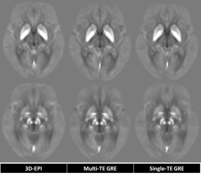 |
Reliability Study of Quantitative Susceptibility Mapping from Three-Dimensional Echo-Planar Imaging with 2 Minutes Scan Time
Krishna Pandu Wicaksono1, Yasutaka Fushimi1, Tomohisa Okada2, Satoshi Nakajima1, Sonoko Oshima1, Yusuke Yokota1, Azusa Sakurama1, Sayo Otani1, and Kaori Togashi1
1Department of Diagnostic Imaging and Nuclear Medicine, Kyoto University, Graduate School of Medicine, Kyoto, Japan, 2Human Brain Research Center, Kyoto University, Graduate School of Medicine, Kyoto, Japan Poster Permission Withheld
3D-EPI QSM has been proposed as one solution of QSM long scan time which hinders its routine application. As its validity has been ascertained in our phantom study, in-vivo reliability study is warranted. This healthy participant study demonstrated excellent reliability of 3D-EPI QSM with 2 minutes’ scan time, compared to the “standard” 5 minutes’ GRE QSM, favoring further study to evaluate its actual clinical performance.
|
|
3251.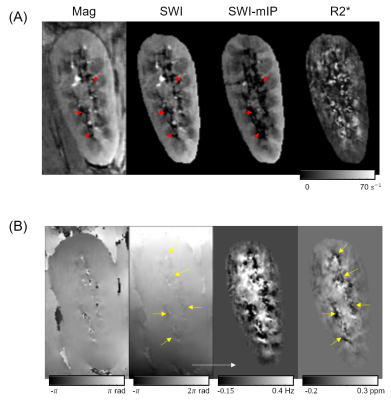 |
Renal Susceptibility Imaging: Analysis of Susceptibility Weighted Imaging and Quantitative Susceptibility Mapping in Transplanted Kidneys
Hyun-Seo Ahn1, Hyo Sung Kwak2, and Sung-Hong Park1
1Bio and Brain Engineering, Korea Advanced Institute of Science and Technology, Daejeon, Republic of Korea, 2Department of Radiology, Bio medical Research Institute of Chonbuk National University Hospital, Jeonju, Republic of Korea
Renal susceptibility imaging is challenging due to the motion artifacts in kidney. In this study, susceptibility weighted imaging (SWI) and quantitative susceptibility mapping (QSM) were performed on transplanted kidney patients, who show less kidney motions. QSM images successfully demonstrated oxygen gradients in kidney, i.e., the hypoxic state in medulla and the fully oxygenated state in cortex. Heterogeneous susceptibility distributions were observed near medullary veins, presumably due to medullary hypoxia and incomplete local phase unwrapping. Further studies are necessary for more complete local phase unwrapping and quantification of oxygenation levels in medullary veins from QSM images.
|
|
3252.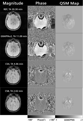 |
Quantitative susceptibility mapping with 1mm isotropic resolution in 2 minutes using compressed sensing
Arun Joseph1,2,3, Tom Hilbert4,5,6, Gabriele Bonanno1,2,3, Emilie Mussard4,5,6, Christoph Forman7, Ashley Stewart8,9, Kieran O’Brien8, and Tobias Kober4,5,6
Video Permission Withheld
1Advanced Clinical Imaging Technology, Siemens Healthcare AG, Bern, Switzerland, 2Translational Imaging Center, Sitem-Insel, Bern, Switzerland, 3Departments of Radiology and Biomedical Research, University of Bern, Bern, Switzerland, 4Advanced Clinical Imaging Technology, Siemens Healthcare AG, Lausanne, Switzerland, 5Department of Radiology, Lausanne University Hospital and University of Lausanne, Lausanne, Switzerland, 6LTS5, École Polytechnique Fédérale de Lausanne (EPFL), Lausanne, Switzerland, 7Siemens Healthcare AG, Erlangen, Germany, 8Siemens Healthcare Pty Ltd, Brisbane, Australia, 9Queensland Brain Institute, The University of Queensland, Brisbane, Australia
Quantitative susceptibility mapping exploits magnetic susceptibility to probe biological tissue. The obtained quantitative information carries additional clinical value, e.g. for the diagnosis of neurodegenerative diseases. However, standard protocols have long acquisition times which impedes their use in clinical routine. Here, we propose a compressed sensing acquisition based on a Cartesian spiral-phyllotaxis readout scheme to drastically reduce the total acquisition times from 20 minutes to 2 minutes for a 1mm isotropic susceptibility map of the whole brain. A preliminary qualitative and quantitative validation is performed on healthy subjects.
|
|
3253.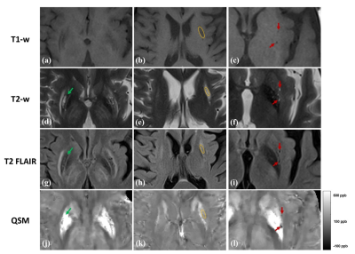 |
Identifying Different Types of Local Lesions in the Lenticular Nucleus of Patients with Wilson’s Disease using Quantitative Susceptibility Maps
Gaiying Li1, Rong Wu2, Yasong Du3, Kelly M Gillen4, Yi Wang4, Xiaoping Wang2, and Jianqi Li1
1East China Normal University, Shanghai, China, 2Shanghai Tong-Ren Hospital, Shanghai, China, 3Shanghai Mental Health Center, Shanghai, China, 4Weill Medical College of Cornell University, New York, NY, United States
The objective of this study was to discriminate signal changes of edema, encephalomalacia and excessive metal deposition in the lenticular nucleus of patients with Wilson’s Disease on quantitative susceptibility maps. Standard T1- weighted, T2- weighted, T2 FLAIR and QSM images were obtained on 14 WD subjects using 3T MRI. The results indicated that QSM could effectively discriminate different types of local lesions, including metal deposition hyperintensity, encephalomalacia hypointensity, and edema medium intensity. Furthermore, our study revealed that UWDRS and neurological symptoms scores of patients with severe abnormal MRI manifestations were significantly higher than that of patients with mild abnormal MRI.
|
|
3254.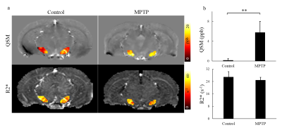 |
Detection of Iron Deposition in Substantia Nigra for Mouse of Parkinson’s Disease: A Comparison of Quantitative Susceptibility Mapping and R2*
Yihao Guo1,2, Jijing Guan1, Jinghui Xu3, Wenbin Si1, Yingjie Mei4, Qianjian Feng1,2, Yunqi Xu3, and Yanqiu Feng1,2
1School of Biomedical Engineering, Southern Medical University, Guagnzhou, China, 2Guangdong Provincial Key Laboratory of Medical Image Processing, Southern Medical University, Guagnzhou, China, 3Department of Neurology, Nanfang Hospital, Southern Medical University, Guangzhou, China, 4Philips Healthcare, Guangzhou, China
Parkinson’s disease (PD), a common neurodegenerative disorder, is associated with iron deposition in the substantial nigra (SN) of both human and animal models. SN iron is a potential biomarker for diagnosing and monitoring PD. The aim of this study was to compare QSM and R2* for quantifying SN iron in mouse brain.
|
|
3255. |
Shielded Electromagnetic Transducer for Liver Magnetic Resonance Elastography
Mohammad Honarvar1, Shahed Khan Mohammed1, Qi Zeng1, Caitlin Schneider1, Robert Rohling1, Piotr Kozlowski2,3, and Septimiu Salcudean1
1Electrical and Computer Engineering, University of British Columbia, Vancouver, BC, Canada, 2UBC MRI Research Center, Vancouver, BC, Canada, 3Department of Radiology, University of British Columbia, Vancouver, BC, Canada
Electromagnetic shielded Lorentz coil actuator allows using the magnetic field of the MRI scanner to induce arbitrary elastic waves in the tissue in a cost-effective way. We developed a Lorentz coil actuator for the liver magnetic resonance elastography (MRE) for providing a larger wave coverage area, where the Lorentz coil is placed outside the imaging area by the use of an additional end-effector. Initial testing for phantom showed an excellent agreement with the reported elasticity, while the liver elasticity study provided promising results in detecting cirrhosis and hepatitis C virus.
|
|
3256.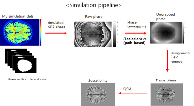 |
Simulation study for investigating the reliability of susceptibility mapping for the superior sagittal sinus from single echo 3D GRE phase
Soohyun Jeon1, Yoonho Nam2, Jinhee Jang3, and Dong-Hyun Kim1
1Department of Electrical & Electronic engineering, Yonsei University, Seoul, Korea, Republic of, 2Department of Radiology, The Catholic University of Korea, Seoul, Korea, Republic of, 3Radiology, The Catholic University of Korea, Seoul, Korea, Republic of Poster Permission Withheld
Quantitative Susceptibility Mapping (QSM) is a technique to describe local susceptibility distribution in the human body . Especially, QSM has the potentials to provide oxygen metabolic information of the venous structure in the brain such as dural sinuses. Among the group of this sinuses, the superior sagittal sinus (SSS) often have been investigated in, neurovascular researches such as Parkinson's disease, headache and stroke . However, the SSS of the susceptibility mapping are often eroded in the conventional QSM pipeline. In this study, we investigate the reliability of QSM for the SSS from single echo 3D GRE phase using the simulated phase data.
|
|
3257.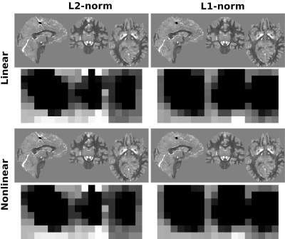 |
QSM streaking suppression with L1 data fidelity terms
Carlos Milovic1,2,3, Mathias Lambert1,2,3, Christian Langkammer4, Kristian Bredies5, Cristian Tejos1,2,3, and Pablo Irarrazaval1,2,3
1Department of Electrical Engineering, Pontificia Universidad Catolica de Chile, Santiago, Chile, 2Biomedical Imaging Center, Pontificia Universidad Catolica de Chile, Santiago, Chile, 3Millennium Nucleus for Cardiovascular Magnetic Resonance, Santiago, Chile, 4Department of Neurology, Medical University of Graz, Graz, Austria, 5Institute of Mathematics and Scientific Computing, University of Graz, Graz, Austria
L2-norm based data fidelity terms in QSM functionals account for Gaussian noise in the phase or complex domain. These approaches are not robust against phase inconsistencies, such as artifacts from previous steps, intra-voxel dephasing, etc. To deal with these errors and to suppress streaking artifacts we present L1-norm algorithms that correct for salt-and-pepper noise. We use simulations and in-vivo acquisitions to show their streaking suppression capabilities. Furthermore, L1 methods do not require magnitude information, and they were able to achieve similar results without ROI masks. This may facilitate susceptibility studies of cortical areas and regions outside the brain.
|