
Digital Poster Session
Contrast Mechanisms: MR Elastography and Perfusion
Contrast Mechanisms
3258 -3273 MR Elastography and Perfusion - Perfusion: Applications & Contrast-Enhanced Methods
3274 -3289 MR Elastography and Perfusion - Arterial Spin Labeling: Acquisition
3290 -3305 MR Elastography and Perfusion - Arterial Spin Labeling: Reconstruction, Analysis & Velocity-Selectivity
3306 -3321 MR Elastography and Perfusion - Elastography & Perfusion
3322 -3337 MR Elastography and Perfusion - Magnetic Resonance Elastography: Applications
3258.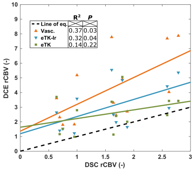 |
Perfusion Estimates of Fast Dynamic Contrast Enhanced MRI Correlates with Dynamic Susceptibility Contrast in Brain Metastases Follow-up
Benoit Bourassa-Moreau1, Réjean Lebel1, Ella Benzaquen2, Ismaël Labbé3, Guillaume Gilbert4, David Mathieu3, and Martin Lepage1
1Centre d’imagerie moléculaire de Sherbrooke, Département de médecine nucléaire et radiobiologie, Université de Sherbrooke, Sherbrooke, QC, Canada, 2Département radiobiologie diagnostique, Université de Sherbrooke, Sherbrooke, QC, Canada, 3Service de neurochirurgie, Département de chirurgie, Université de Sherbrooke, Sherbrooke, QC, Canada, 4MR Clinical Science, Philips Healthcare Canada, Markham, ON, Canada
Perfusion MRI is routinely used for brain tumor diagnosis and treatment follow-up. This work compares the performance of the standard dynamic susceptibility contrast sequence against a new fast dynamic contrast-enhanced sequence for brain metastasis follow-up after stereotactic radiosurgery. This new sequence enables the simultaneous measurement of blood brain barrier permeability that could help differentiate tumor recurrence and pseudo-progression. Our preliminary results show a correlation between relative cerebral blood volume estimates from both methods. A trend towards significance after analysis of thirteen lesions is expected to reach significance after inclusion of the remainder of our cohort.
|
|
3259.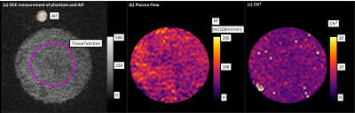 |
Influence of different temporal resolutions by varying parallel imaging factors on DCE perfusion parameters studied in a phantom
Tanja Uhrig1, Frank Zöllner1, and Lothar Schad1
1Computer Assisted Clinical Medicine, Medical Faculty Mannheim, Heidelberg University, Mannheim, Germany
No uniform consensus on the optimal sampling resolution for quantitative DCE MRI exists. In order to increase the sampling resolution, accelerated parallel imaging techniques (PAT) can be used. The inter- and intra-measurement precision of the plasma flow (PF) was determined in a phantom depending on the sampling resolution (1.9-3.8s), achieved by varying the PAT factor (1-6). A significant influence of the PAT factors and the sampling resolution on the PF itself and the robustness and reproducibility of the fit could be observed, even with small sampling rates as recommended in literature.
|
|
3260.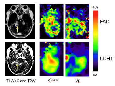 |
A Novel Approach Using First-Pass Adjusted DTR-DCE-MRI (FAD) for Accurate Super-Spatial Resolution Brain Pharmacokinetic Parametric Mapping
Xiaoping Zhu1, Daniel Lewis2, Sha Zhao1, Alan Jackson1, and Ka-Loh Li1
1DIIDS, University of Manchester, Manchester, United Kingdom, 2Salford Royal NHS Foundation Trust, Manchester Academic Health Science Centre, Manchester, United Kingdom
This study describes a new dual-injection, dual-temporal resolution (DTR) DCE-MRI mapping technique, which performs first-pass adjustment (FAD) of pre-bolus uptake curves using enhancement patterns measured from a main-dose, high-spatial resolution DCE acquisition. This new technique permits derivation of whole brain, super-spatial resolution kinetic parameter maps and its clinical applicability in vivo was assessed through application to retrospective DTR-DCE-MRI data from 12 NF2 patients undergoing Avastin therapy. Compared to the classical DTR-DCE-FDHS, method, baseline Ktrans derived using the FAD technique demonstrated superior ability in the prediction of 90 day volumetric response in Avastin treated NF2 related VS.
|
|
3261.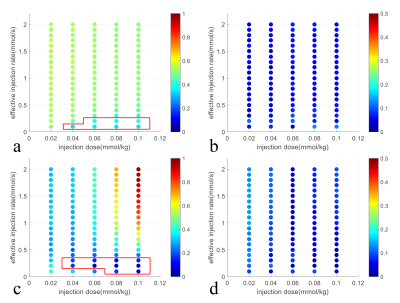 |
Optimization of the Injection Protocol for Carotid Black-blood and Interleaved Black-bright-blood Dynamic Contrast-Enhanced MRI
Yajie Wang1, Yishi Wang2, Xian Liu1, and Huijun Chen1
1Center for Biomedical Imaging Research, School of Medicine, Tsinghua University, Beijing, China, 2Philips Healthcare, Beijing, China
The injection protocol used in carotid dynamic contrast-enhanced (DCE) MRI varies among different studies. The effect of the injection protocol on carotid black-blood and interleaved black-bright-blood DCE sequences were evaluated in this study. The results demonstrated that high injection dose (~ 0.1mmol/kg) with relative low effective injection rate (< 0.3mmol/s) was recommended for the simulated black-blood and interleaved black-bright-blood sequences. These two sequences were also shown to be insensitive to the uncertain time gap between the contrast injection and the post-contrast image acquisition.
|
|
3262.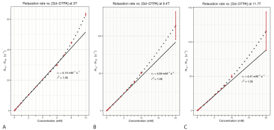 |
Range of Gd-DTPA concentration where $$$T_1$$$ shortening effect remains linear at 3T, 9.4T and 11.7T
Anh Nguyen Tuan Tran1,2, Xing Qi Teo2, Septian Hartono1, Philip Teck Hock Lee2, Choon Hua Thng1, and Tong San Koh1
1Oncologic Imaging, National Cancer Center Singapore, Singapore, Singapore, 2Functional Metabolism Group, Singapore BioImaging Consortium, Singapore, Singapore
In $$$T_1$$$-weighted perfusion studies, the relationship between the contrast concentration and $$$T_1$$$ shortening effect is assumed to be linear within the clinical range. However, this assumption may not hold at higher concentration observed in the arterial input functions (AIFs). We set out to investigate this relationship by measuring the $$$R_1$$$ relaxation rate of various Gd-DTPA concentrations in saline. We find that the change in $$$R_1$$$ relaxation rate of Gd-DTPA is linear up to 4 mM at 9.4T and 11.7T, and up to 5 mM at 3T. At higher concentration, $$$R_1$$$ relaxation rate increases faster than a linear relationship.
|
|
3263.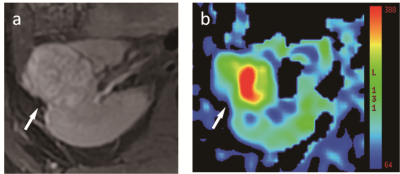 |
Use of arterial spin labeling MRI in differentiating fat-poor angiomyolipoma from clear cell renal cell carcinoma
Qing Xu1, Weiqiang Dou2, and Jing Ye1
1Department of Radiology, Clinical Medical School of Yangzhou University, Northern Jiangsu People’s Hospital, Yangzhou, 457, China, 2GE Healthcare, MR Research China, Beijing, China
In this study, we aimed to test if arterial spin labeling (ASL) MRI can be used to differentiate renal fat-poor angiomyolipoma (AML) from the clear cell renal cell carcinoma (ccRCC). To achieve this goal, we compared the ASL derived parameters including tumor blood flow (TBF), tumor-to-cortex ratio, and tumor-to-medulla ratio between fat-poor AML and ccRCC. Our results showed that TBF, tumor-to-cortex and tumor-to-medulla ratios were notably higher in ccRCC group than in fat-poor AML group (270.49±78.88ml/100g/min vs. 146.68±47.21ml/100g/min, 1.22±0.26 vs. 0.74±0.14, 3.13±0.94 vs. 1.77±0.55; p<0.05), indicating that ASL MRI can be an effective tool in differentiating fat-poor AML from ccRCC.
|
|
3264.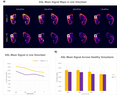 |
Optimization of Pseudo Continuous Arterial Spin Labeling for measuring perfusion in CKD patients
Rebeca Echeverria-Chasco1, Marta Vidorreta2, Verónica Aramendia-Vidaurreta1, Nuria Garcia-Fernandez3, Gorka Bastarrika1, and Maria A. Fernandez-Seara1
1Radiology, Clinica Universidad de Navarra, Pamplona, Spain, 2Siemens Healthineers, Madrid, Spain, 3Nephrology, Clinica Universidad de Navarra, Pamplona, Spain
The main goal of this work was to optimize the robustness of pseudo continuous Arterial Spin Labeling (pCASL) to acquire renal images in CKD patients. The optimization was firstly carried out through numerical simulations, and a pCASL experiment in healhty volunteers to assess the veracity of the simulations. The results were in agreeement with the simulations (p-value = 0.04). For CKD patients, the numerical simulations were made to select the parameters that maximize the efficiency. The results for CKD patients and controls that show highest efficiencies are ratios 6-7 and Gave of 0.5-0.7 mT/m.
|
|
3265.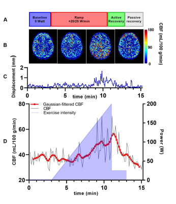 |
Dynamic arterial spin labeling MRI of cerebrovascular perfusion during exercise
Isa Mast1,2, Koen P.A. Baas2, Aart J. Nederveen2, and Adrianus J. Bakermans2
1Human Movement Sciences, Vrije Universiteit van Amsterdam, Amsterdam, Netherlands, 2Radiology and Nuclear Medicine, Amsterdam UMC, Amsterdam, Netherlands
Pathophysiological changes in cerebrovascular reactivity can remain undetectable at rest, and may only become apparent during a cerebrovascular challenge. We evaluated the feasibility of dynamically measuring the cerebrovascular response to exercise using pseudo-continuous arterial spin labeling (pCASL) at 3 Tesla during a bicycle exercise-recovery stress test. We observed a transient increase in cerebrovascular blood flow (CBF) during exercise in four volunteers, demonstrating that pCASL-MRI can capture dynamic changes in CBF during physiological bicycle exercise. This approach may become an important quantitative tool to noninvasively investigate the cerebrovascular reactivity in health and disease.
|
|
3266.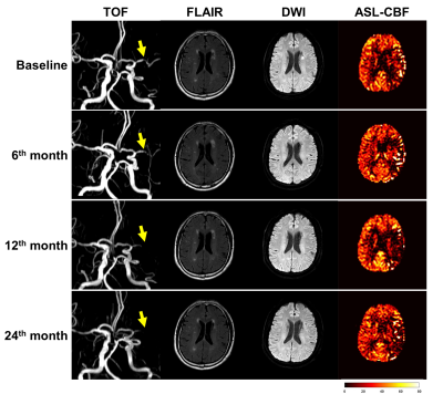 |
Time Course of Cerebral Blood Flow in Patients with Symptomatic Middle Cerebral Artery Stenosis during Lipid-lowering Treatment
Hualu Han1, Dandan Yang1, Huiyu Qiao1, Zihan Ning1, and Xihai Zhao1
1Tsinghua University, Beijing, China
Statin treatment is considered as an effective method to stabilize atherosclerosis and potentially improve cerebral perfusion. This study evaluated the changes of artery stenosis and CBF on arterial spin labeling during statin treatment with two-years’ follow-up. We found that CBF improved in the symptomatic territory among all subjects at 6th month (rCBF: 0.92±0.07 vs. 0.95±0.07, P=0.001). After 6 months, however, CBF decreased in patients with stenosis progression whereas maintained at a stable level in patients with stenosis regression, suggesting that changes of symptomatic MCA stenosis may have an effect on the cerebral hemodynamic improvements during statin treatment in different stages.
|
|
3267.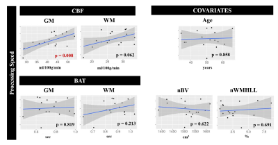 |
ASL perfusion imaging predicts cognitive impairment in cerebral small vessel disease
Joana Pinto1, Ana Fouto1, Rita G. Nunes1, Luísa Alves2, Sofia Calado2, Carina Gonçalves2, Margarida Rebolo3, Miguel Viana Baptista2, Pedro Vilela4, and Patrícia Figueiredo1
1ISR-Lisboa/LARSyS and Department of Bioengineering, Instituto Superior Técnico – Universidade de Lisboa, Lisboa, Portugal, 2Neurology Department, Hospital Egas Moniz, Centro Hospitalar de Lisboa Ocidental; CEDOC - Nova Medical School, New University of Lisbon, Lisboa, Portugal, 3Faculdade de Medicina, Universidade de Lisboa, Lisboa, Portugal, 4Imaging Department, Hospital da Luz, Lisboa, Portugal Poster Permission Withheld
Cerebral small vessel disease (SVD) is one of the most common vascular causes of dementia, and a major contributor of age-related cognitive decline. We evaluated cerebral perfusion using arterial spin labeling (ASL), in terms of its predictive power of cognitive impairment in a group of SVD patients. We employed a multiple-delay pulsed ASL acquisition and fitted an extended kinetic model to the signal in order to derive cerebral blood flow (CBF) as well as bolus arrival time (BAT) maps. Regression analysis demonstrated that CBF in gray matter (GM) significantly contributed to explaining cognitive impairments in processing speed.
|
|
3268.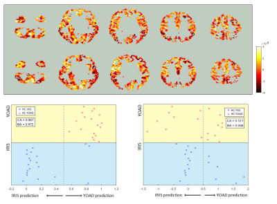 |
Machine Learning to Improve Compatibility of Multi Centre Arterial Spin Labelling Data to Characterise Dementia Spatial Perfusion Abnormality
Jack Highton1, Rebecca Steketee2, Rozanna Meijboom3, Marion Smits2, Ross Paterson4, Nick Fox4, Alexander Foulkes5, Catherine Slattery5, Jonathan Schott5, Enrico De Vita6, and David L Thomas7,8,9
1Department of Medical Physics and Biomedical Engineering, University College London, London, United Kingdom, 2Department of Radiology and Nuclear Medicine, Erasmus Medical Centre, Rotterdam, Netherlands, Rotterdam, Netherlands, 3Centre for Clinical Brain Sciences, University of Edinburgh, Edinburgh, United Kingdom, 4Institute of Neurology, University College London, London, United Kingdom, 5Dementia Research Centre, University College London, London, United Kingdom, 6King's College London, London, United Kingdom, 7University College London, London, United Kingdom, 8Neuroradiological Academic Unit, Department of Brain Repair and Rehabilitation, Institute of Neurology,, University College London, London, United Kingdom, 9Wellcome Centre for Human Neuroimaging, Institute of Neurology, University College London, London, United Kingdom
Arterial Spin Labelling (ASL) is a non-invasive MRI method to measure cerebral blood flow (CBF). Here, Support Vector Machine (SVM) based machine learning was used to identify spatial patterns of CBF abnormality in patients with Alzheimer’s Disease from two cohorts scanned at different centers. Support Vector Machine Regression models were found to be more accurate than conventional SVMs previously used for dementia classification. Then, motivated by the lack of ASL standardisation, an SVM based method was used to improve the compatibility of the two studies by removing differences in spatial patterns likely caused by differences in hardware and acquisition protocol.
|
|
3269.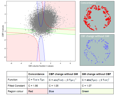 |
Untangling Perfusion from Atrophy in Fronto-Temporal Dementia using Multimodal Correlation and Machine Learning
Jack Highton1, Rebecca Steketee2, Rozanna Meijboom3, Marion Smits2, Enrico De Vita4, Jonathan Schott5, and David L Thomas6,7,8
1Department of Medical Physics and Biomedical Engineering, University College London, London, United Kingdom, 2Department of Radiology and Nuclear Medicine, Erasmus Medical Centre, Rotterdam, Netherlands, Rotterdam, Netherlands, 3Centre for Clinical Brain Sciences, University of Edinburgh, Edinburgh, United Kingdom, 4King's College London, London, United Kingdom, 5University College London, London, United Kingdom, 6Dementia Research Centre, University College London, London, United Kingdom, 7Neuroradiological Academic Unit, Department of Brain Repair and Rehabilitation, Institute of Neurology, University College London, London, United Kingdom, 8Wellcome Centre for Human Neuroimaging, Institute of Neurology,, University College London, London, United Kingdom Partial volume correction (PVC) is often applied to analyse cerebral blood flow (CBF) in dementia, in order to isolate perfusion changes from atrophy of grey matter. But PVC inevitably results in spatial smoothing of CBF, which may weaken the informative spatial pattern of CBF changes. Here, machine learning was used to detect Fronto-Temporal Dementia (FTD), considering spatial patterns in CBF from Arterial Spin Labelling, with and without PVC. Also, multimodal voxel concordance analysis untangled the spatial relationship between GM atrophy and CBF. This information was used with CBF PVC to improve the FTD detection accuracy without the atrophy signature. |
|
3270.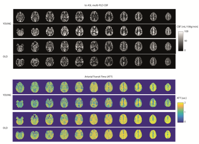 |
Time-encoded ASL provides added value in differentiating healthy older from younger individuals compared to single time-point methods
Lena Vaclavu1, Carles Falcon 2, Paula Montesinos 3, Kim van de Ven4, Juan Domingo Gispert 2, and Matthias J.P. van Osch1
1Department of Radiology, Leiden University Medical Center, Leiden, Netherlands, 2Pasqual Maragall Foundation, Barcelonaβeta Brain Research Center (BBRC), Barcelona, Spain, 3Philips Iberia, Madrid, Spain, 4Philips, Best, Netherlands
Age is an important risk factor for cerebrovascular disease. ASL can lend different insights depending on the choice of read-out (3D or 2D) and post-label-delay (PLD) for instance. We assessed differences in cerebral blood flow (CBF), and investigated the added value of arterial transit time (ATT) for differentiating older from younger healthy volunteers. Single-PLD 2D-pCASL and time-encoded (te-)ASL had almost identical CBF, while multi-PLD te-ASL offered the additional option to estimate ATT and blood volume. We found prolonged ATT despite unaffected CBF in older versus younger volunteers. TE-ASL could therefore provide ‘free’ information aside from perfusion in clinical settings.
|
|
3271. |
Arterial spin labeling signal in the Sagittal Sinus as hemodynamic proxy parameter in patients with sickle cell disease.
Liza Afzali-Hashemi1, Lena Vaclavu2, Jan Petr3, John Wood4, Bart J Biemond5, Aart J Nederveen6, and Henk JMM Mutsaerts7
1Radiology and Nuclear Medicine, Amsterdam University Medical Center, location AMC, Amsterdam, Netherlands, 2Radiology, Leiden University Medical Center, Leiden, Netherlands, 3Helmholtz-Zentrum Dresden-Rossendorf, Institute of Radiopharmaceutical Cancer Research, Dresden, Germany, 4Department of Cardiology and Radiology, Children’s Hospital of Los Angeles, Los Angeles, CA, United States, 5Department of Hematology, Amsterdam UMC, location AMC, Amsterdam, Netherlands, 6Department of Radiology & Nuclear Medicine, Amsterdam UMC, location AMC, Amsterdam, Netherlands, 7Amsterdam UMC, Amsterdam, Netherlands
Higher sagittal sinus signal is present in the ASL images of patients with sickle cell disease (SCD). The purpose of this study was to assess if the signal in the sagittal sinus is correlated with clinical parameters and if this is affected by the vasoactive stimulus. The sagittal sinus signal was measured in patients with SCD and in healthy controls. Signal in sagittal sinus of the SCD patients were significantly correlated with clinical parameters including hemolysis markers. Our results show that sagittal sinus signal can be used as a hemodynamic proxy parameter in patients with SCD.
|
|
3272.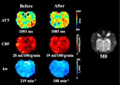 |
DW-pCASL measure of BBB water permeability is sensitive to pharmacological manipulation of AQP4 function
Hsiao-Ying Wey1, Xingfeng Shao2, and Danny JJ Wang2
1Athinoula A. Martinos Center for Biomedical Imaging, Department of Radiology, Massachusetts General Hospital, Harvard Medical School, Charlestown, MA, United States, 2USC Stevens Neuroimaging and Informatics Institute, University of Southern California, Los Angeles, CA, United States
The blood-brain barrier (BBB) is a tightly regulated structure that protect the central nervous system (CNS). BBB impairment is implicated in several brain disorders, such as Alzheimer’s disease. Aquaporin-4 (AQP4) water channels are critically involved in regulating brain water transport across the BBB. We have recently developed a diffusion-weighted pCASL (DW-pCASL) technique allowing measurement of water permeability across the BBB. In this study, we implemented and optimized DW-pCASL in nonhuman primates. We directly validated that the underlying signal mechanism of DW-pCASL is related to AQP4-mediated BBB water exchange using a pharmacological challenge.
|
|
3273.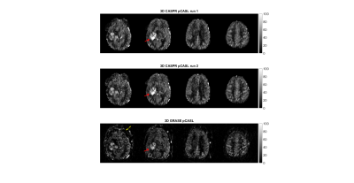 |
Reliability and Reproducibility of ASL-MRI Measured Perfusion in GBM at 3T
Limin Zhou1, Yiming Wang1, Marco Da Cunha Pinho1,2, Edward Pan3,4,5, Joseph Maldjian1,2, Yin Xi1,6, and Ananth Madhuranthakam1,2
1Radiology, University of Texas Southwestern Medical Center, Dallas, TX, United States, 2Advanced Imaging Research Center, University of Texas Southwestern Medical Center, Dallas, TX, United States, 3Harold C. Simmons Comprehensive Cancer Center, University of Texas Southwestern Medical Center, Dallas, TX, United States, 4Neurological Surgery, University of Texas Southwestern Medical Center, Dallas, TX, United States, 5Neurology and Neurotherapeutics, University of Texas Southwestern Medical Center, Dallas, TX, United States, 6Population and Data Sciences, University of Texas Southwestern Medical Center, Dallas, TX, United States
We have assessed the reliability of pCASL using 3D TSE-CASPR in healthy volunteers and glioblastoma (GBM) patients. “Excellent” agreement was observed for intrasession reliability in both normal appearing grey matter (NAGM) and tumors with intraclass correlation coefficient (ICC) of 0.945 (95% CI: 0.916-0.964) and 0.948 (95% CI: 0.883-0.978) respectively. Across multiple ROIs, the within-subject coefficient of variation (wsCV) was measured to be 6.1 ± 1.2% across all subjects in NAGM, and 5.1% in tumors. The lower wsCV in tumors allow 3D TSE pCASL using CASPR to be used for longitudinal monitoring of GBM patients.
|
 View the Poster
 Watch the Video
3274.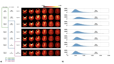 |
Reducing the effect of cerebrospinal fluid pulsation on arterial spin labeled perfusion MRI acquired with fast spin echo readouts
Jianxun Qu1, Tianye Lin1,2, Karthik Prabhakaran3, M. Dylan Tisdall1, and John A. Detre1,3
1Radiology, Perelman School of Medicine, University of Pennsylvania, Philadelphia, PA, United States, 2Chinese Academy of Medical Sciences, Peking Union Medical College, PUMCH, Beijing, China, 3Neurology, Perelman School of Medicine, University of Pennsylvania, Philadelphia, PA, United States
Fast spin echo is widely used for ASL acquisition. However, if the flip angle reduced to minimize SAR and/or stabilize signal evolution over the echo train, the fluctuation of CSF increases markedly. This work explored the influence of minimum flip angle, and crusher strength in ASL acquisition. A CSF-prioritized background suppression (BS) planning was also performed. We found that increasing minimum flip angle together with reducing crusher in superior-inferior direction benefits the stability of ASL acquisitions. Prioritizing CSF suppression in BS was also helpful.
|
|
3275.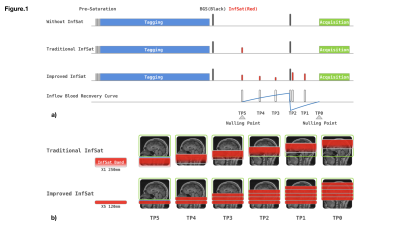 |
Improved inflow saturation markedly reduces inflow artifacts in background-suppressed 3D arterial spin labeling
Jianxun Qu1, Tianye Lin1,2, Priti Balchandani3, M. Dylan Tisdall1, and John A. Detre1,4
1Radiology, Perelman School of Medicine, University of Pennsylvania, Philadelphia, PA, United States, 2Chinese Academy of Medical Sciences, Peking Union Medical College, PUMCH, Beijing, China, 3Icahn School of Medicine at Mount Sinai, New York, NY, United States, 4Neurology, Perelman School of Medicine, University of Pennsylvania, Philadelphia, PA, United States
Inflow blood during the post labeling delay is a significant source of physiological noise in arterial spin labeling. This study utilized a novel inflow saturation method to address this issue. Compared to traditional inflow saturation approach where a 90o thick saturation slab is used to null blood within the interval of background suppression inversion pulses, the proposed method employs serial of thin slab sub-bolus saturations, with the saturation strength planned adaptively to return the blood to nulling point at the excitation point. The proposed method reduces the inflow artifacts markedly and mitigates global signal fluctuations.
|
|
3276.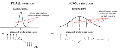 |
Off-resonance and flow-velocity immune ASL at low-power using pseudo-continuous saturation labeling
Manuel Taso1 and David C. Alsop1
1Division of MRI research, Department of Radiology, Beth Israel Deaconess Medical Center, Harvard Medical School, Boston, MA, United States
While widely used for perfusion imaging, the flow-driven inversion labeling commonly used with pseudo-continuous ASL (PCASL) presents a significant off-resonance sensitivity as well as high power deposition potentially limiting high-field applications. We therefore investigated an alternate labeling scheme that takes advantage of the multiple aliased labeling planes that arise within the labeling RF envelope when reducing the peak-to-average gradient ratio. First numerical simulations and experiments show the potential for low-SAR, B0 and flow-velocity robust saturation labeling.
|
|
3277.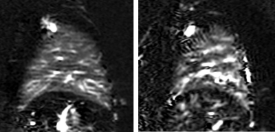 |
Robust non-contrast pulmonary perfusion imaging using pseudo-continuous arterial spin labeling with background suppression
Joshua S. Greer1,2, Yiming Wang2, Limin Zhou2, Tarique Hussain1,2, and Ananth J. Madhuranthakam2,3
1Pediatrics, UT Southwestern Medical Center, Dallas, TX, United States, 2Radiology, UT Southwestern Medical Center, Dallas, TX, United States, 3Advanced Imaging Research Center, UT Southwestern Medical Center, Dallas, TX, United States
Pulmonary perfusion imaging using ASL has been demonstrated using the pCASL technique with varying degrees of reproducibility. In this study, a pCASL labeling strategy is proposed to improve the stability of perfusion signal in the lungs using cardiac triggering to reduce flow-induced signal variations, and background suppression to improve SNR. With the proposed technique, high SNR perfusion images are created with a limited number of signal averages. This optimized approach may allow full coverage of the lungs in clinically reasonable scan times.
|
|
3278.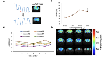 |
High-resolution whole brain perfusion mapping in mice by a drift-corrected steady pulsed imaging and labeling sequence
Wenjing Xu1,2, Jiadi Xu3,4, and Xiaoyong Zhang1,2
1Institute of Science and Technology for Brain-Inspired Intelligence, Fudan University, Shanghai, China, 2Key Laboratory of Computational Neuroscience and Brain-Inspired Intelligence (Fudan University), Ministry of Education, Shanghai, China, 3Russell H. Morgan Department of Radiology and Radiological Science, Johns Hopkins University School of Medicine, Baltimore, MD, United States, 4F.M. Kirby Research Center for Functional Brain Imaging, Kennedy Krieger Institute, Baltimore, MD, United States
We developed a drift-corrected steady pulsed imaging and labeling (dSPIL) to measure CBF with high-resolution and whole brain coverage. We applied a metric of low band pass to remove MRI signal drift using interleaved label-control images. We also optimized several key imaging parameters in an effort to minimize scan time while covering the whole brain. With the optimized dSPIL scheme, quantification of high-resolution CBF in 22 mouse brain regions can be obtained. As a demonstration and validation of the method, the volume of ischemic lesion regions in mice was quantified by calculating CBF reductions.
|
|
3279.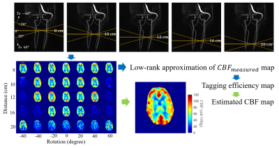 |
Tagging Error Robust pseudo-Continuous Arterial Spin Labeling using Multiplane Labeling and Low Rank Methods
Chenwei Tang1, Mu-Lan Jen1, Laura Eisenmenger2, and Kevin M Johnson1,2
1Department of Medical Physics, University of Wisconsin-Madison, Madison, WI, United States, 2Department of Radiology, University of Wisconsin-Madison, Madison, WI, United States
While
pseudo-continuous ASL (pCASL) is still the recommended technique for clinical and research applications, it suffers from subject vascular anatomy dependent variations in tagging efficiency which may lead to inaccurate quantification of perfusion, or gross misrepresentations of perfusion heterogeneity. In this work, we explore a strategy in which a span of tagging plane offsets and rotations is performed and cerebral blood flow (CBF) is robustly estimated retrospectively from the series of images with differential tagging efficiency. Our results demonstrate significant fluctuation of measured CBF for different relative labeling plane locations and robust recovered CBF against these fluctuations. |
|
3280. |
Comparison of k-space filtering and variable flip angle schemes for reduced T2 blurring in 3D ASL MRI
Yiming Wang1, Limin Zhou1, Sheng Qing Lin1, Joshua S. Greer1,2, and Ananth J. Madhuranthakam1,3
1Radiology, UT Southwestern Medical Center, Dallas, TX, United States, 2Pediatrics, UT Southwestern Medical Center, Dallas, TX, United States, 3Advanced Imaging Research Center, UT Southwestern Medical Center, Dallas, TX, United States
3D arterial spin labeled (ASL) MRI using turbo spin echo (TSE) based acquisitions suffer from image blurring, due to T2 decay along the echo train. To date, several methods have been proposed to reduce T2 blurring, including k-space filtering and variable flip angle schemes. In this study, the performances of k-space filtering was compared with variable flip angle scheme to compensate or reduce T2 blurring of 3D ASL images acquired with Cartesian TSE. Results showed k-space filtered images provided similar sharpness improvement compared with variable flip angle method.
|
|
3281. |
Variable Density Sampling of 3D TSE Cartesian Acquisition for Improved Robustness and SNR of ASL-MRI
Yiming Wang1, Limin Zhou1, Joshua S. Greer1,2, Marco C. Pinho1,3, Joseph A. Maldjian1,3, and Ananth J. Madhuranthakam1,3
1Radiology, UT Southwestern Medical Center, Dallas, TX, United States, 2Pediatrics, UT Southwestern Medical Center, Dallas, TX, United States, 3Advanced Imaging Research Center, UT Southwestern Medical Center, Dallas, TX, United States
3D Cartesian turbo spin echo (TSE) acquisition has shown robustness for arterial spin labeled (ASL) MRI. However, 3D Cartesian TSE are acquired with single average due to relatively long TR and longer scan times, which may lead to suboptimal signal to noise ratio (SNR). In this study, we implemented variable density sampling of 3D Cartesian TSE that acquires the center of the k-space with higher signal averages, and improves the robustness and SNR without significantly prolonging the scan time. We further combined this with M0 images using partial k-space to compensate for increased scan time, but without compromising perfusion quantification.
|
|
3282.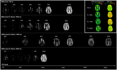 |
Optimal subject-specific pCASL settings by automated inner-scan timing adaption
Nora-Josefin Breutigam1, Daniel Christopher Hoinkiss1, Mareike Alicja Buck1, Amnah Mahroo1, Federico von Samson-Himmelstjerna1, and Matthias Günther1,2
1MR Physics, Fraunhofer MEVIS, Bremen, Germany, 2MR-Imaging and Spectroscopy, Faculty 01 (Physics/Electrical Engineering, University of Bremen, Bremen, Germany
The presented automated inner-scan timing adjustment for subject-specific pCASL measurements can reduce uncertainties in perfusion quantification. The high variability of arterial transit times often leads to undesired arterial transit delay (ATD) artifacts. The method evaluated here allows the optimal FL-bolus duration to be individually adjusted by analyzing intermediate decoded perfusion-weighted images and directly adjusting the sub-bolus durations without increasing the measurement time. The aim is to reduce the FL-bolus duration as much as necessary, but to keep it as long as possible in order to obtain a maximum signal.
|
|
3283.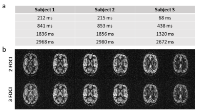 |
Subject-specific background suppression in 3D pseudo-continuous arterial spin labeling perfusion imaging
Jörn Huber1, Daniel Christopher Hoinkiss1, and Matthias Günther1,2
1Fraunhofer MEVIS, Bremen, Germany, 2University of Bremen, Bremen, Germany
This study introduces a subject-specific automated approach for optimization of background suppression (BS) in 3D pseudo-continuous arterial spin labeling perfusion imaging. Optimised timing of BS related FOCI pulses is calculated from the T1 spectrum, obtained from a M0 scan of the organ of interest. Results show strong suppression of brain tissue, when magnetization is nulled at the time of excitation as well as high quality perfusion images, when a slight delay between nullpoint and excitation is introduced for different numbers of FOCI pulses.
|
|
3284.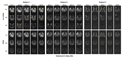 |
Mitigating spurious transit time artifacts in background-suppressed multi-delay 3D pseudo-continuous ASL using inflow suppression
Tianye Lin1,2, Jianxun Qu2, and John A. Detre2
1Chinese Academy of Medical Sciences, Peking Union Medical College, PUMCH, Beijing, China, 2University of Pennsylvania, Perelman School of Medicine, Philadelphia, PA, United States
The purpose of our study was to demonstrate that the inflow of arterial blood in post-labeling delay can cause intravascular high signal in multi-PLD ASL that resembles arterial transit artifact and potentially interfere with clinical interpretation. An optimized inflow-saturation method was used to suppress this artifact and improve the image quality of Hadamard-encoded and sequential multi-PLD ASL.
|
|
3285. |
Separating spin-compartment in arterial spin labeling using delays alternating with nutation for tailored excitation (DANTE) pulse
Shota Ishida1, Hirohiko Kimura2, Naoyuki Takei3, Yasuhiro Fujiwara4, Tsuyoshi Matsuda5, Yuki Matta1, Masayuki Kanamoto1, Nobuyuki Kosaka2, and Eiji Kidoya1
1Radiological center, University of Fukui Hospital, Eiheiji, Japan, 2Department of Radiology, Faculty of Medical Sciences, University of Fukui, Eiheiji, Japan, 3Global MR Applications and Workflow, GE Healthcare Japan, Hino, Japan, 4Department of Medical Image Sciences, Faculty of Life Sciences, Kumamoto University, Kumamoto, Japan, 5Division of Ultra-high Field MRI, Institute for Biomedical Science, Iwate Medical University, Shiwa-gun, Japan
Delays alternating with nutation for tailored excitation (DANTE) pulse is capable of uniform and flow velocity related signal suppression. DANTE-ASL was compared to T2-mesurement in ASL (T2-ASL) to validate what spin-compartment is suppressed by DANTE. T2-ASL showed labeled signal remained in vascular compartment even at a PLD of 2.0 s. However, ASL signals using DANTE were approximately similar value in all the PLDs (0.5, 1.0, 2.0, and 3.0 s), because ASL signals in vascular compartment were suppressed selectively and sufficiently with concurrently preserving signals in tissue compartment. We could eliminate vascular compartment from total ASL signal using DANTE.
|
|
3286.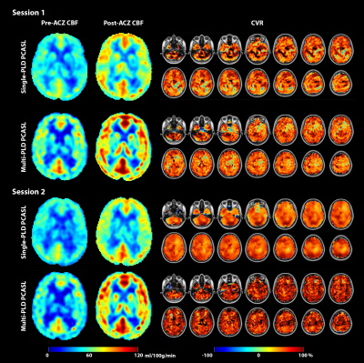 |
Investigating the Repeatability of Cerebrovascular Reactivity Using Single and Multi-PLD PCASL
Moss Y Zhao1, Audrey P Fan1, David Yen-Ting Chen1,2, David D Shin3, Mohammad Mehdi Khalighi1, Dawn Holley1, Kim Halbert1, Andrea Otte1, Brittney Williams1, Jun-Hyung Park1, Bin Shen1, and Greg Zaharchuk1
1Department of Radiology, Stanford University, Stanford, CA, United States, 2Department of Radiology, Shuang-Ho Hospital, Taipei Medical University, Taipei, Taiwan, 3GE Healthcare, Menlo Park, CA, United States
This work investigates the repeatability of CVR (induced by acetazolamide) using ASL on a healthy cohort. Single and multi-PLD PCASL data were collected before and after the administration of acetazolamide in two identical sessions. Results showed that the mean CVR of both ASL techniques were between 20% and 60% and not significantly different. Measurement using single-PLD PCASL demonstrated higher repeatability based on a lower coefficient of variation and a higher intraclass correlations.
|
|
3287.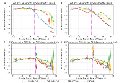 |
Examination of optimised PLD protocols for pCASL: accounting for dispersion and macrovascular contamination over a prolonged ATT range
Xin Logan Zhang1, Joseph G. Woods1,2, Flora Kennedy McConnell1, Thomas W. Okell1, and Michael A. Chappell1
1Institute of Biomedical Engineering & Wellcome Centre for Integrative Neuroimaging, University of Oxford, Oxford, United Kingdom, 2Department of Radiology, University of California San Diego, La Jolla, CA, United States
A general framework has been proposed for optimising PLD sampling protocols in pCASL under an ideal kinetic model. In this study, the performance of CBF and ATT estimation of two optimised protocols CBF-ATTopt and CBFopt, along with a reference multi-PLD and single-PLD protocol, was investigated using simulations and in vivo when accounting for dispersion and macrovascular contamination (MVC) over a prolonged ATT range. CBFopt showed the least sensitivity among all protocols for CBF estimation when ATT was prolonged and when MVC was present. Future work can optimise for both macrovascular component and tissue component with a prolonged ATT range.
|
|
3288.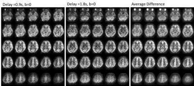 |
Volumetric, multi B-value, multi-delay ASL using 3D single-shot spirals, expanded encoding models, and low rank reconstruction
Mu-Lan Jen1, James H Holmes2, Laura Eisenmenger2, and Kevin M Johnson1,2
1Medical Physics, University of Wisconsin-Madison, Madison, WI, United States, 2Radiology, University of Wisconsin-Madison, Madison, WI, United States
In this work, we investigate a strategy to acquire single-shot, multi b-value, multi-delay ASL scans using the combination of an undersampled spiral in-out 3D fast spin echo readout with reconstructions enforcing a low rank constraint with an expanded encoding model. Images are demonstrated in healthy volunteers, acquiring single 3D ASL images in as short as 11s.
|
|
3289.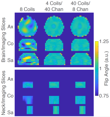 |
Optimizing an Array-Compressed Coil for Arterial Spin Labeling at 7T
Xinqiang Yan1,2, Manus J. Donahue1,2, and William A. Grissom1,2,3
1Department of Radiology, Vanderbilt University Medical Center, Nashville, TN, United States, 2Institute of Imaging Science, Vanderbilt University, Nashville, TN, United States, 3Department of Biomedical Engineering, Vanderbilt University, Nashville, TN, United States
Arterial spin labeling (ASL) is a primary approach for non-invasive perfusion measurements with MRI. Theoretically, performing ASL at ultrahigh magnetic field strength can be beneficial because of the increased SNR as well as the lengthened T1-relaxation time. However, ASL is still not widely used at 7T due to the obstacles of B1+ inhomogeneity and high SAR associated with labeling. To overcome this problem, we apply array compressed parallel transmission (acpTx) in ASL and find that the optimized coil achieved 2x better B1+ homogeneity, and 4x lower SAR than a conventional 8-channel coil, which could enable 7T ASL with neck labeling.
|
3290.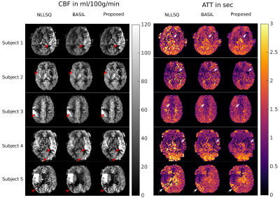 |
Robust Perfusion Parameter Quantification from 3D Single-Shot Multi-Delay ASL measurements
Oliver Maier1, Stefan M Spann1, Daniela Pinter2, Thomas Gattringer2, Lukas Pirpamer2, Christian Enzinger2, Josef Pfeuffer3, and Rudolf Stollberger1,4
1Institute of Medical Engineering, Graz University of Technology, Graz, Austria, 2Deptartment of General Neurology, Medical University of Graz, Graz, Austria, 3Application Development, Siemens Healthcare, Erlangen, Germany, 4Biotechmed, Graz, Austria
Multi-Delay single-shot ASL imaging provides accurate CBF and, in addition, ATT maps but the inherent low SNR can be challenging. State-of-the-art fitting techniques can improve the SNR in the estimated maps but typically suffer from spatial blurring. To this end, we propose a new reconstruction method with a joint TGV regularization on CBF and ATT to reconstruct sharp maps with improved noise suppression. Validation of the proposed method on a healthy subject and five stroke patients showed preservation of even small features in CBF and ATT while increasing SNR and sharpness over recent approaches.
|
|
3291.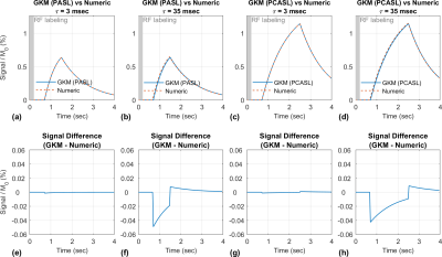 |
Numerical Approximation to the General Kinetic Model for ASL Quantification
Nam Gyun Lee1, Ahsan Javed2, Terrence R. Jao1, and Krishna S. Nayak2
1Biomedical Engineering, University of Southern California, Los Angeles, CA, United States, 2Electrical and Computer Engineering, University of Southern California, Los Angeles, CA, United States
We propose a numerical approximation to Buxton's general kinetic model (GKM) for ASL quantification that will enable greater flexibility in ASL acquisition methods. The proposed method combines the Bloch-McConnell equations with the flow effects and hence model the effects of flow simultaneously with magnetization transfer, T2 effects, off-resonance, and irregular timing of labeling. These can be solved using Jaynes’ matrix formalism. The proposed approximation is compared with GKM using simulations for PASL, PCASL, steady-pulse ASL, and MR fingerprinting ASL. Accuracy of the approximation is studied as a function of a key “time interval” parameter using Monte-Carlo simulations.
|
|
3292. |
Partial volume effect correction of arterial spin labelling data using surface segmentations
Thomas Kirk1,2, Flora Kennedy McConnell1,2, Dimo Ivanov3, Sriranga Kashyap3, Martin Craig1,2, and Michael Chappell1,2
1Institute of Biomedical Engineering, University of Oxford, Oxford, United Kingdom, 2Wellcome Centre for Integrative Neuroimaging, University of Oxford, Oxford, United Kingdom, 3Faculty of Psychology and Neuroscience, Maastricht University, Maastricht, Netherlands
We have recently developed a new approach to partial volume estimation that uses surface segmentations (for example, those produced by FreeSurfer). We compare this approach to conventional volumetric PV estimation for the application of PV correction to two ASL datasets and find evidence in support of using surface-derived PV estimates.
|
|
3293.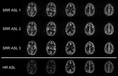 |
Super-resolution reconstruction of single-PLD pseudo-continuous ASL images
Piet Bladt1, Quinten Beirinckx1, Merlijn C.E. van der Plas2, Sophie Schmid2, Wouter M Teeuwisse2, Ben Jeurissen1, Arnold J den Dekker1, Jan Sijbers1, and Matthias J.P. van Osch2,3
1imec - Vision Lab, Department of Physics, University of Antwerp, Antwerp, Belgium, 2C.J. Gorter Center for High Field MRI, Department of Radiology, Leiden University Medical Center, Leiden, Netherlands, 3Leiden Institute of Brain and Cognition, Leiden University, Leiden, Netherlands
Super-resolution reconstruction (SRR) allows for high-resolution image reconstruction from a set of low-resolution multi-slice images with different orientations. Arterial spin labeling (ASL) is an interesting albeit complicated candidate for SRR, as it relies on subtraction. SRR-ASL can be performed on low-SNR subtracted or on low-contrast unsubtracted ASL data. Different ASL-SRR implementations were applied to single-PLD PCASL data and validated against traditional ASL-scans. Combining motion correction, super-resolution post-processing and pairwise subtraction of label-control pairs in a single framework yielded comparable CBF maps as with traditional HR-ASL. Furthermore, in certain slices, SRR-ASL appears to reconstruct the underlying anatomical structure with higher fidelity.
|
|
3294.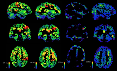 |
Measurement of intra- and extra-neurite perfusion by combining ASL with the NODDI DWI model
Iris Asllani1,2, Alicia Plaindoux3, Jan Petr4, Joseph G. Woods5,6, Matthias J. P. van Osch7, and Mara Cercignani1
1Neuroscience, University of Sussex, Brighton, United Kingdom, 2Biomedical Engineering, Rochester Institute of Technology, Rochester, NY, United States, 3Grenoble Institute of Technology, Grenoble, France, 4Helmholtz-Zentrum Dresden-Rossendorf, Dresden, Germany, 5University of California San Diego, San Diego, CA, United States, 6University of Oxford, Oxford, United Kingdom, 7Leiden University Medical Center, Leiden, Netherlands
Intra- and extra-neurite perfusion in gray and white matter were estimated by applying a spatial linear regression algorithm on ASL images using the micro-structural anatomical information derived from the NODDI analysis of the DWI data. Baseline ASL images were acquired with 4 post-labeling delay (PLD) values in order to test the hypothesis of redistribution of ASL signal across the micro-compartments with increasing PLD. Motor activation was used to investigate the sensitivity of the method for detecting changes in perfusion at the micro-structural level.
|
|
3295. |
Perfusion Quantification from Multi-delay Arterial Spin Labeling (ASL): Validation on Numerical Ground Truth
Qihao Zhang1, Liangdong Zhou2, John Morgan3, Thanh D Nguyen4, Pascal Spincemaille3, and Yi Wang2
1Cornell University, Ithaca, NY, NY, United States, 2Weill Cornell Medicine, New York, NY, United States, 3Radiology, Weill Cornell Medicine, New York, NY, United States, 4Weill Cornell Medicine College, New York, NY, United States
Tissue flow vector field is generated by solving the inverse problem of a voxelized transport equation using multi-delay ASL data and an optimization method. The accuracy of the flow estimation is validated on a vasculature model. For calculating flow vector field in vivo, time resolved 3D (4D) tracer concentration data is acquired using a multi-delay pseudo continuous ASL sequence. The background suppression pulse is interleaved into labeling pulse to generate short post label delay. The output flow magnitude map shows a more anterior-poster-uniform CBF than the traditional Kety’s method.
|
|
3296.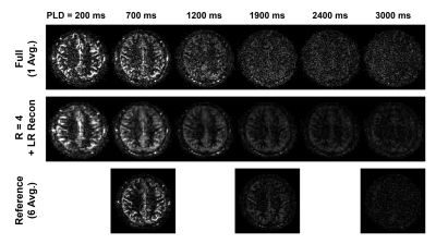 |
Low-Rank Reconstruction for Multi-Delay Arterial Spin Labeling
Paul Kyu Han1, Yanis Djebra1,2, Thibault Marin1, Georges El Fakhri1, Jinsong Ouyang1, and Chao Ma1
1Radiology, Massachusetts General Hospital and Harvard Medical School, Boston, MA, United States, 2LTCI, Télécom Paris, Institut Polytechnique de Paris, Palaiseau, France
Multi-delay arterial spin labeling (ASL) is a powerful MRI technique that allows to non-invasively measure both cerebral blood flow and arterial transit time quantitatively, which has great potential for clinical applications. However, usage of multi-delay ASL is limited by the prolonged scan time with the number of post-labeling delay times used for acquisition. Up to date, low-rank property has been observed in signals of various MR applications where low-rank approach has demonstrated promising results. This work shows that the dynamics of ASL signal display low-rank property and the scan time of multi-delay ASL can be accelerated using low-rank image reconstruction.
|
|
3297.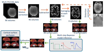 |
Estimation of cortical perfusion from arterial spin labelling data on the cortical surface
Flora A. Kennedy McConnell1,2, Jack Toner1, Thomas Kirk1,2, Martin Craig1,2, Andrew R. Segerdahl2, Michael P. Harms3, and Michael A. Chappell1,2
1Institute of Biomedical Engineering, Department of Engineering Science, University of Oxford, Oxford, United Kingdom, 2Wellcome Centre for Integrative Neuroimaging, University of Oxford, Oxford, United Kingdom, 3Department of Psychiatry, School of Medicine, Washington University, St Louis, MO, United States
Surface-based analysis is a popular approach to fMRI analysis as, compared with volumetric analysis, it allows easier registration of functional regions between individuals. This work sought to move towards surface-based perfusion analysis of arterial spin labelling data. It presents a comparison of the results of a surface-based analysis pipeline with results from a conventional volumetric perfusion analysis pipeline projected onto the cortical surface. Bayesian inference of cortical surface perfusion is demonstrated here and is shown to produce substantially lower perfusion estimates than conventional voxelwise analysis. Future work will investigate to origins of these perfusion differences.
|
|
3298.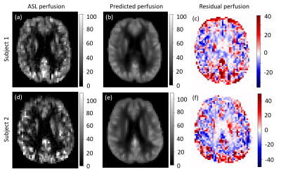 |
Use of population-derived perfusion information in predicting subject-specific perfusion references based on tissue partial volumes
Flora Kennedy McConnell1,2, Sana Suri2,3, Eniko Zsoldos2,3, Klaus Ebmeier3, Clare MacKay2,3, and Michael Chappell1,2
1Institute of Biomedical Engineering, Department of Engineering Science, University of Oxford, Oxford, United Kingdom, 2Wellcome Centre for Integrative Neuroimaging, University of Oxford, Oxford, United Kingdom, 3Department of Psychiatry, University of Oxford, Oxford, United Kingdom
Within and between subject variability in ASL perfusion data can obscure subtle perfusion changes indicative of neurodegenerative disease. It prohibits the production of a 'healthy' population-average perfusion atlas to which individuals can be compared. This work used a GLM to produce subject-specific perfusion references based on tissue partial volumes. Results show that such a linear model can successfully decompose perfusion, at the population level, into components related to tissue partial volumes. However, the model struggles to capture variability at the individual subject level and thus, the problem will in future be addressed with non-linear modelling techniques.
|
|
3299. |
A Learning-From-Noise Dilated Wide Activation Network for Denoising Arterial Spin Labeling (ASL) Perfusion Images
Danfeng Xie1, Yiran Li1, Hanlu Yang1, Li Bai1, and Ze Wang2
1Temple University, Philadelphia, PA, United States, 2University of Maryland School of Medicine, Philadelphia, MD, United States
In this study, we showed that without a noise-free reference, Deep Learning based ASL denoising network can produce cerebral blood flow images with higher signal-to-noise-ratio (SNR) than the reference. In this learning-from-noise training scheme, cerebral blood flow images with very high noise level can be used as reference during network training. This will remove any deliberate pre-processing step for getting the quasi-noise-free reference when training deep learning neural networks. Experimental results this learning-from-noise training scheme preserved the genuine cerebral blood flow information of individual subjects while suppressed noise.
|
|
3300.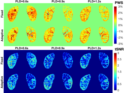 |
Free Breathing Renal Perfusion Imaging at 3T Using Velocity-Selective Inversion prepared Arterial Spin Labeling
Dan Zhu1,2, Dapeng Liu2,3, Wenbo Li2,3, Michael Schär2, and Qin Qin2,3
1Department of Biomedical Engineering, Johns Hopkins University School of Medicine, Baltimore, MD, United States, 2Russell H. Morgan Department of Radiology and Radiological Science, Johns Hopkins University School of Medicine, Baltimore, MD, United States, 3. F.M. Kirby Research Center for Functional Brain Imaging, Johns Hopkins University School of Medicine, Baltimore, MD, United States
Renal perfusion has clinical significance in diagnosis of renal diseases. Velocity-selective arterial spin labeling (VSASL) has minimal sensitivity to arterial transit time delay and velocity-selective inversion (VSI) labeling could improve SNR of perfusion signal. Here renal VSASL images were acquired using different post-labeling delay time and under different B1 shimming conditions. The feasibility of VSI based ASL for renal perfusion mapping at 3T under free breathing is demonstrated on healthy volunteers.
|
|
3301. |
Improving Robustness to Field Imperfections for Velocity-Selective Inversion prepared Arterial Spin Labeling through Dynamic Phase-Cycling
Dapeng Liu1,2, Wenbo Li1,2, Feng Xu1,2, Dan Zhu3, Taehoon Shin4,5, and Qin Qin1,2
1Department of Radiology, Johns Hopkins University School of Medicine, Baltimore, MD, United States, 2F.M. Kirby Research Center for Functional Brain Imaging, Kennedy Krieger Institute, Baltimore, MD, United States, 3Department of Biomedical Engineering, Johns Hopkins University School of Medicine, Baltimore, MD, United States, 4Division of Mechanical and Biomedical Engineering, Ewha Womans University, Seoul, Korea, Republic of, 5Department of Medicine, Case Western Reserve University, Cleveland, OH, United States
In Fourier-transform based velocity-selective inversion (FT-VSI) arterial spin labeling (ASL), either velocity-insensitive or velocity-compensated waveforms could be applied for control modules. Under poor B0/B1 conditions with inefficient refocusing, the scheme with velocity-insensitive control is susceptible to strong false signal but maintained high labeling efficiency, while the scheme with velocity-compensated control has poor velocity-selective labeling profiles. This study overcome these problems by proposing a dynamic phase-cycling scheme based on velocity-insensitive waveform and improve the robustness to B0/B1 field inhomogeneities for VSI ASL. Simulation, phantom and brain ASL scans were conducted for demonstration.
|
|
3302.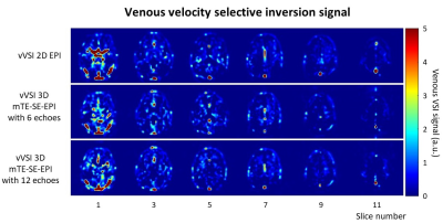 |
Comparison of 2D EPI and 3D multi-echo spin echo EPI readout for Venous Velocity Selective Inversion to measure venous blood T2.
Sophie Schmid1,2, Leonie Petitclerc1,2, André Paschoal3, Suzanne Franklin1,4, Merlijn Van der Plas1,2, and Matthias Van Osch1,2
1Radiology, Leiden University Medical Center, Leiden, Netherlands, 2Leiden Institute of Brain and Cognition (LIBC), Leiden, Netherlands, 3Physics, InBrain Lab, University of Sao Paulo, Ribeirao Preto, Brazil, 4University Medical Center Utrecht, Center for Image Sciences, Utrecht, Netherlands
In this study the variation in the measured apparent T2 was assessed over different slices for venous velocity selective inversion (vVSI) with 2D EPI readout in the sagittal sinus and we compared vVSI with 2D EPI and 3D multi-echo spin echo EPI (mTE-SE-EPI) readout to the gold standard TRUST. With the 2D EPI readout the T2 increases significantly over the slices.
|
|
3303.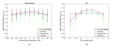 |
Comparison of Velocity-Selective Arterial Spin Labeling using multiple VS Saturation and FT-VS inversion – a Theoretical Study
Shaurov Das1, Luis Hernandez-Garcia2, Ryan Daniel Hernandez3, and Jia Guo1
1Bioengineering, University of California, Riverside, Riverside, CA, United States, 2Biomedical Engineering, University of Michigan, Ann Arbor, MI, United States, 3Physics, University of California, Riverside, Riverside, CA, United States
Velocity selective ASL (VSASL) is the latest addition to the family of ASL techniques, with good promise on resolving transit delay sensitivity in ASL. Though an improvement in SNR efficiency has been reported by using two-module VS saturation and VS inversion pulse, the comparison between them is yet to be done. This study aims to compare their theoretical performance with Bloch simulation and a few realistic scenarios and look for knowledge about existing limitations which can, in turn, lead to further improvement of these techniques. Additionally, a few variants of VSI pulses including most recent improvement were explored and compared.
|
|
3304.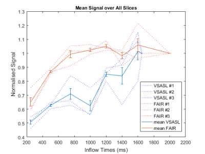 |
Velocity selective arterial spin labeling at 7Tesla: a comparison of the temporal bolus width with FAIR
Juliet Polkey1, Lydiane Hirschler2, Suzanne Franklin2, Matthias van Osch2, and Sophie Schmid2
1University College London Hospital, London, United Kingdom, 2Leiden University Medical Center, Leiden, Netherlands
To the best of our knowledge this work is the first implementation of VSASL at 7T. The VSASL technique was compared with FAIR in 3 healthy volunteers. The temporal bolus width was sampled in each case and compared between the two ASL methods.
|
|
3305.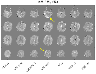 |
Experimental comparison of velocity selective ASL schemes
Luis Hernandez-Garcia1 and Jia Guo2
1FMRI Laboratory, University of Michigan, Ann Arbor, MI, United States, 2University of California at Riverside, Riverside, CA, United States
This work presents an experimental comparison of different velocity selective (VS) labeling strategies for non-contrast perfusion MRI, including new variants of VS saturation and VS inversion, in relation to pseudo-continuous labeling pulses (PCASL).
|
3306. |
Implementation of Liver MR Elastography Tutorial Module to Improve Exam Quality and Reduce Patient Callback Rate
Kelly Tung Smith1, Alvin Silva1, Jonathan Flug1, Justin Yu1, and Anshuman Panda1
1Radiology, Mayo Clinic Arizona, PHOENIX, AZ, United States
In this age of specialized imaging sequences such as liver MR elastography, inexperience with the technical image acquisition and unawareness of artifacts and pitfalls confound diagnostic accuracy due to poor image quality. We feel there is a knowledge gap in those at the front line involved with image acquisition, and that by defining and filling in this knowledge gap with an educational tutorial module, MR elastography image quality can be optimized and callback rates can be decreased. In turn, decreased callbacks will translate into reduced cost for unnecessary repeated scans; reduced patient time and frustration; and increased MR scheduling access.
|
|
3307.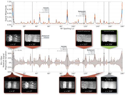 |
Optimal RF-Spoiling for Bias-Free Displacement Field Estimation in Fast 3D MR Elastography Sequences
Christian Guenthner1 and Sebastian Kozerke1
1University and ETH Zurich, Zurich, Switzerland
RF-spoiling is employed in order to reduce transverse coherences. While spoiling angles are typically optimized for contrast, efficacy for bias-free phase-contrast measurements is yet to be proven. Here, we investigate the influence of the RF-spoiling angle on displacement estimates in fast GRE-MR Elastography sequences using Extended Phase Graph simulations. Theoretical findings are validated using a novel RF-spoiling test sequence as well as conventional MRE phantom experiments. An optimal spoiling angle of 158° degrees is identified allowing for bias-free MRE acquisitions at repetition times as low as 5ms.
|
|
3308.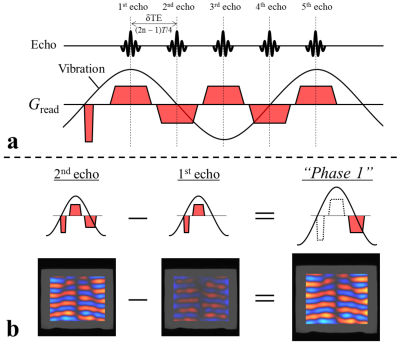 |
Simultaneous acquisition of multi-phase offset for rapid imaging in MR elastography (SAMURAI-MRE) using a multi-echo sequence
Daiki Ito1,2,3, Tomokazu Numano1,3, Tetsushi Habe1,2, Toshiki Maeno1, Kazuyuki Mizuhara3,4, Surendra Maharjan1, Kouichi Takamoto5, and Hisao Nishijo6
1Department of Radiological Sciences, Graduate School of Human Health Sciences, Tokyo Metropolitan University, Tokyo, Japan, 2Office of Radiation Technology, Keio University Hospital, Tokyo, Japan, 3Health Research Institute, National Institute of Advanced Industrial Science and Technology, Ibaraki, Japan, 4Department of Mechanical Engineering, Tokyo Denki University, Tokyo, Japan, 5Department of sport and Health Sciences, Faculty of Human Sciences, University of East Asia, Yamaguchi, Japan, 6System Emotional Science, Graduate School of Medicine and Pharmaceutical Sciences, University of Toyama, Toyama, Japan
MR elastography (MRE) requires acquisitions of multiple phase offset images. Patient movement between acquisitions of phase offset images is not only difficult to be detected, but also leads to calculate shear modulus incorrectly. To resolve this problem, we present a novel MRE technique that can acquire multiple phase offset images simultaneously (SAMURAI-MRE) using a motion-encoding gradient (MEG)-less multi-echo sequence. MRE images obtained at spin-echo (SE)- and gradient-echo (GRE)-based SAMURAI-MRE were compared with those at conventional SE- and GRE-based MEG-less multi-echo MRE. The results demonstrate that it is possible to perform SAMURAI-MRE, and SE-based SAMURAI-MRE was superior to GRE-based SAMURAI-MRE.
|
|
3309.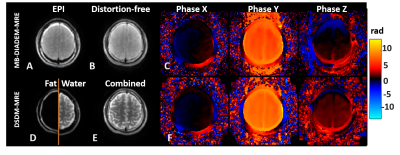 |
Simultaneous Fat-Water MR Elastography Imaging using Distortion-free DIADEM-EPI
Yi Sui1, MyungHo In1, Armando Manduca2, Matthew A. Bernstein1, Richard L. Ehman1, John III Huston1, and Ziying Yin1
1Radiology, Mayo Clinic, Rochester, MN, United States, 2Physiology and Biomedical Engineering, Mayo Clinic, Rochester, MN, United States
Simultaneous fat-water EPI-MR elastography imaging was developed for acquisition of the brain (water) and skull (bone marrow fat) MRE motion. Compared to our previous dual-saturation and dual-sensitivity motion encoding (DSDM) technique, the proposed method minimize the EPI distortion and misalignment between skull and brain. This technique could offer a unique opportunity to quantify the skull-brain mechanical decoupling performance modulated by the pia-arachnoid complex, and its potential alterations in response to repetitive head impacts (RHI) in our future studies.
|
|
3310.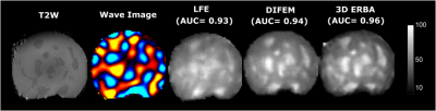 |
Joint Elasticity Reconstruction and Displacement Filtering for 3D Magnetic Resonance Elastography
Shahed Khan Mohammed1, Mohammad Honarvar1, Davood Karimi1, Piotr Kozlowski2,3, and Septimiu Salcudean1
1Electrical and Computer Engineering, University of British Columbia, Vancouver, BC, Canada, 2UBC MRI Research Center, Vancouver, BC, Canada, 3Department of Radiology, University of British Columbia, Vancouver, BC, Canada
Iterative reconstruction methods in magnetic resonance elastography (MRE) inversion can allow incorporating sparsity prior and can provide displacement filtering. However the problem is difficult to converge as MRE inversion is a non-convex and ill-conditioned problem. Here, we presented 3D ERBA with total variation prior on the elasticity, which showed good convergence by utilizing a bi-convex optimization of 3D finite element modeling of elastic wave. Extensive experiments on numerical phantom, agar phantom, and in-vivo liver study showed promising indications in detecting inclusion and abnormality, and improving the diagnostic efficacy of MRE.
|
|
3311.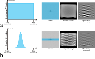 |
Dynamic MR elastography using MR magnitude images
Tomokazu Numano1,2, Daiki Ito2,3,4, Kazuyuki Mizuhara5, Toshikatsu Washio2, Tetsushi Habe3,4, Toshiki Maeno3, Masaki Misawa2, and Naotaka Nitta2
1Radiological Sciences, Tokyo Metropolitan University, Tokyo, Japan, 2National Institute of Advanced Industrial Science and Technology (AIST), Tsukuba, Japan, 3Tokyo Metropolitan University, Tokyo, Japan, 4Keio University Hospital, Tokyo, Japan, 5Tokyo Denki University, Tokyo, Japan
We developed a new technique for dynamic MR elastography (MRE) using a MR magnitude image. A general MRE uses a MR phase image as a wave image. Proposed technique was used a MR magnitude image instead of MR phase image as a wave image by using a special vibration. Proposed technique (special vibration MRE) performance was comparable to that of a continuous vibration MRE. Since special vibration MRE need an only few second vibration, it could dramatically eliminated the patients' vibration-related discomfort.
|
|
3312.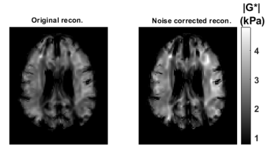 |
Improving the SNR and Correcting the Bias in Elastograms in Helmholtz Inversion for MR Elastography
Cemre Ariyurek1,2, Yusuf Ziya Ider1, and Ergin Atalar1,2
1Department of Electrical and Electronics Engineering, Bilkent University, Ankara, Turkey, 2National Magnetic Resonance Research Center (UMRAM), Ankara, Turkey
In Helmholtz inversion an equation is obtained for each MR elastography displacement data collected for each motion direction and each excitation frequency. Under high SNR, SNR of the estimated elasticity map by the Helmholtz inversion can be derived. Prior to the inversion, each equation can be weighted by the SNR of the elasticity estimate to improve SNR of the elastogram and low SNR ones can be thresholded to reduce bias in the elasticity estimation. Simulation and experiment results demonstrate that the original inversion underestimates the elasticity when there is noise and this bias can be corrected by the proposed reconstruction.
|
|
3313.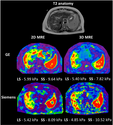 |
Comparison of multi-platform 2D and 3D MR elastography in vivo and in vitro
Paul Kennedy1,2, Daniel Stocker1,2, Octavia Bane1,2, Stefanie Hectors1,2,3, Bradley D Bolster Jr. 4, and Bachir Taouli1,2
1BioMedical Engineering and Imaging Institute, Icahn School of Medicine at Mount Sinai, New York, NY, United States, 2Department of Diagnostic, Molecular and Interventional Radiology, Icahn School of Medicine at Mount Sinai, New York, NY, United States, 3Department of Radiology, Weill Cornell Medicine, New York, NY, United States, 4Siemens Medical Solutions USA, Inc., Salt Lake City, UT, United States
In this study we assess the variation in 2D and 3D MR elastography (MRE) measures in patients, volunteers and a phantom across two MR system vendors. Interobserver variability was also examined. 2D and 3D MRE liver measurements were not significantly different across systems and showed excellent intraclass correlation coefficient (ICC) between readers. Spleen measurement showed excellent ICC between readers but significant variation in 3D MRE spleen stiffness was found between systems. Liver 3D MRE is stable across platforms however further study is required to assess the bias in 3D MRE spleen measurements from multiple vendors.
|
|
3314. |
Noninvasive identification of renal perfusion in renal fibrosis of Adriamycin nephropathy by using ASL and IVIM-DWI imaging
Shudan Min1, Jilei Zhang2, and Shenghong Ju1
1Jiangsu Key Laboratory of Molecular Imaging and Functional Imaging,, Southeast University, Nanjing, Jiangsu, China, 2Clinical Science, Philips Healthcare, Shanghai, China
Renal fibrosis is the final common consequence of chronic kidney disease (CKD) and is the mechanism of CKD progressing to end-stage renal failure. Here we employed ASL and IVIM to assess longitudinal changes of kidney perfusion to reflect renal fibrosis in an animal model of ADR nephropathy. A significant decline in renal microcirculation was detected in the ADR group compared with the sham group. Administration of VCP979 could prevent the renal perfusion, and the treatment response was also detected by the two imaging technology.
|
|
3315.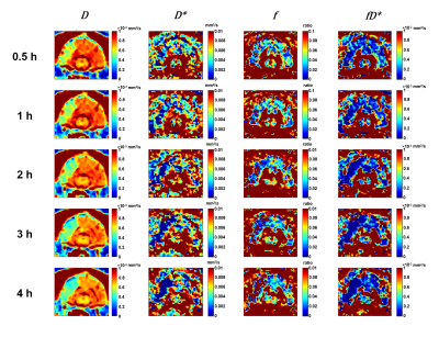 |
Quantification of collateral blood flow using intravoxel incoherent motion MRI in a canine large vessel occlusion model of slow and fast evolvers
Mohammed Salman Shazeeb1,2,3, Robert King1,2, Karl Helmer3, Josephine Kolstad1, Christopher Raskett1, Natacha Le Moan4, Jonathan A. Winger4, Lauren Kelly4, Ana Krtolica4, Nils Henninger1, and Matthew Gounis1
1University of Massachusetts Medical School, Worcester, MA, United States, 2Worcester Polytechnic Institute, Worcester, MA, United States, 3Massachusetts General Hospital & Harvard Medical School, Boston, MA, United States, 4Omniox Inc., San Carlos, CA, United States
In acute ischemic stroke due to large vessel occlusion (LVO), variability of infarct evolution observed in humans is closely captured by the canine LVO model, which can predict dogs to be slow or fast evolvers. The extent of collateral blood supply has shown good correlations with infarct growth rate. This study investigated the use of intravoxel incoherent motion (IVIM) MRI to quantify microvascular perfusion. Longitudinal IVIM parameters clearly differentiated between slow and fast evolvers depicting collateral blood flow changes, which can determine the severity of ischemic injury and also track longitudinal changes in response to therapeutic treatments/interventions in preclinical studies.
|
|
3316.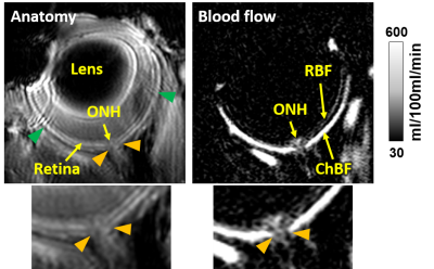 |
Blood flow MRI of the mouse optic nerve head
Eric R. Muir1 and Timothy Q. Duong1
1Radiology, Stony Brook University, Stony Brook, NY, United States
Different retinal diseases likely affect the retinal, choroidal, and optic nerve head (ONH) circulations differently. Methods to quantitatively measure ONH blood flow with depth resolution are lacking. In this study, a blood flow MRI method was optimized to image blood flow of the ONH, retina, and choroid in the mouse eye at 42x42x275µm. This method could be useful to study models of retinal disease, such as glaucoma.
|
|
3317.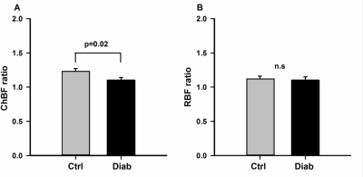 |
Impaired blood flow and vascular reactivity in the choroid in diabetic mice
Eric R. Muir1, Saurav B. Chandra2, Divya Narayanan3, Nikolay P. Akimov4, René C. Rentería4,5, and Timothy Q. Duong1
1Radiology, Stony Brook University, Stony Brook, NY, United States, 2Icon Clinical Research Inc, North Wales, PA, United States, 3Ora Clinical, Andover, MA, United States, 4Ophthalmology, University of Texas Health Science Center, San Antonio, TX, United States, 5School of Osteopathic Medicine, University of the Incarnate Word, San Antonio, TX, United States
Diabetic retinopathy is a microvascular disease of the retina, in which basal blood flow and the vascular responses to metabolic conditions in diabetic eyes are perturbed. To determine if diabetic mice have impaired retinal/choroidal blood flow and vascular reactivity, we measured ocular blood flow using MRI during hypercapnia. In diabetic mice, basal choroidal blood flow was significantly decreased. In both control and diabetic mice, hypercapnia caused a significant increase in retinal and choroidal blood flow, but the choroidal response was significantly reduced in diabetic mice, indicating impaired vascular reactivity.
|
|
3318.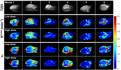 |
Parametric maps of murine cancers from dynamic contrast enhanced MRI: low versus high doses of contrast agent
Xiaobing Fan1, Xueyan Zhou2, Devkumar Mustafi1, Federico Pineda1, Erica Markiewicz1, Marta Zamora1, Deepa Sheth1, Olufunmilayo I. Olopade3, and Gregory S. Karczmar1
1Radiology, The University of Chicago, Chicago, IL, United States, 2School of Technology, Harbin University, Harbin, China, 3Medicine, The University of Chicago, Chicago, IL, United States
We compared dynamic contrast enhanced (DCE) MRI with low (0.04mmol/kg) and high doses (0.2mmol/kg) of contrast agent on C3H mice at 9.4T with 1.5s temporal resolution before and after a bolus injection. The standard Tofts model was used to extract physiological parameters (Ktrans and ve) with arterial input function derived from muscle reference tissue. On average, rate constants were larger for low dose than high dose data. There were strong correlations for Ktrans and ve between low and high dose data. Therefore, low dose may be as effective as a high dose of contrast agent for cancer diagnosis.
|
|
3319.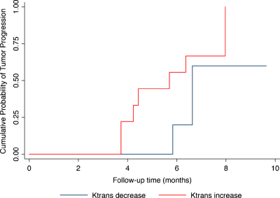 |
Parameters From Dynamic Contrast-Enhanced MRI Are Biomarkers Predicting Response after Radiotherapy to Brain Metastases
Zhuo Shi1, Lizhi Xie2, Peng Wang1, Xinming Zhao1, and Han Ouyang1
1National Cancer Center/Cancer Hospital, Chinese Academy of Medical Sciences, Beijing, China, 2GE Healthcare, MR Research China, Beijing, Beijing, China
Dynamic contrast-enhanced (DCE) MRI provides additional information on blood-brain barrier integrity. The Ktrans is a measure of capillary permeability obtained using DCE MRI that is directly proportional to the level of permeability of the blood-brain barrier. The purpose of this study was to evaluate the effect of SRS on cerebral metastases using Ktrans derived from DCE MRI and to explore the ability of Ktrans measurements on predicting midterm tumor outcomes after Stereotactic radiosurgery (SRS).
|
|
3320. |
Reducing TSM by Oxygen Inhalation: effective for pleural effusion but not ascites
Xiaoqian Jia1, Jianxin Guo2, Xiaocheng Wei3, and Jian Yang2
1Ridiology, The First Affiliated Hospital of Xi'an Jiao Tong University, xi'an, China, 2The First Affiliated Hospital of Xi'an Jiao Tong University, xi'an, China, 3MR Research China, GE Healthcare, xi'an, China
In this study, we try to provide a wider cohort of subjects with continuous low flow oxygen inhalation during dynamic contrast-enhancedMR liver imaging to reduce “Transient Severe Motion” (TSM) after administration of gadoxetate disodium.In addition, separate comparison of arterial phase motion scores was conducted among: category of combined Pleural Effusion, category of combined Ascites, and category of None.The results show that continuous oxygen can significantly reduce the occurrence of TSM; and 1) pleural effusion may reduce image quality, and can be alleviated by oxygen inhalation, and 2) oxygen inhalation is less effective under the condition of ascites.
|
|
3321.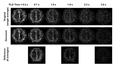 |
Compartmental Low-Rank Denoising for Multi-Delay ASL
Yanis Djebra1,2, Thibault Marin1, Jinsong Ouyang1, Georges El Fakhri1, Chao Ma1, and Paul Kyu Han1
1Gordon Center for Medical Imaging, Department of Radiology, Massachusetts General Hospital, Harvard Medical School, Boston, MA, United States, 2LTCI, Télécom Paris, Institut Polytechnique de Paris, Palaiseau, France
Arterial spin labeling (ASL) is a powerful magnetic resonance imaging (MRI) technique that allows to measure tissue perfusion, potentially an important biomarker for assessing cerebral tissue viability, without the use of contrast agent. However, the main drawback of ASL is in the intrinsically low signal-to-noise ratio (SNR). In this work, we investigate the low-rank property of ASL signal and explore the feasibility of improving the SNR of ASL signal using low-rank denoising approach. Results show that the ASL signal indeed display low-rank property and that the SNR in multi-delay ASL imaging can be improved using compartmental low-rank denoising method.
|
3322. |
Mechanical Property Recovery in the Cerebral Cortex using Magnetic Resonance Elastography
Lucy V Hiscox1, Matthew DJ McGarry2, and Curtis L Johnson1
1University of Delaware, Newark, DE, United States, 2Dartmouth College, Hanover, NH, United States
Magnetic resonance elastography of the brain has shown promise as a sensitive neuroimaging biomarker for neurodegenerative disorders; however, the feasibility and reliability of performing MRE of the cerebral cortex warrants investigation due to the unique challenges of studying thinner and more complex geometries. In this work, we found good agreement between realistic simulated MRE data versus recovered displacement data of an older adult participant at various levels of applied Gaussian noise. Future work should consider additional and more advanced imaging and inversion approaches to improve the reliability and sensitivity of cortical MRE measures.
|
|
3323.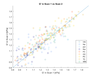 |
Robustness of MR Elastography in the brain – Test-retest reliability and effect of different reconstruction methods
Siri Flogstad Svensson1, Jorunn Fraser-Green2, Omar Darwish3, José Rodriguez de Arcos3, Tryggve Holck Storås1, Sverre Holm4, Ralph Sinkus3,5, and Kyrre Eeg Emblem1
1Department for Diagnostic Physics, Oslo University hospital, Oslo, Norway, 2The Intervention Center, Oslo University hospital, Oslo, Norway, 3Division of Imaging Sciences & Biomedical Engineering, King's College, London, United Kingdom, 4Department of Informatics, University of Oslo, Oslo, Norway, 5INSERM U1148, LVTS, University Paris Diderot, Paris, France
We assessed the test-retest reliability of MR Elastography, and compared the estimates of the shear storage modulus G’ (tissue elasticity) from two different reconstruction methods; a localized divergence-free finite-element approach and a local curl approach. The median coefficient of variation for two scans of the brain was 2.8 %, whereas the intraclass correlation coefficient between scan 1 and scan 2 was 0.80. G’ values estimated using the local curl method were 19% lower than with the divergence-free method. MRE of the human brain appears robust, while absolute values are dependent on the reconstruction method and should be used with care.
|
|
3324.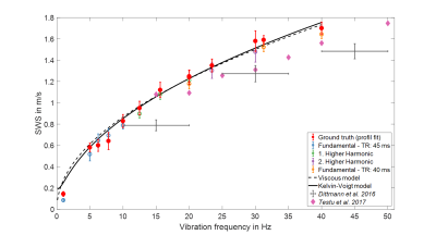 |
Ultra-low frequency MR elastography of the in vivo human brain from endogenous pulsation to 40 Hz time-harmonic vibrations.
Helge Herthum1, Heiko Tzschätzsch1, Felix Schrank1, Mehrgan Shahryari1, Gergely Bertalan1, Carsten Warmuth1, Jürgen Braun2, and Ingolf Sack1
1Department of Radiology, Charité Universitätsmedizin Berlin, Berlin, Germany, 2Institute of Medical Informatics, Charité Universitätsmedizin Berlin, Berlin, Germany
In this work, we applied single-shot steady-state MR elastography with spiral readout for quantifying shear wave speed (SWS) from endogenous pulsation to 40 Hz external vibrations with high sampling rate for first-time measurement of SWS dispersion of in vivo human brain in the ultra-low frequency regime. Ground truth SWS was determined by profile fitting approaches and reproduced by SWS mapping based on frequency-adaptive multi-component wave-number inversion. The obtained SWS dispersion was fitted by two-parameter viscoelastic models showing that brain tissue is dominated by fluid properties in this ultra-low frequency regime. Our method is potentially sensitive to vessel-related neurological disorders.
|
|
3325.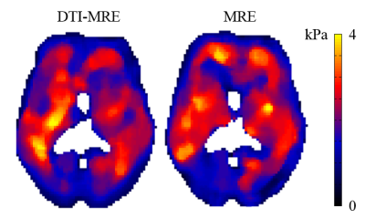 |
Development of in vivo human brain DTI-MRE
Shujun Lin1, Brad Sutton2, Richard Magin1, and Dieter Klatt1
1Richard and Loan Hill Department of Bioengineering, University of Illinois at Chicago, Chicago, IL, United States, 2Department of Bioengineering, University of Illinois at Urbana-Champaign, Urbana-Champaign, IL, United States
Simultaneous acquisition of diffusion tensor imaging (DTI) and magnetic resonance elastography (MRE) has been proven feasible in previous pre-clinical studies. DTI-MRE requires the identification of experimental parameter ranges that depend on the specific target. Here we identify valid experimental parameter sets in simulations for in vivo human brain applications and the DTI-MRE technique is implemented by modifying a single-shot spin-echo EPI sequence. A good agreement was found between DTI-MRE and conventional acquisitions. DTI-MRE may increase the clinical acceptance of MRE and DTI by providing co-registered diffusion and tissue mechanical property maps within the scan time of a conventional DTI acquisition.
|
|
3326.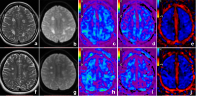 |
The Investigation of Three-Dimensional MR elastography of the Brain in Multiple Sclerosis
Ling Fang1, Matthew C. Murphy2, Sujuan Liu1, Qiuxia Luo1, Jingbiao Chen1, Bingjun He1, Wei Qiu3, Jun Chen2, Meng Yin2, Kevin J. Glaser2, Richard L. Ehman2, and Jin Wang1
1Department of Radiology, the Third Affiliated Hospital, Sun Yat-sen University, Guangzhou, China, 2Department of Radiology, Mayo Clinic, Rochester, MN, United States, 3Department of Neurology, the Third Affiliated Hospital, Sun Yat-sen University, Guangzhou, China Poster Permission Withheld
Multiple sclerosis (MS) is a chronic inflammatory demyelinating disease of the central nervous system. MR Elastography (MRE) can quantitatively measure biomechanical tissue properties in vivo noninvasively. In this study, we investigated the potential value of MRE for the evaluation of centrum ovale viscoelasticity (including shear stiffness, storage modulus, shear loss modulus, and damping ratio) in Southern Chinese MS patients and to try to analyze its relevance to clinical manifestations. Our results showed that only the damping ratio of the centrum ovale was significantly decreased in MS patients and may provide a potential quantitative biomarker to evaluate MS.
|
|
3327. |
Free Breathing Magnetic Resonance Elastography in Patients with Idiopathic Pulmonary Fibrosis
Faisal S Fakhouri1,2, Vincent Esguerra3, and Arunark Kolipaka1,2
1Department of Biomedical Engineering, The Ohio State University, Columbus, OH, United States, 2Department of Radiology, The Ohio State University Wexner Medical Center, Columbus, OH, United States, 3Division of Pulmonary, Critical Care, and Sleep Medicine, The Ohio State University Wexner Medical Center, Columbus, OH, United States
Interstitial lung diseases alter the mechanical properties of the lung parenchyma. Lung stiffness is a potential diagnostic marker. Standard breath hold techniques are difficult for patient with pulmonary disease. By using a free breathing Spin-Echo Dual-Density Spiral (SE-DDS) MRE sequence, a feasibility study was performed on 6 idiopathic pulmonary fibrosis (IPF) patients. It was found that the shear stiffness of IPF diseased lung is 2±0.48kPa for the right lung and 1.99±0.42kPa for the left lung which is significantly higher (P=0.0108 and P=0.0072) than healthy lungs, which has shear stiffness of 1.29±0.35kPa and 1.43±0.16kPa for right and left lungs, respectively.
|
|
3328.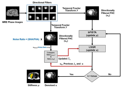 |
Helmholtz Inversion using Unconstrained Optimization for MR Elastography of the Lung: A Comparison to Direct Inversion
Huiming Dong1,2, Rizwan Ahmad2, and Arunark Kolipaka1,2
1Department of Radiology, The Ohio State University Wexner Medical Center, Columbus, OH, United States, 2Department of Biomedical Engineering, The Ohio State University, Columbus, OH, United States
Lung stiffness is a potential biomarker for multiple lung diseases. MR elastography (MRE) allows non-invasive measurement of lung stiffness. However, it is challenging to estimate stiffness using direct Helmholtz inversion due to low signal-to-noise ratio (SNR) from lung MRE. In this work, a compressed-sensing-based Helmholtz inversion is proposed where noise is reduced via Laplacian of Gaussian (LoG) and Morozov’s discrepancy principle, while the sparsity of stiffness map is explored in a wavelet domain. Results demonstrated that the proposed inversion yielded robust stiffness estimation and successfully detected higher stiffness at total lung capacity (TLC) compared to residual volume (RV).
|
|
3329.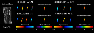 |
In Vivo Aortic MR Elastography using Free-Breathing and Single-Breath-Hold SE-EPI: Inter-Scanner Reproducibility at 1.5T and 3T
Huiming Dong1,2, Richard D White1,3, and Arunark Kolipaka1,2,3
1Department of Radiology, The Ohio State University Wexner Medical Center, Columbus, OH, United States, 2Department of Biomedical Engineering, The Ohio State University, Columbus, OH, United States, 3Internal Medicine-Division of Cardiology, The Ohio State University Wexner Medical Center, Columbus, OH, United States
In vivo MR elastography (MRE) allows non-invasive estimation of aortic stiffness. Recently, the feasibility of multi-slice free-breathing (FB) spin-echo echo-planar imaging (SE-EPI) aortic MRE was investigated. In this study, a single breath-hold (SBH) SE-EPI aortic MRE protocol was proposed and compared to FB SE-EPI aortic MRE. Moreover, in vivo FB and SBH SE-EPI aortic MRE were studied using both1.5T and 3T scanners. The main aim was to assess inter-scanner reproducibilities of FB and SBH SE-EPI aortic MRE.
|
|
3330.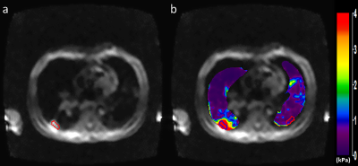 |
Magnetic Resonance Elastography and Computational Modeling of Indeterminate Pulmonary Nodules Mechanical Properties.
Faisal S Fakhouri1,2, Youjin Cho1,3, Joshua Englert4, Samir Ghadiali1,4, and Arunark Kolipaka1,2
1Department of Biomedical Engineering, The Ohio State University, Columbus, OH, United States, 2Department of Radiology, The Ohio State University Wexner Medical Center, Columbus, OH, United States, 3College of Medicine, The Ohio State University, Columbus, OH, United States, 4Division of Pulmonary, Critical Care, and Sleep Medicine, The Ohio State University Wexner Medical Center, Columbus, OH, United States
Many pulmonary diseases alter lung mechanical properties making lung stiffness an important biomarker. By using a Spin-Echo Echo Planar Imaging (SE-EPI) MRE sequence, a feasibility study was performed on 6 indeterminate pulmonary nodules (IPN) patients to determine the shear stiffness within the nodule and compare it with stiffness in the contralateral lung. It was found that in all the patients the median shear stiffness within the IPN was significantly higher than in the contralateral lung (1.77kPa vs 0.86kPa, p=0.008) Computational models based on MRE stiffness measurements also indicate decreased strain and increased strain gradients within the IPN.
|
|
3331.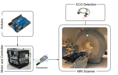 |
In Vivo Cardiac MR Elastography using Simultaneous Multi Slice Imaging
Ayse Sila Dokumaci1, Torben Schneider2, Daniel Fovargue1, Anthony Price1, Omar Darwish1, George Jolly1, Stefan-Heinz Hoelzl1, Jelizaveta Sudakova1, Ralph Sinkus1,3, and David Nordsletten1,4
1Imaging Sciences and Biomedical Engineering, King's College London, London, United Kingdom, 2Philips Healthcare, Guildford, United Kingdom, 3Inserm U1148, University Paris, Paris, France, 4Departments of Biomedical Engineering and Cardiac Surgery, University of Michigan, Ann Arbor, Ann Arbor, United Kingdom
MR Elastography is a technique to evaluate the biomechanical properties of soft tissues noninvasively. Its application in the heart is very challenging due to several factors including actuator hardware, robust MR sequences, and suitable reconstruction strategies. One of the biggest limitations for data acquisition of cardiac MRE scans is the breath hold duration which is up to 21 heart beats, repeated several times for a full dataset. The main goal of this study was the implementation of an MR sequence with simultaneous multi slice acquisition capability to reduce the breath hold duration and increase comfort and compliance for the subjects.
|
|
3332.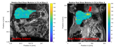 |
Actuator-Free MR Elastography of the Liver: Imaging Liver Tumors with Viscoelastic Intrinsic MRE
Elijah Van Houten1, Guillaume Gilbert2, and An Tang3
1Génie mécanique, Université de Sherbrooke, Sherbrooke, QC, Canada, 2MR Clinical Science, Philips Healthcare, Markham, ON, Canada, 3Département de radiologie, Université de Montréal, Montréal, QC, Canada
A new intrinsic activation MR Elastography method is presented through a proof of concept for imaging the viscoelasticity of a liver tumor. Results are presented for a healthy volunteer and a focal nodular hyperplasia subject. Relative stiffness, damping and compressibility maps clearly indicate the location of the tumor and are in agreement with mechanical property values previously reported in the literature. The method shows promise for the diagnosis of focal liver lesions and provides insight into new physiologically related biomarkers for these lesions.
|
|
3333.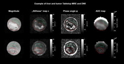 |
High-resolution multifrequency MR Elastography of liver tumors using a compact tabletop MRI Scanner
Mehrgan Shahryari1, Gergely Bertalan1, Helge Herthum1, Heiko Tzschätzsch1, Jürgen Braun2, Marcus R. Makowski1, Sarah Keller1, and Ingolf Sack1
1Department of Radiology, Berlin, Germany, 2Institute of Medical Informatics, Berlin, Germany Poster Permission Withheld
MR elastography is a clinical imaging technique for the mapping of viscoelastic tissue properties on the millimeter scale. However, basic studies in tissue samples require a compact MRE setup which provides sub-millimeter resolution. We here introduce compact 0.5-Tesla tabletop multifrequency MRE for high-resolution mapping of viscoelasticity and water diffusion in VX2-liver tumors and surrounding liver tissue. Ex-vivo tumors were stiffer, more viscous and showed higher water diffusivity than adjacent tissue. In tumors, viscosity was inversely correlated with water diffusion while in liver tissue, stiffness was inversely correlated with diffusion.
|
|
3334.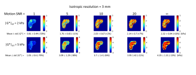 |
Validation of the Accuracy of Magnetic Resonance Elastography of the Liver via 3D Finite Element Modelling Simulation
Deirdre McGrath1,2, Christopher Bradley1,2, and Susan Francis1,2
1NIHR Nottingham Biomedical Research Centre, Radiological Sciences, Division of Clinical Neuroscience, Queens Medical Centre, Nottingham, United Kingdom, 2Sir Peter Mansfield Imaging Centre, University of Nottingham, Nottingham, United Kingdom
In this novel study, magnetic resonance elastography (MRE) of the liver is simulated in a detailed 3D anthropomorphic finite element model of the human torso, to explore the influence of imaging spatial resolution and motion-based signal to noise ratio (SNR) on the accuracy of elastograms calculated by direct inversion. It was found that MRE accuracy was highly dependent on imaging voxel dimensions, motion-SNR and the ground truth material properties. These findings will guide the optimisation and development of future liver MRE methodology.
|
|
3335.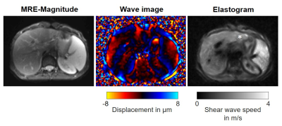 |
Tomoelastography for Quantification of Heterogeneity in Primary Sclerosing Cholangitis-related Hepatic Fibrosis
Rolf Reiter1,2, Heiko Tzschätzsch1, Matthias Haas1, Christian Bayerl1, Dieter Klatt2, Meltem Uyanik2, Shreyan Majumdar2, Marion Muche3, Bernd Hamm1, Jürgen Braun4, Ingolf Sack1, and Patrick Asbach1
1Radiology, Charité - Universitätsmedizin Berlin, Berlin, Germany, 2Bioengineering, University of Illinois at Chicago, Chicago, IL, United States, 3Hepatology, Charité - Universitätsmedizin Berlin, Berlin, Germany, 4Medical Informatics, Charité - Universitätsmedizin Berlin, Berlin, Germany Poster Permission Withheld Primary sclerosing cholangitis (PSC) is a rare chronic liver disease that leads to stricturing of bile ducts and a heterogeneous distribution of hepatic fibrosis. Despite the success of MR cholangiopancreaticogram and liver biopsy for the detection of PSC, they do not correlate with the severity of disease. Currently, there is no accurate diagnostic reference standard for monitoring the disease activity in PSC which limits patient management. Tomoelastography has the potential to quantify heterogeneity in PSC-related hepatic fibrosis using a normalized standard deviation index as a new biomarker besides overall stiffness. |
|
3336.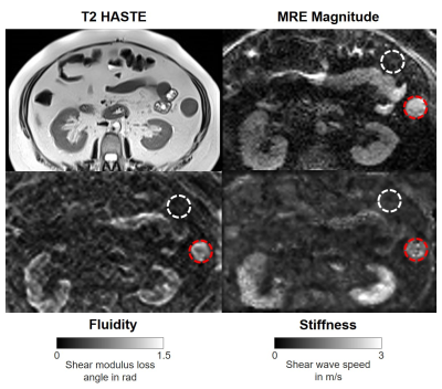 |
Feasibility of Tomoelastography in Inflammatory Bowel Disease: Preliminary Results
Rolf Reiter1, Florian Nino Loch2, Carsten Kamphues2, Stephan Rodrigo Marticorena Garcia1, Bernd Hamm1, Jürgen Braun3, Ingolf Sack1, and Patrick Asbach1
1Radiology, Charité - Universitätsmedizin Berlin, Berlin, Germany, 2Surgery, Charité – Universitätsmedizin Berlin, Berlin, Germany, 3Medical Informatics, Charité - Universitätsmedizin Berlin, Berlin, Germany Despite its success in the detection of inflammatory bowel disease (IBD), standard clinical MRI and other imaging modalities do not correlate with disease severity and cannot differentiate fibrotic and inflammatory bowel strictures. Anatomical details such as the descending colon and other parts of the intestine were demarcated on tomoelastography stiffness and fluidity maps, even without superimposed morphological images. Preliminary results demonstrate the feasibility of investigating IBD using tomoelastography. This study motivates larger trials and the assessment of fibrosis and inflammation in active IBD. |
|
3337.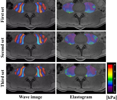 |
Evaluation of precision of the psoas major muscle MR elastography
Tetsushi Habe1,2, Tomokazu Numano1,3, Daiki Ito1,2,3, Toshiki Maeno1, Kazuyuki Mizuhara4, Kouichi Takamoto5, and Hisao Nishijo6
1Department of Radiological Sciences, Tokyo Metropolitan University, Tokyo, Japan, 2Office of Radiation Technology, Keio University Hospital, Tokyo, Japan, 3Human Technology Research Institute, National Institute of Advanced Industrial Science and Technology (AIST), Ibaraki, Japan, 4Department of Mechanical Engineering, Tokyo Denki University, Tokyo, Japan, 5Department of Sport and Health Sciences, University of Ease Asia, Yamaguchi, Japan, 6Department of System Emotional Science, Toyama University, Toyama, Japan
We developed psoas major muscle (PM) MR elastography (MRE) technique and reported shear modulus of the PM for the first time in the world. However, measurement precision (repeatability and reproducibility) of this technique was not assessed. In this study, repeatability with three repetitions and reproducibility with different three operators were evaluated. Repeatability of this technique was about the same as required for clinical liver MRE and reproducibility was almost perfect. The original vibration pad was focused on generating shear wave in the PM efficiently. The development of the vibration pad specialized for PM MRE resulted in acceptable repeatability and reproducibility.
|