
Digital Poster Session
Acquisition, Reconstruction & Analysis: Mitigating Motion, Artifacts and Imperfections
Acquisition, Reconstruction & Analysis
3338 -3353 Mitigating motion, artifacts and imperfections - Motion Correction: Non-Brain
3354 -3369 Mitigating motion, artifacts and imperfections - Motion Correction: Brain
3370 -3383 Mitigating motion, artifacts and imperfections - Acquisition, Reconstruction & Motion Artefact Correction
3384 -3398 Mitigating motion, artifacts and imperfections - Measuring & Correcting System Imperfections
3399 -3414 Mitigating motion, artifacts and imperfections - Mitigating Sample-Induced Artifacts
3415 -3429 Mitigating motion, artifacts and imperfections - Artifact Correction: Miscellaneous
3338.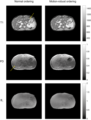 |
Quantitative abdominal imaging using 3D motion-robust MR fingerprinting
Max van Riel1,2, Zidan Yu2,3, Shota Hodono2,3, Ding Xia2, Hersh Chandarana2, and Martijn A. Cloos2,3
1Department of Biomedical Engineering, Eindhoven University of Technology, Eindhoven, Netherlands, 2Center for Advanced Imaging Innovation and Research and Bernard and Irene Schwartz Center for Biomedical Imaging, Department of Radiology, New York University School of Medicine, New York, NY, United States, 3Sackler Institute of Graduate Biomedical Sciences, New York University School of Medicine, New York, NY, United States
We demonstrate a 3D MR fingerprinting sequence for quantitative abdominal imaging. The sequence was made robust to motion by modifying the order of acquisition to allow for free-breathing imaging. Furthermore, the flip angle pattern was optimized using the Cramér-Rao Lower Bound to increase the efficiency of the sequence. A phantom was used to validate the new sequence and it was shown that the motion-robust ordering reduced motion-related artefacts. In vivo results showed reduced artefacts as well. We conclude that it is possible to generate B1-robust quantitative T1 and proton density maps at a clinically usable resolution within 5 minutes.
|
|
3339. |
Motion Correction of T2SE Multi-Pass Acquisitions for Super-Resolution in the Slice Direction
Eric A. Borisch1, Soudabeh Kargar1, Akira Kawashima2, and Stephen J. Riederer1
1Radiology, Mayo Clinic, Rochester, MN, United States, 2Radiology, Mayo Clinic, Phoenix, AZ, United States
A new method for retrospectively correcting subtle transverse slice-to-slice motion in a set of overlapped axial slices is introduced. Such motion can be problematic for high through-plane resolution imaging of the pelvis due to peristalsis of the rectum. The method exploits the known slice-to-slice correlation to determine offsets as well as characteristics of the multi-pass acquisition. When applied to 16 T2-weighted fast spin-echo exams of the prostate the method shows significant improvement in prostate-rectum margin clarity and overall image quality as assessed on sagittal reformats from axial images.
|
|
 |
3340. |
Non-rigid “image” registration in k-space
Thomas Küstner1,2,3, Christopher Gilliam4, Thierry Blu5, Martin Schwartz3,6, Gastao Cruz1, Jiazhen Pan3, Christian Würslin2, Nina F Schwenzer7, Holger Schmidt7, Bin Yang3, Konstantin Nikolaou8, René M Botnar1, Claudia Prieto1,
and Sergios Gatidis2,8
1Biomedical Engineering Department, School of Biomedical Engineering and Imaging Sciences, King's College London, London, United Kingdom, 2Medical Image and Data Analysis (MIDAS), University Hospital Tübingen, Tübingen, Germany, 3Institute of Signal Processing and System Theory, University of Stuttgart, Stuttgart, Germany, 4RMIT University, Melbourne, Australia, 5Chinese University Hong Kong, Hong Kong, Hong Kong, 6Section on Experimental Radiology, University Hospital Tübingen, Tübingen, Germany, 7University of Tübingen, Tübingen, Germany, 8Department of Radiology, University Hospital Tübingen, Tübingen, Germany
Non-rigid motion estimation is an important task in correction of respiratory and cardiac motion. Usually this problem is formulated in image space via diffusion, parametric-spline or optical flow methods. For these applications non-rigid motion commonly needs to be estimated from cardiac or respiratory states which are highly subsampled in k-space. Image-based registration can be impaired by aliasing artefacts or by estimation from low-resolution images. In this work, we propose a novel non-rigid registration technique directly in k-space based on optical flow. The proposed method is compared against image-based registrations and tested on fully-sampled and highly-accelerated 3D motion-resolved MR imaging.
|
3341. |
Self-navigated abrupt Motion Correction for SNAP with 3D golden angle radial k-space sampling (GOAL-SNAP) in Carotid Artery T1 mapping
Jiaqi Dou1, Yajie Wang1, Chunyao Wang1, Haikun Qi2, Yu Wang1, and Huijun Chen1
1CBIR, Center for Biomedical Imaging Reserch, Tsinghua University, Beijing, China, 2School of Biomedical Engineering and Imaging Sciences, King's College London, London, United Kingdom
GOAL-SANP is a promising technique for carotid atherosclerotic plaque T1 mapping using 3D golden angle radial acquisition. However, sever patient abrupt motion is still a challenge for this sequence. In this study, a self-navigated retrospective motion correction scheme based on independent components analysis (ICA) and peak filtering was developed for GOAL-SNAP utilizing the advantage of its 3D golden angle radial trajectory. The results validated the feasibility of the proposed method to detect the abrupt motion (cough) and correct the related artifacts for carotid vessel wall imaging.
|
|
3342. |
Impact of cardiac function on objective evaluation of motion correction on cardiovascular magnetic resonance T1 mapping
Sixian Hu1, Wanlin Peng1, Huayan Xu2, Xiaoyue Zhou3, Chunchao Xia1, and Zhenlin Li1
1Department of Radiology, West China Hospital of Sichuan University, Chengdu 610041, China, Chengdu, China, 2Department of Radiology, Key Laboratory of Obstetric & Gynecologic and Pediatric Diseases and Birth Defects of Ministry of Education, West China Second University Hospital, China, Chengdu, China, 3MR Collaboration, Siemens Healthineers Ltd., Shanghai, China, Shanghai, China
Quantitative evaluation of whether cardiac function affects the motion correction effect on cardiovascular magnetic resonance T1 mapping was conducted. Pre- and post-contrast T1 values, ECV values, SNR, and CNR were measured and compared between MOCO and non-MOCO images in groups with either preserved left ventricular function or impaired left ventricular function. Motion-corrected images showed good T1 and ECV value agreement compared to images without correction. The group with preserved left ventricular function showed greater improvement after motion correction than the impaired left ventricular function group.
|
|
|
3343.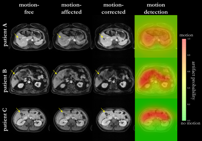 |
Evaluation of motion correction capability in retrospective motion correction with MoCo MedGAN
Thomas Küstner1,2,3, Friederike Gänzle3, Tobias Hepp2, Martin Schwartz3,4, Konstantin Nikolaou5, Bin Yang3, Karim Armanious2,3, and Sergios Gatidis2,5
1Biomedical Engineering Department, School of Biomedical Engineering and Imaging Sciences, King's College London, London, United Kingdom, 2Medical Image and Data Analysis (MIDAS), University Hospital Tübingen, Tübingen, Germany, 3Institute of Signal Processing and System Theory, University of Stuttgart, Stuttgart, Germany, 4Section on Experimental Radiology, University Hospital Tübingen, Tübingen, Germany, 5Department of Radiology, University Hospital Tübingen, Tübingen, Germany
Motion is the main extrinsic source for imaging artifacts which can strongly deteriorate image quality and thus impair diagnostic accuracy. Numerous motion correction strategies have been proposed to mitigate or capture the artifacts. These methods require some a-priori knowledge about the expected motion type and appearance. We have recently proposed a deep neural network (MoCo MedGAN) to perform retrospective motion correction in a reference-free setting, i.e. not requiring any a-priori motion information. In this work, we propose a confidence-check and evaluate the correction capability of MoCo MedGAN with respect to different motion patterns in healthy subjects and patients.
|
3344.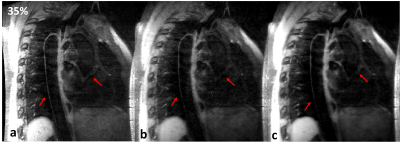 |
High efficiency Free-Breathing 3D Thoracic Aorta Imaging with Self-Navigated Image Reconstruction
Caiyun Shi1,2, Congcong Liu1, Shi Su1, Haifeng Wang1, Xin Liu1, Hairong Zheng1, Dong Liang1, and Yanjie Zhu1
1Shenzhen Institutes of Advanced Technology, Chinese Academy of Sciences, Shenzhen, China, 2Shenzhen College of Advanced Technology, University of Chinese Academy of Sciences, Shenzhen, China
Self-gating techniques can be used to solve and compensate for cardiac or respiratory motion during MRI with free-breathing. In this work, a self-gating based motion correction scheme is proposed, and combined with a 3D variable-flip-angle TSE sequence for high-efficiency thoracic aorta imaging. Specifically, the slab-selective SPACE sequence is modified to acquire self-gating signals, which are used for detecting the respiratory motion. The image data is subsequently corrected based on the binning motion correction and image registration approach. The comparison was conducted on healthy volunteers and compared against a conventional diaphragmatic navigator-gated acquisition to assess the feasibility of the proposed scheme.
|
|
3345.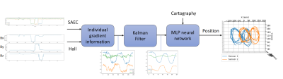 |
Machine learning based Magnetic tracking technique using a Mapping of the Magnetic Gradient Fields on a 3T MRI scanner
Benjamin Roussel1,2, Joris Pascal3, Nicolas Weber1,2, Philip Keller4, Antoine Daridon4, Jacques Felblinger1,2, and Julien Oster1,2
1IADI, U1254, INSERM, Nancy, France, 2Université de Lorraine, Nancy, France, 3FHNW, University of Applied Sciences and Arts Northwestern Switzerland, Muttenz, Switzerland, 4Metrolab Technology SA, Plan-les-Ouates, Switzerland
This paper presents a magnetic tracking method for the localization of a three-axis magnetometer within an MRI bore (Prisma, Siemens, Erlangen, Germany). The unique relationship between the Magnetic Gradient Fields (MGF) and the position within the bore allows locating the sensor through the use of a mapping of the MGF. This mapping is used for the training of a multi-layer perceptron neural network, which estimates the position of the sensor when measuring the MGF. Our technique was experimentally validated by moving the sensors within the MRI bore while playing a customized pulse sequence and by reconstructing the movements during post-processing.
|
|
3346.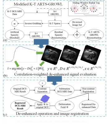 |
A Synthetic Combination of Accurate De-enhanced Registration and Dynamic Artificial Sparsity for Robust High-Resolution Liver DCE-MRI
Zhifeng Chen1, Yujia Zhou1, Xinyuan Zhang1, Peiwei Yi1, Zhongbiao Xu2, Jian Gong1, Zhenguo Yuan3, Xia Kong4, Yaohui Wang5, Ling Xia6, Wufan Chen1, Yanqiu Feng1, and Feng Liu7
1School of Biomedical Engineering, Guangdong Provincial Key Laboratory of Medical Image Processing, Southern Medical University, Guangzhou, China, 2Department of Radiotherapy, Cancer Center, Guangdong Provincial People's Hospital & Guangdong Academy of Medical Science, Guangzhou, China, 3Shandong Medical Imaging Research Institute, Shandong University, Jinan, China, 4School of Computer and Information Science, Hubei Engineering University, Wuhan, China, 5Division of Superconducting Magnet Science and Technology, Institute of Electrical Engineering, Chinese Academy of Sciences, Beijing, China, 6Department of Biomedical Engineering, Zhejiang University, Hangzhou, China, 7School of Information Technology and Electrical Engineering, The University of Queensland, Brisbane, Australia
High spatiotemporal DCE-MRI is a valuable tool in liver disease diagnoses and treatments. Recently, there is a growing research trend which focuses on the motion-robustness of liver DCE-MRI. However, current techniques cannot simultaneously solve the motion problem when pursuing high spatiotemporal resolution. In this work, we propose to combine an accurate registration technique with dynamic artificial sparsity for high spatiotemporal resolution DCE-MRI of liver. The experiments indicated that the proposed framework results in better image quality than iGRASP due to de-enhanced image registration. Compared to motion-sorting techniques, the proposed framework generates better temporal resolution.
|
|
3347. |
MR-Based Cardio-Respiratory Motion Correction of Simultaneously Acquired PET Images Through Coil Fingerprints
David Rigie1, Thomas Vahle2, Tiejun Zhao3, Klaus Schaffers4, and Fernando Boada5
1New York University, New York, NY, United States, 2Siemens Healthineers, Erlangen, Germany, 3Siemens Medical Solutions, New York, NY, United States, 4European Institute for Molecular Imaging, Munster, Germany, 5Radiology, New York University, New York, NY, United States
We present of a real-time, motion correction methodology for MR/PET acquisitions. Our approach utilizes self-refocused navigators that could be integrated into any pulse sequence for providing cardio-respiratory motion information after each excitation. We demonstrate that this approach improves small lesion detection sensitivity and quantitative accuracy for simultaneously acquired MR/PET scans.
|
|
3348. |
MR compatible 4D Ultrasound based validation of respiratory motion compensation strategies
Zachary Miller1, James Holmes1, Sydney Jupitz1, Ty Cashen2, Frank Ong3, Michael Lustig4, Peder Larson5, Bryan Bednarz6, and Kevin Johnson1
1Department of Medical Physics, University of Wisconsin-Madison, Madison, WI, United States, 2GE Healthcare, University of Wisconsin-Madison, Madison, WI, United States, 3Department of Electrical Engineering, Stanford University, Stanford, CA, United States, 4Department of Electrical Engineering, University of California-Berkely, Berkeley, CA, United States, 5Department of Radiology and Biomedical Imaging, University of California San Francisco, San Francisco, CA, United States, 6Department of Engineering Physics, University of Wisconsin-Madison, Madison, WI, United States
Imaging during respiratory and cardiac motion remains a major challenge for body and cardiac MR. A number of motion correction approaches have been proposed to address this challenge including use of external gating signals, navigator acquisitions, and motion estimation from the image data itself. However, the accuracy of these motion estimation techniques has not been validated in-vivo. In this work, we use a recently developed MR compatible 4D ultrasound probe combined with feature tracking to evaluate the performance of MR based navigation algorithms in the setting of free-breathing 3D pulmonary imaging.
|
|
3349.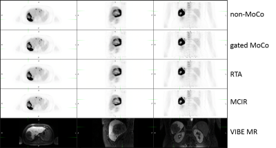 |
Quantitative Assessment MR-Assisted PET Respiratory Motion Correction in Colorectal Liver Metastases using PET/MR
Sihao Chen1,2, Cihat Eldeniz2, Yasheng Chen3, and Hongyu An2
1Biomedical Engineering, Washington University in St. Louis, Saint Louis, MO, United States, 2Mallinckrodt Institute of Radiology, Washington University in St. Louis, Saint Louis, MO, United States, 3Department of Neurology, Washington University in St. Louis, Saint Louis, MO, United States
Respiratory motion causes signal blurring and reduced tumor-to-background ratio (TBR). Simultaneous PET/MR imaging uniquely allows for MR-assisted motion correction in PET imaging, potentially leading to improved PET images for detection of lesions. In this study, we implemented several MR-assisted PET motion correction methods, including gated reconstruction (gated MoCo), reconstruct-transform-average (RTA) and motion-compensated image reconstruction (MCIR), to FDG imaging of colorectal liver metastases. We quantitatively compared TBR and CNR in FDG avid liver lesions. Our results demonstrated improvement of TBR and CNR using MCIR.
|
|
3350.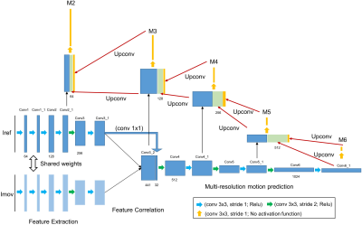 |
Non-rigid Respiratory Motion Estimation of Coronary MR Angiography using Unsupervised Fully Convolutional Neural Network
Haikun Qi1, Gastao Cruz1, Thomas Kuestner1, Niccolo Fuin1, René Botnar1, and Claudia Prieto1
1School of Biomedical Engineering and Imaging Sciences, King's College London, London, United Kingdom
Non-rigid motion corrected coronary MR angiography (CMRA) in combination with 2D image-based navigators has been proposed to account for the complex respiratory-induced motion of the heart in undersampled acquisitions. However, this framework requires the efficient and accurate estimation of non-rigid bin-to-bin motion from undersampled respiratory-resolved images. In this study, we aim to investigate the feasibility of using an unsupervised fully convolutional network to estimate non-rigid motion from undersampled respiratory-resolved CMRA. The performance of the proposed approach was evaluated on 5-fold accelerated free-breathing CMRA and validated against a widely used conventional non-rigid registration method.
|
|
3351.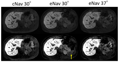 |
Effective Flip Angle of Enhanced Navigator-Gated 3D Spoiled Gradient-Recalled Echo Sequence
Yuji Iwadate1, Atsushi Nozaki1, Keisuke Sato2, Kengo Yoshimitsu2, Ryotaro Jingu3, Ryuji Nakamuta3, and Hiroyuki Kabasawa1
1Global MR Applications and Workflow, GE Healthcare Japan, Hino, Japan, 2Department of Radiology, Fukuoka university, Fukuoka, Japan, 3Radiology Center, Fukuoka University Hospital, Fukuoka, Japan
An enhanced navigator-gated 3D-SPGR (eNAV-3D-SPGR) enables free-breathing T1-weighted abdominal MRI with navigator echo signal enhancement. In this study, we calculated effective flip angle of eNav-3D-SPGR in simulation and examined its validity for obtaining desired contrast. Simulation shows that the effective flip angle of 30° was achieved with eNav-3D-SPGR when the flip angle was set to 36.8°. Actual scan resulted in the similar signal ratios between the conventional method with flip angle 30° and eNav-3D-SPGR at flip angle of 37°. eNav-3D-SPGR with the desired effective flip angle can be useful for accurate motion detection when the liver MRI signal is low.
|
|
3352.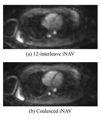 |
Segmented Twin-resolution Acquisition of Cones Trajectory (STACY) and Coalesced Image Navigator for Free-breathing Cardiac MRI
Kwang Eun Jang1,2, Dwight G Nishimura3, and Shreyas S Vasanawala4
1Magnetic Resonance Systems Research Lab (MRSRL), Stanford University, Stanford, CA, United States, 2Department of Bioengineering, Stanford University, Stanford, CA, United States, 3Magnetic Resonance Systems Research Lab (MRSRL), Department of Electrical Engineering, Stanford University, Stanford, CA, United States, 4Department of Radiology, Stanford University, Stanford, CA, United States
3D image-based navigators (iNAVs) are effective for tracking motion in free-breathing cardiac MRI, though they require a long acquisition window. In this work, we present Segmented Twin-resolution Acquisition of Cones trajectorY (STACY) and the concept of coalesced image navigators. In STACY, different segments of a 3D low-resolution cones trajectory are collected across heartbeats as navigators. Coalesced image navigators are reconstructed by merging data from similarly translated heartbeatsh, leading to better conditioned images than iNAVs from undersampled data collected over individual heartbeats.
|
|
3353.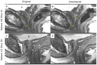 |
Reduced Motion Artifact in Super Resolution T2 FSE Multislice MRI: Application to Prostate
Soudabeh Kargar1, Eric A Borisch2, Adam T Froemming2, Roger C Grimm2, Akira Kawashima3, Bernard F King2, Eric G Stinson2, and Stephen J Riederer2
1Mayo Graduate School, Rochester, MN, United States, 2Mayo Clinic, Rochester, MN, United States, 3Mayo Clinic, Scottsdale, AZ, United States
T2-weighted fast spin echo is acquired in virtually all clinical MRI exams; however, it has coarse through-plane vs. in-plane resolution and is thus often acquired in multiple orientations. This extends the exam time. We have shown improved through-plane resolution in T2 FSE prostate MRI by acquiring overlapped slices and accounting for the slice profile in the reconstruction. Due to acquisition of overlapped slices in several passes, an artifact was observed in the reformatted images, due to subtle peristaltic motion. We propose acquiring the phase encoding lines in several segments to suppress the motion artifact observed in the reformatted images.
|
 Watch the Video
 View the Poster
3354.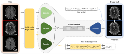 |
Motion Correction for a Multi-Contrast Brain MRI using a Multi-Input Neural Network
Jongyeon Lee1, Byungjai Kim1, Namho Jeong1, and Hyunwook Park1
1Korea Advanced Institute of Science and Technology, Daejeon, Republic of Korea
Numerous motion correction methods have been developed to reduce motion artifacts and improve image quality in MRI. Conventional techniques utilizing motion measurement required a prolonged scan time or intensive computational costs. Deep learning methods have opened up a new way for motion correction without motion information. A proposed method using a multi-input neural network with the structural similarity loss takes an advantage of a common clinical setting of multi-contrast acquisition to clearly correct motion artifacts in brain imaging. Motion artifacts can be fully retrospectively and greatly reduced without any motion measurement by the proposed method.
|
|
3355.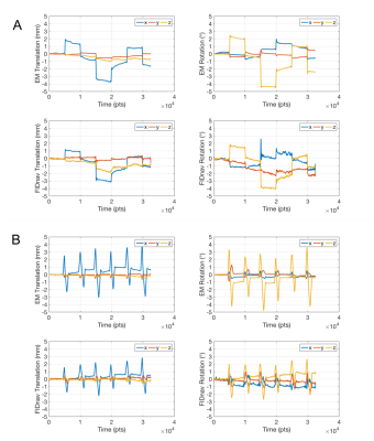 |
Head motion estimation and correction using slab-selective FIDnavs
Nurten Ceren Askin Incebacak1, Tess E. Wallace2, Tobias Kober3,4,5, Francois Lazeyras1, Simon K. Warfield2, and Onur Afacan2
1Department of Radiology and CIBM, University of Geneva, Geneva, Switzerland, 2Department of Radiology, Boston Children's Hospital and Harvard Medical School, Boston, MA, United States, 3Advanced Clinical Imaging Technology, Siemens Healthcare, Lausanne, Switzerland, 4Department of Radiology, UNIL and CHUV, Laussanne, Switzerland, 5LTS5, EPFL, Lausanne, Switzerland
In this work, we propose to measure and correct rigid body head motion using slab-selective FID navigators. It is possible to estimate motion without relying on any
|
|
3356.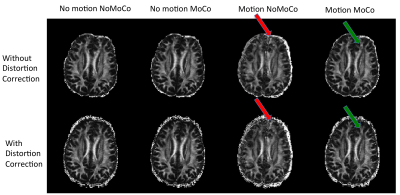 |
Prospectively corrected dual-echo EPI using optical tracking
Onur Afacan1, W. Scott Hoge2, Tess E. Wallace1, Tobias Kober3,4,5, Daniel Nicolas Splitthoff6, Ali Gholipour1, Sila Kurugol1, Camilo J. Cobos1, Richard L. Robertson1, and Simon K. Warfield1
1Department of Radiology, Boston Children's Hospital and Harvard Medical School, Boston, MA, United States, 2Brigham and Women's Hospital and Harvard Medical School, Boston, MA, United States, 3Advanced Clinical Imaging Technology, Siemens Healthcare, Lausanne, Switzerland, 4Department of Radiology, Lausanne University Hospital and University of Lausanne, Lausanne, Switzerland, 5École Polytechnique Fédérale de Lausanne (EPFL), Lausanne, Switzerland, 6Siemens Healthcare, Erlangen, Germany
Large head motion induces different local magnetic-field inhomogeneities, even if the field of view is corrected prospectively. In this work, we implemented and evaluated a diffusion-weighted dual-echo EPI sequence that prospectively corrects for motion using real-time measurements from an optical tracker and uses echoes acquired with reversed phase-encoding to correct for the distortions resulting from the induced local magnetic field inhomogeneities. We evaluated our motion and distortion correction framework in volunteer experiments undergoing controlled motion, and in pediatric patients undergoing routine MRI. Prospective motion correction using our proposed method produced high-quality diffusion parameter maps in all volunteer and patient scans.
|
|
3357.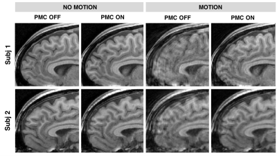 |
Prospective motion correction using real-time FID navigator motion measurements
Tess E. Wallace1,2, Onur Afacan1,2, Tobias Kober3,4,5, and Simon K. Warfield1,2
1Computational Radiology Laboratory, Boston Children's Hospital, Boston, MA, United States, 2Harvard Medical School, Boston, MA, United States, 3Advanced Clinical Imaging Technology, Siemens Healthcare, Lausanne, Switzerland, 4Radiology, Lausanne University Hospital and University of Lausanne, Lausanne, Switzerland, 5LTS5, École Polytechnique Fédérale de Lausanne (EPFL), Lausanne, Switzerland
FIDnavs can be acquired extremely rapidly using standard scanner hardware and are consequently an attractive tracking strategy for prospective motion correction (PMC), which requires accurate pose updates to be passed to the sequence with minimal delay. In this work, we demonstrate for the first time the efficacy of PMC using FIDnav motion estimates in a moving phantom and in volunteers performing deliberate head motion. Real-time pose updates from measured FIDnavs enabled substantial improvements in image quality in structural scans acquired with motion. FID-navigated PMC is a promising method for motion-robust imaging of patients who have difficulty staying still during imaging.
|
|
3358.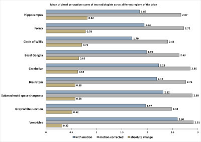 |
Clinical Utility of Deep Learning Motion Correction for Neuroimaging
Kamlesh Pawar1, Jarrel Seah2, Meng Law3, Tom Close4, Zhaolin Chen1, and Gary Egan1
1Monash Biomedical Imaging, Monash University, Melbourne, Australia, 2Department of Neuroscience, Monash University, Melbourne, Australia, 3Radiology and Nuclear Medicine, Alfred Health, Melbourne, Australia, 4Monash Biomedical Imging, Monash University, Melbourne, Australia
The deep learning techniques have been shown to reduce the motion artifact in simulated motion scenarios and a few volunteer scans, the validation of it during routine clinical scans remains an unanswered question. In this study, we focus on evaluating the quality of images from the DL motion correction approach on a cohort of 27 actual patient motion cases that were obtained from the routine clinical scans. Two board-certified radiologists evaluated 9 anatomical regions in the 3D MPRAGE brain images and rated them on the 3-point scale.
|
|
3359.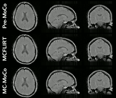 |
Motion Correction Strategy for Multi-Contrast based 3D parametric imaging: Application to Inhomogeneous Magnetization Transfer (ihMT)
Lucas Soustelle1,2, Julien Lamy3, Arnaud Le Troter1,2, Patrick Viout1,2, Lauriane Pini1,2, Claire Costes1,2, Jean-Philippe Ranjeva1,2, Maxime Guye1,2, Adil Maarouf1,2,4, Bertrand Audoin1,2,4, Clémence Boutière1,2,4, Audrey Rico1,2,4, Gopal Varma5,
David C Alsop5, Jean Pelletier1,2,4, Olivier M Girard1,2, and Guillaume Duhamel1,2
1Aix-Marseille Univ, CNRS, CRMBM, Marseille, France, 2APHM, Hôpital Universitaire Timone, CEMEREM, Marseille, France, 3Université de Strasbourg, CNRS, ICube, FMTS, Strasbourg, France, 4APHM, Hôpital Universitaire Timone, Service de neurologie, Marseille, France, 5Division of MR Research, Radiology, Beth Israel Deaconess Medical Center, Harvard Medical School, Boston, MA, United States Multi-contrast-based parametric MRI techniques provide parametric maps intrinsically sensitive to voxel misalignment upon images combination. In this work, we propose an efficient retrospective motion correction method adapted to this problem, with an application to inhomogeneous Magnetization Transfer (ihMT) imaging. The proposed method is confronted with the MCFLIRT (FSL) method used as reference, and residual motion is quantified and analysed across native and motion corrected images. |
|
3360.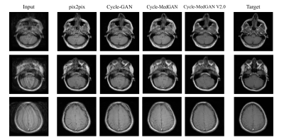 |
MoCo Cycle-MedGAN: Unsupervised correction of rigid MR motion artifacts
Tobias Hepp1, Karim Armanious1,2, Aastha Tanwar2, Sherif Abdulatif2, Thomas Küstner1, Bin Yang2, and Sergios Gatidis1
1University Hospital Tübingen, Tübingen, Germany, 2University of Stuttgart, Stuttgart, Germany
Motion is one of the main sources for artifacts in magnetic resonance (MR) imaging and can affect the diagnostic quality of MR images significantly. Previously, supervised adversarial approaches have been suggested for the correction of MR motion artifacts. However,supervised approaches require paired and co-registered datasets for training, which are often hard or impossible to acquire. We introduced a new adversarial framework for the unsupervised correction of severe rigid motion artifacts in the brain region. Quantitative and qualitative comparisons with other supervised and unsupervised translation approaches showed the enhanced performance of the introduced framework.
|
|
3361.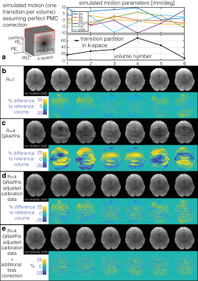 |
Modelling RF coil-sensitivity induced artefacts in prospective motion-corrected accelerated 3D-EPI fMRI
Nadine N Graedel1, Yael Balbastre1, Nadège Corbin1, Oliver Josephs1, and Martina F Callaghan1
1Wellcome Centre for Human Neuroimaging, UCL Queen Square Institute of Neurology, University College London, London, United Kingdom
The increased SNR offered by 3D EPI makes it well-suited for high-resolution fMRI but this benefit needs to be weighed against the increased motion sensitivity. Even when prospective motion correction is used to create a consistent excitation volume and image encoding, residual artefacts remain, caused for example by the apparent motion of coil sensitivities relative to the head after PMC. Using simulated fMRI data we examined the impact of this effect on fMRI and a potential correction using motion-adjusted GRAPPA calibration combined with volume-wise bias field removal which in simulations achieved improved sensitivity and specificity of detection of activation.
|
|
3362.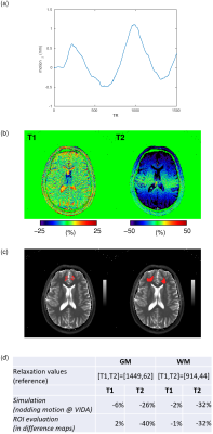 |
The impact of through-plane motion on 2D FISP-MR Fingerprinting: a simulation study based on realistic patient and healthy volunteer movements
Pedro Lima Cardoso1, Gregor Körzdörfer2, Eva Hečková1, Mathias Nittka2, Siegfried Trattnig1,3, and Wolfgang Bogner1,3
1High Field MR Centre, Department of Biomedical Imaging and Image-guided Therapy, Medical University of Vienna, Vienna, Austria, 2Siemens Healthcare GmbH, Erlangen, Germany, 3Christian Doppler Laboratory for Clinical Molecular MR Imaging, MOLIMA, Vienna, Austria
Motion-induced artifacts in quantitative MRI may not be visually detectable. MRF has shown to be robust to in-plane movement, and methods to mitigate it were made available. However, the impact of clinically prevalent motion patterns has not yet been investigated. The effect of realistic motion data from patients with neurodegenerative disorders and young/elderly controls on 2D FISP-MRF is therefore investigated in this simulation study. Results are validated with in-vivo motion-tracked measurements. Although through-plane motions were shown to significantly impact MRF parametric maps (particularly T2), MRF may preserve its sensitivity in clinical cases in which expected lesion-related differences are large enough.
|
|
3363. |
Using NMR field probes to flag significant head movement at 7T
Laura Bortolotti1 and Richard Bowtell1
1Physics, Sir Peter Mansfield Imaging Centre, University of Nottingham, Nottingham, United Kingdom
A fixed array of NMR field probes has proved to be a valid tool for monitoring the effects of a wide range of head movements in a 7T scanner. Here, we use this set up to detect significant field changes in order to give early feedback on head motion. This information could be used in deciding to stop a scan or to reacquire all or part of the k-space data in order to avoid acquisition of motion corrupted image data. This method can be implemented without requiring image sequence modification nor rigid coupling of a motion marker to the head.
|
|
3364. |
A deep network for continuous motion detection during MRI scanning
Isabelle Heukensfeldt Jansen1, Sangtae Ahn1, Rafi Brada2, Michael Rotman2, and Christopher J. Hardy1
1GE Global Research Center, Niskayuna, NY, United States, 2GE Global Research Center, Herzliya, Israel
We introduce ISHMAPS, a method for detecting and adapting to patient motion in real time during an MR scan. The method uses a neural network trained on motion-corrupted data to detect and score motion using as little as 6% of k-space. Once motion is detected, multiple separate complex sub-images from different motion states can be reconstructed and combined into a motion-free image, or the scan can adaptively re-acquire sections of k-space taken before motion occurred.
|
|
3365. |
Optical prospective motion correction enables improved quantitative susceptibility mapping (QSM) of the brain at 7T
Phillip DiGiacomo1, Mackenzie Carlson1, Julian Maclaren1, Murat Aksoy1, Yi Wang2, Pascal Spincemaille2, Brian Burns3, Roland Bammer1, Brian Rutt1, and Michael Zeineh1
1Stanford University, Stanford, CA, United States, 2Cornell University, Ithaca, NY, United States, 3GE Healthcare, San Francisco, CA, United States
Recent literature has shown the potential of high‐resolution quantitative susceptibility mapping (QSM) with ultra‐high field (7T) MRI for investigating the magnetostatic properties of brain structures and disease pathology. Higher spatial resolutions, however, require longer scans resulting in a higher likelihood of subject movement. Here, we apply a novel prospective real-time optical motion tracking and correction system using a camera integrated between the Tx and Rx coils of a commercial 7T head coil to demonstrate the feasibility of acquiring high-resolution R2* and QSM images robust to subject motion.
|
|
3366.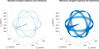 |
Towards Optimal Design of Orbital K-Space Navigators for 3D Rigid-Body Motion Estimation
Thomas Ulrich1, Franz Patzig1, Bertram Jakob Wilm1, and Klaas Paul Pruessmann1
1Institute for Biomedical Engineering, ETH Zurich and University of Zurich, Zurich, Switzerland
A single-shot k-space navigator trajectory and a corresponding motion estimation algorithm is proposed. They allow for 3D rigid-body motion estimation. Their performance in terms of accuracy and precision is studied and a cross-validation experiment is conducted to show that the method is suitable for in-vivo use. The accuracy and precision of the method depend on the orbital radius of the navigator. A simulation study is conducted to determine the best choice of the navigator radius.
|
|
3367.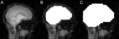 |
Real-time brain masking algorithm improves motion tracking accuracy in scans with volumetric navigators (vNavs)
Malte Hoffmann1,2, Robert Frost1,2, David Salat1,2, M Dylan Tisdall3, Jonathan Polimeni1,2, and André van der Kouwe1,2
1Athinoula A. Martinos Center for Biomedical Imaging, Charlestown, MA, United States, 2Department of Radiology, Harvard Medical School, Boston, MA, United States, 3Radiology, Perelman School of Medicine, University of Pennsylvania, Philadelphia, PA, United States
Volumetric navigators (vNavs) interleaved within longer MRI sequences are an effective method for dynamically detecting and correcting head motion during the acquisition. However, motion is estimated by registering each vNav back to the first, and bias can be introduced by non-rigid deformations of, e.g., the mouth, since the head is taken to be a rigid body. We demonstrate that this bias introduces correction-related artifacts. We present robust real-time brain extraction (0.02 s per vNav) and demonstrate offline that the bias and associated artifacts can be removed by using motion tracks from brain-masked registration.
|
|
3368.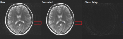 |
Sensitivities Constrained Phase Update (sCPU) for Ghost Artifacts Reduction
Hai Luo1, Meining Chen1, Ziyue Wu2, Bei Lv1, Fei Peng1, Shijie Wang1, Wenkui Hou1, Weiqian Wang1, and Gaojie Zhu1
1AllTech Medical Systems, Chengdu, China, 2Marvel Stone Healthcare, Wuxi, China
Compared with magnitude value, phase of MRI signal is more prone to be influenced by motion. A novel method, sensitivity constrained phase update (sCPU) was proposed for robust and efficient ghost artifacts reduction. Using coil sensitivities as constraints, a synthetic image can be generated in which the ghost is reduced due to phase cancelation. Phase error was first estimated from the raw image and the synthetic image, and then was used to update the phase of raw k-space. The results with simulated and in-vivo data show that the ghost artifacts can be efficiently reduced after several iterations.
|
|
3369.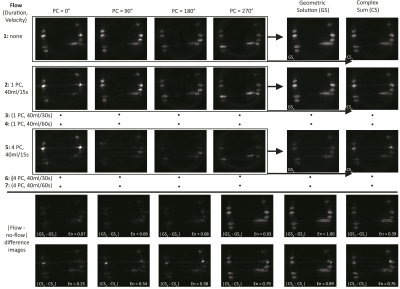 |
The geometric solution to banding mitigates motion artifact in bSSFP imaging
Michael Nicholas Hoff1, Nathan M Cross2, Qing-San Xiang3, Daniel S Hippe2, Charles G Colip2, and Jalal B Andre2
1Radiology, University of Washington, Seattle, WA, United States, 2Radiology, University of Washington, SEATTLE, WA, United States, 3Radiology, University of British Columbia, Vancouver, BC, Canada
Widespread use of phase-cycled bSSFP imaging is hampered by problematic sensitivity to artifacts caused by susceptibility and motion. The geometric solution (GS), which can eliminate susceptibility-related banding and signal modulation, has previously show relative insensitivity to motion. Here, GS-bSSFP is evaluated using a phantom and in humans, imaging the skull base. The GS mitigates motion artifact in both paradigms and is particularly resilient when one of its four phase cycles is corrupted by motion.
|
3370.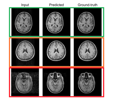 |
Evaluation of Deep Learning Techniques for Motion Artifacts Removal
Alessandro Sciarra1,2, Soumick Chatterjee2,3, Max Dünnwald1,4, Oliver Speck2,5,6,7, and Steffen Oeltze-Jafra1,5
1MedDigit, Department of Neurology, Medical Faculty, Otto von Guericke University, Magdeburg, Germany, 2BMMR, Biomedical Magnetic Resonance, Otto von Guericke University, Magdeburg, Germany, 3Data & Knowledge Engineering Group, Otto von Guericke University, Magdeburg, Germany, 4Faculty of Computer Science, Otto von Guericke University, Magdeburg, Germany, 5Center for Behavioral Brain Sciences (CBBS), Magdeburg, Germany, 6German Center for Neurodegenerative Disease, Magdeburg, Germany, 7Leibniz Institute for Neurobiology, Magdeburg, Germany
Removing motion artifacts in MR images remains a challenging task. In this work, we employed 2 convolutional neural networks, a conditional generative adversarial network (c-GAN), also known as pix2pix, as well as a network based on the residual network (ResNet) architecture, to remove synthetic motion artifacts for phantom images and T1-w brain images. The corrected images were compared with the ground-truth ones in order to assess the performance of the chosen neural networks quantitatively and qualitatively.
|
|
| 3371. | QUIQI – using a QUality Index for the analysis of Quantitative Imaging data
Giulia Di Domenicantonio1, Nadège Corbin2, John Ashburner2, Martina Callaghan2, and Antoine Lutti1
1Department for Clinical Neuroscience, Lausanne University Hospital and University of Lausanne, Lausanne, Switzerland, 2Wellcome Centre for Human Neuroimaging, Queen Square Institute of Neurology, UCL, London, London, United Kingdom
Image degradation due to head motion is ubiquitous in MRI, reduces sensitivity, hinders clinical diagnosis and increases the risk of spurious findings. The few existing objective measures of degradation are often used sub-optimally, to remove the most degraded datasets from analysis. Using a large dataset (N~1400) we show how to incorporate an validated index of degradation into the analysis of group studies. The benefit is demonstrated for the case of healthy age-related difference in brain relaxometry data using the SPM software. However, the proposed framework is flexible with broad potential, including the analysis of other metrics and body regions.
|
|
3372.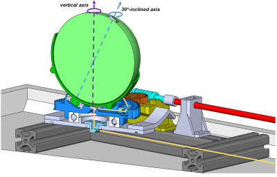 |
Remotely controllable phantom rotation device for cross-calibration at 7T
Hendrik Mattern1, Robert Odenbach2, Niklas Thoma3, Frank Godenschweger1, and Oliver Speck1,4,5,6
1Biomedical Magnetic Resonance, Otto-von-Guericke University, Magdeburg, Germany, 2Institute for Medical Engineering, Otto-von-Guericke University, Magdeburg, Germany, 3Department of Mechanical Engineering, Otto-von-Guericke University, Magdeburg, Germany, 4German Center for Neurodegenerative Disease, Magdeburg, Germany, 5Center for Behavioral Brain Sciences, Magdeburg, Germany, 6Leibniz Institute for Neurobiology, Magdeburg, Germany
A low-cost rotation device was designed and built to enable remotely controllable phantom movements. With the device, motions are highly reproducible. For cross-calibrations of external tracking systems, it prevents table movement or the need to move the phantom by hand from inside the scanner during the calibration, reduces the overall calibration duration, and provides similar calibration performance compared to the freehand approach performed by an expert. The CAD model was made publically available.
|
|
3373. |
Relaxation-Time Selective Imaging Using the Inverse Z-transform
Seong-Min Kim1, Phuong Anh Chu Dang1, Sehong Oh2,3, and Jang-Yeon Park1,4
1Department of Biomedical Engineering, Sungkyunkwan University, Suwon, Korea, Republic of, 2Division of Biomedical Engineering, Hankuk University of Foregn Studies, Yongin, Korea, Republic of, 3Imaing Institute, Cleveland Clinic Foundation, Cleveland, OH, United States, 4Center for Neuroscience Imaging Research, Suwon, Korea, Republic of
There have been many attempts to separate mixed relaxation-time decays. Among them, some approaches do not assume the number of exponential decays a priori for the analysis of multiple-component decay signals. However, they are sensitive to ill conditions and have a poor resolving ability in terms of decay constants. In this study, a new method is proposed that can analyze multiple T2 decays with high resolution using the inverse Z-transform, which was demonstrated in simulation and in vivo human brain experiment. T2-selective images were also presented and used for myelin-water fraction mapping and deep brain-tissue segmentation.
|
|
3374. |
Noise Reduction and Ghosting Alleviation in ASL Perfusion Measurement using Multi-dimensional Integration (MDI)
Yichen Hu1, Qing Wei2, Zhongyang Zhou2, Jun Xie2, and Yongquan Ye1
1UIH America, Inc., Houston, TX, United States, 2United Imaging Healthcare, Shanghai, China
3D GRASE (GRAdient and Spin Echo) pulse sequence has been widely employed as the readout in arterial spin labeling (ASL) applications, given its efficient acquisition and relatively long lasting signal intensities. However, the inherent weakness of GRASE, such as vulnerability to motions, can induce ghosting artifacts to perfusion imaging and quantitative cerebral blood perfusion (CBF) maps. Herein, we propose applying the Multi-Dimensional Integration (MDI) algorithm to processing the perfusion and CBF maps, by which method noticeable alleviation of motion ghosting can be obtained, and the imaging noise is reduced as well.
|
|
3375.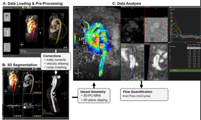 |
Highly-accelerated Compressed Sensing 4D Flow MRI for Quantification of Whole Heart Hemodynamics
Daniel Gordon1, Allison Blake1, Liliana Ma1, Yoshihiro Tanaka1, Kelvin Chow1,2, Ning Jin3, Philip Greenland1, Rod Passman1, Daniel Kim1, and Michael Markl1
1Northwestern University, Chicago, IL, United States, 2Siemens Medical Solutions USA, Inc., Chicago, IL, United States, 3Siemens Medical Solutions USA, Inc., Cleveland, OH, United States
The purpose of this study was to evaluate an efficient, free-breathing, whole-heart 4D flow CS protocol in a cohort of 21 healthy controls. The CS technique was evaluated for internal self-consistency by comparing net flow through various cardiac structures. In addition, cine imaging was performed and used to calculate left ventricular (LV) stroke volume (SV) for comparison to net flow in the ascending aorta. CS whole-heart 4D flow was acquired in 5:23 ± 0:51 minutes. Input and output flow, and LV stroke volume and ascending aorta net volume with 4D flow were significantly (p<0.05) correlated for all comparisons.
|
|
3376. |
Methods and challenges for body fat composition and hepatic fat fraction measurement in a multi-site study using MRI
SHILPY CHOWDHURY1, SPENCER LOONG1, JOAN SABATE1, and SAMUEL BARNES1
1LOMA LINDA UNIVERSITY, LOMA LINDA, CA, United States
Running a large longitudinal multi-center study to measure visceral fat and hepatic fat fraction present unique challenges. We scanned fat/water and ex vivo liver phantoms across all sites and over time to show consistency. SliceOmatic and ImageJ were used for quantification of visceral fat and LCModel for hepatic fat fraction. Visceral fat showed a good correlation with hepatic fat and demographics. The use of phantoms and careful design of the protocol can help address challenges of a longitudinal study and help maintain consistency across sites.
|
|
3377. |
Brain regional hemodynamic alterations in obstructive sleep apnea using dynamic susceptibility contrast MRI
Ruyi Zhang1, Hea Ree Park2, Hosung Kim3, Gele Qin1, Eun Yeon Joo4, and Lirong Yan3
1Department of Electric Engineering, University of Southern California, Los Angeles, CA, United States, 2Inje University College of Medicine, GoyangSouth, Korea, Republic of, 3Stevens Neuroimaging and Informatics Institute, Keck School of Medicine, University of Southern California, Los Angeles, CA, United States, 4Sungkyunkwan University School of Medicine, Seoul, Korea, Republic of
In this study, we comprehensively studied the regional hemodynamic measures including CBF, cerebral blood volume (CBV), and time-related hemodynamic parameters in OSA patients using Gadolinium contrast agent (Gd)-base dynamic susceptibility contrast MRI.
|
|
3378.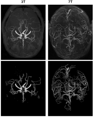 |
Performance of Automatic Cerebral Arterial Segmentation of MRA Images Improves at Ultra-high Field.
Alexander Saunders1,2, Tales Santini3, Tiago Martins3, Howard Aizenstein3, John C. Wood1, Matthew Borzage (co-corresponding author)1, and Tamer S. Ibrahim (co-corresponding author)3
1Children's Hospital Los Angeles, University of Southern California, Los Angeles, CA, United States, 2Rudi Schulte Research Institute, Santa Barbara, CA, United States, 3Swanson School of Engineering and School of Medicine, University of Pittsburgh, Pittsburgh, PA, United States
Ultra-high field time of flight MRA can generate images with higher signal and resolution but image quality may suffer from increased field inhomogeneity. Because we would like to extract arteries for further analysis, we sought to evaluate automatic segmentation performance compared with standard field strength. Five segmentation algorithms were applied to two MRA images (one 3T, one 7T) and performance was measured against manually segmented ground truth data. We found that automatic segmentation performs better in 7T images but confounds in acquisition and image processing need to be further investigated.
|
|
3379.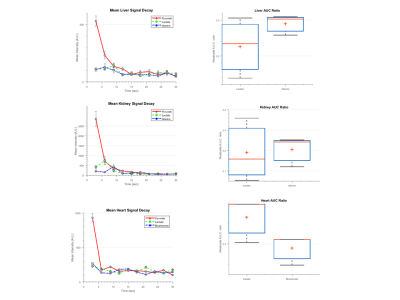 |
Hyperpolarized 13C imaging with multi-shot multi-echo echo planar imaging
Kofi Deh1, Kristin Granlund1, Roozbeh Eskandari1, Arsen Mamakhanyan1, Nathaniel Kim1, and Kayvan Keshari,1
1Memorial Sloan Kettering Cancer Center, New York, NY, United States
We demonstrate the use of a use of a broadband RF excitation with a multi-shot EPI readout for robust separation of metabolites in hyperpolarized 13C imaging. The approach places less demands on scanner hardware and compensates for local B0 inhomogeneity, making it possible to obtain high resolution time-resolved multi-slice imaging over a large region of interest which can be reformatted into 3D volumetric images to facilitate the study diseases such as cancer metastasis using HP probes.
|
|
3380. |
Validation of Small Motion Quantification by aMRI Using a Digital Phantom
Zhiyue J Wang1,2 and Youngseob Seo3
1University of Texas Southwestern Medical Center, Dallas, TX, United States, 2Children's Health, Dallas, TX, United States, 3Korea Research Institute of Standards & Science, Daejeon, Republic of Korea
In MRI, small signal intensity changes not perceivable by naked eyes are routinely analyzed to extract valuable information, such as in fMRI. However, a small movement was difficult to detect. Recently, image amplification methods have been introduced for visualizing small sub-pixel motions that are too small to be discerned by naked eyes, based on methods developed for video processing. We use computer simulations to explore the range of parameters for the technique to work optimally.
|
|
3381. |
Filter-pipeline based algorithm to find the AIF in DCE-MRI images for perfusion calculation of rectal cancer.
Christian Tönnes1, Sonja Janssen2, Alena-Kathrin Schnurr1, Tanja Uhrig1, Khanlian Chung1, Lothar R. Schad1, and Frank Gerrit Zöllner1
1Computer Assisted Clinical Medicine, Mannheim Institute for Intelligent Systems in Medicine, Medical Faculty Mannheim, Heidelberg University, Mannheim, Germany, 2Institute of Clinical Radiology and Nuclear Medicine, Medical Faculty Mannheim, Heidelberg University, Mannheim, Germany
Perfusion calculation is highly dependent on the expertise and input of the physician and therefore reproduction of the results is difficult. A filter pipeline based algorithm for selecting the AIF ROI was implemented and evaluated. Our results show that such an algorithm can be used to determine the AIF ROI for perfusion calculation in dynamic contrast enhanced MRI images with a high accuracy. A fully automatic and deterministic algorithm removes the need for manual interaction and standardizes clinical perfusion measurements while simultaneously reducing the time requirement for the ROI determination by 86%.
|
|
3382. |
Design of deep neural network in time-phase encoding plane for compressed sensing cardiovascular CINE MRI
Chang-Beom Ahn1 and Seong-Jae Park1
1Kwangwoon University, Seoul, Republic of Korea
We build a deep neural network in time-phase encoding plane (t-y) for compressed sensing cardiovascular CINE MRI. Previously neural networks were developed in cross-sectional image planes (x-y). A hierarchical convolutional neural network (CNN), known as U-net is used. By adopting the t-y plane, instead of the x-y plane, simultaneous restoration in time and space is effectively achieved. By computer simulation, the proposed deep neural network based on the t-y plane shows better cross-sectional images, clearer temporal profiles, and less normalized mean square errors compared to that of the x-y plane.
|
|
3383. |
Clinical Image Quality Evaluation For Field Strength and Contrast Independence of Deep Learning Reconstruction
Erin J Kelly1, Hung P Do2, Dawn M Berkeley2, and Jonathan K Furuyama2
1Canon Medical Systems USA, Inc, Tustin, CA, United States, 2Canon Medical Systems USA, Inc., Tustin, CA, United States
dDLR algorithms with two path CNN architecture that are designed to be noise adaptive are robust against differences in image contrast and field strength. This study shows clinical image quality improvement on 1.5T brain and knee datasets using an algorithm trained on 3T data.
|
3384.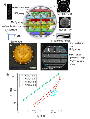 |
MRI Scanner Characterization with the ISMRM/NIST System Phantom
Stephen E. Russek1, Michael A. Boss2, H. Cecil Charles3, Andrew M. Dienstfrey1, Jeffrey L. Evelhoch4, Jeffrey L. Gunter5, Derek L. G. Hill6, Edward F. Jackson7, Kathryn E. Keenan1, Guoying Liu8, Michele Martin1, Nikki S. Rentz1, Karl F. Stupic1,
Chun Yuan9, and Zydrunas Gimbutas1
1National Institute of Standards and Technology, Boulder, CO, United States, 2American College of Radiology, Philadelphia, PA, United States, 3Duke Image Analysis Laboratory, Durham, NC, United States, 4Merck Research Laboratories, West Point, PA, United States, 5Mayo Clinic, Rochester, MN, United States, 6University College London, London, United Kingdom, 7University of Wisconsin, Madison, WI, United States, 8National Institute of Biomedical Imaging and Bioengineering, Bethesda, MD, United States, 9University of Washington, Seattle, WA, United States
We describe basic scanner characterization and determination of MR-parameter measurement accuracy using the ISMRM/NIST system phantom. The phantom provides a convenient method to measure geometric distortion; the efficacy of non-linear gradient corrections; slice profiles and associated measurement uncertainties; protocol and system dependent resolution contributions; SNR according to NEMA protocols; accuracy of relaxation time; and proton density measurements. The phantom is unique in having SI-traceability, a high level of precision, long-term stability, and monitoring by a national metrology institute.
|
|
3385. |
Characterization and elimination of X-nuclear eddy current artifacts on clinical MR systems
Mary A McLean1,2, Scott Hinks3, Joshua Kaggie1, Ramona Woitek1, Frank Riemer4, Martin Graves1, Dominick McIntyre2, Ferdia Gallagher1, and Rolf Schulte5
1Dept of Radiology, University of Cambridge, Cambridge, United Kingdom, 2Cancer Research UK Cambridge Institute, University of Cambridge, Cambridge, United Kingdom, 3GE Healthcare, Waukesha, WI, United States, 4MMIV, Dept of Radiology, Haukeland University Hospital, Bergen, Norway, 5GE Healthcare, Munich, Germany
Using pulse-acquire spectroscopy, line-shape distortions characteristic of eddy currents were demonstrated for X-nuclei, which were not seen for 1H, on systems from multiple vendors. The severity of these appeared correlated with the amplitude of the f0 eddy current frequency compensation term applied by the system along the axis of the applied spoiler gradient. A proposed correction to eddy current compensation taking account of the X-nuclear gyromagnetic ratio was shown to dramatically reduce these distortions on a GE system. The same correction was also shown to improve the quality of non-Cartesian imaging (spirals and cones).
|
|
3386. |
Optimized gradient spoiling for B1 mapping with UTE AFI that is less sensitive to the RF-spoiling phase increment
Marta Brigid Maggioni1, Martin Krämer1, and Jürgen R. Reichenbach1,2,3,4
1Medical Physics Group, Institute of Diagnostic and Interventional Radiology, Jena University Hospital - Friedrich Schiller University, Jena, Germany, 2Michael Stifel Center for Data-driven and Simulation Science Jena, Friedrich Schiller University, Jena, Germany, 3Abbe School of Photonics, Friedrich Schiller University, Jena, Germany, 4Center of Medical Optics and Photonics, Friedrich Schiller University, Jena, Germany
B1 mapping is a major challenge for reliable T1 quantification, and it is especially difficult for UTE sequences, because most conventional B1 mapping methods are unable to perform on short T2* tissues. Recently, an implementation of the actual flip angle imaging (AFI) method was proposed for UTE sequences. The AFI method requires ideal spoiling of residual transverse magnetization, which is typically achieved by fine-tuning RF-spoiling phase increments as well as the strength of spoiler gradients. In this work, we propose an improved gradient spoiling scheme, which reduces the effect of the RF-spoiling phase increment on the AFI results.
|
|
3387.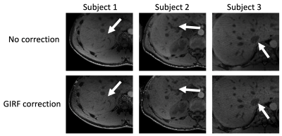 |
Trajectory correction in high-resolution gated golden-angle radial Dixon imaging using the gradient impulse response function
Christoph Zöllner1, Sophia Kronthaler1, Stefan Ruschke1, Jürgen Rahmer2, Johannes M. Peeters3, Holger Eggers2, Peter Börnert2, Rickmer F. Braren1, and Dimitrios C. Karampinos1
1Department of Diagnostic and Interventional Radiology, Technical University of Munich, München, Germany, 2Philips Research Laboratory, Hamburg, Germany, 3Philips Healthcare, Best, Netherlands
Stack-of-stars-type radial k-space trajectories employing golden-angle ordering have been becoming popular for either free breathing or navigator-gated volumetric T1-weighted imaging of the abdomen and heart. Most methods for compensating radial k-space trajectory errors induced by eddy currents and system delays are based either on the acquisition of calibration lines with opposite polarity or on the processing of approximately anti‐parallel spokes from the actual radial acquisition. This work shows that a trajectory correction based on a gradient system impulse response function improves image quality in high-resolution gated golden-angle radial Dixon imaging.
|
|
3388.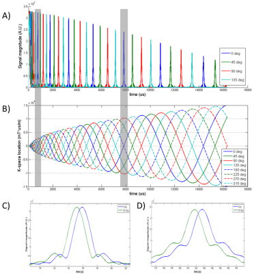 |
Spiral Gradient Waveform Correction Using Multipoint Gradient Anchoring (MGA)
Qi Liu1, Yuan Zheng1, Yu Ding1, Jian Xu1, and Weiguo Zhang1
1UIH America, Inc., Houston, TX, United States
A new comprehensive spiral gradient waveform correction strategy is proposed that features multipoint gradient anchoring (MGA). By measuring multiple gradient delays in spiral waveforms at different rotation angles, it effectively ‘anchors’ the waveform at a series of locations and improves gradient waveform fidelity. Application of this innovative design on phantom and volunteer imaging indicates it is an effective and promising technique.
|
|
3389.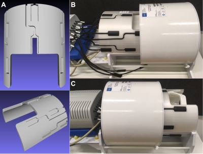 |
Replaceable field probe holder for the Nova coil on a 7 Tesla Siemens scanner
Sascha Brunheim1, Christian Mirkes2, Benjamin E Dietrich2, Jolanda M Schwarz1, Rüdiger Stirnberg1, Sebastian Ismar3, Alexander Christen3, and Tony Stöcker1,3
1MR Physics, German Center for Neurodegenerative Diseases (DZNE), Bonn, Germany, 2Skope Magnetic Resonance Technologies Inc., Zurich, Switzerland, 3Department of Physics and Astronomy, University of Bonn, Bonn, Germany
An optimized holder for fast mounting of the Skope clip-on field camera into the common Nova Tx/Rx head coil on a 7 Tesla Siemens scanner is presented. Field probe positioning was based on transceiver coil architecture constraints and conditioning for best approximation of field dynamics. The conditioning results for the proposed probe holder positions show very good agreement with the corresponding simulation. They approximate the optimal spherical probe arrangement as well as possible. In comparison, the probe holder outperforms the manual mounting situation conditioning-wise, which corresponds to arbitrary placement onto the receive part of the head coil.
|
|
3390.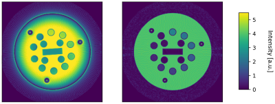 |
Data-Driven non-Cartesian trajectory correction based on Cartesian reference data
Felix Horger1, Mathias Nittka1, and Gregor Körzdörfer1
1Siemens Healthcare, Erlangen, Germany
Trajectory deformations in non-Cartesian scans can lead to significant image artifacts, while in comparison the effect on Cartesian scans is evidentially minor. We propose a novel approach for trajectory correction relying on the minimization of a suitable measure between a non-Cartesian image and a Cartesian reference image, given a trajectory deformation model. Our proof of concept is applied on simulated as well as measured data. The approach is fast, accurate and easily extendable. Given the right use-case, no extra scan time is required (e.g. mixed spiral-Cartesian MR Fingerprinting).
|
|
3391.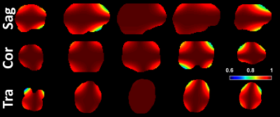 |
The impact of gradient non-linearity on Maxwell compensation when using asymmetric gradient waveforms for tensor-valued diffusion encoding
Filip Szczepankiewicz1,2,3, Cornelius Eichner4, Alfred Anwander4, Carl-Fredrik Westin1,2, and Michael Paquette4
1Harvard Medical School, Boston, MA, United States, 2Radiology, Brigham and Women's Hospital, Boston, MA, United States, 3Clinical Sciences Lund, Lund University, Lund, Sweden, 4Neuropsychology, Max Planck Institute for Human Cognitive and Brain Sciences, Leipzig, Germany
Gradient non-linearity distorts the shape of the gradient waveform used for diffuison encoding. This distortion also compromises Maxwell compensation in asymmetric gradient waveforms. We show that one of two strategies for Maxwell compensation fails under non-linear gradiets (K-nulling) whereas M-nulling is immune to this effect.
|
|
3392. |
Formulating EPI ghost correction as a convolution: Insights into Dual-Polarity GRAPPA by examining kernels of limited extent
W Scott Hoge1,2,3 and Jonathan R Polimeni2,4,5
1Radiology, Brigham and Women's Hospital, Boston, MA, United States, 2Athinoula A. Martinos Center for Biomedical Imaging, Massachusetts General Hospital, Charlestown, MA, United States, 3Harvard Medical School, Boston, MA, United States, 4Department of Radiology, Harvard Medical School, Boston, MA, United States, 5Massachusetts Institute of Technology, Cambridge, MA, United States We examine the relationship between traditional 1D linear phase-error correction methods and Dual-Polarity GRAPPA (DPG) for EPI ghost correction. We first show that conventional ghost correction methods based on 1D linear phase errors can be implemented with convolution kernels in k-space, and then generalized in several steps to replicate a typical DPG kernel. We identify that employing multiple phase-encoded lines is important for ghost correction kernel calibration, while incorporating data from multiple channels is critical. Surprisingly, having a kernel extent along the phase-encoded direction is less critical, demonstrating the utility of DPG kernels with limited extent for ghost correction. |
|
3393. |
Correcting Gradient Delays in Multi-Echo Rosette Trajectories with RING
Volkert Roeloffs1, Adam Michael Bush2, Suma Anand1, and Michael Lustig1
1EECS, UC Berkeley, Berkeley, CA, United States, 2Radiology, Stanford, Stanford, CA, United States
Rosette trajectories are sensitive to gradient delays as caused by eddy currents. Here, we use the RING method recently proposed for radial trajectories to correct for these delays. To this end, we approximate the center portion of the Rosette trajectory to be radial-like, determine delay constants, and correct the entire trajectory in a second step. First phantom and patient studies show that this simple correction method improves image quality in multi-echo Rosette imaging and leads to prolonged T2* values in derived maps. The small amount of required data renders echo-adaptive corrections feasible.
|
|
3394.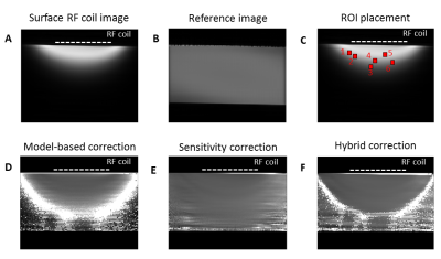 |
Comparison of three B1 correction methods for RARE imaging with transceive surface RF coils
Paula Ramos Delgado1, Andre Kuehne2, Jason M. Millward1, Joao Periquito1, Thoralf Niendorf1,3, Sonia Waiczies1, and Andreas Pohlmann1
1Berlin Ultrahigh Field Facility (B.U.F.F), Max Delbrück Center for Molecular Medicine in the Helmholtz Association, Berlin, Germany, 2MRI.tools GmbH, Berlin, Germany, 3Experimental and Clinical Research Center, a joint cooperation between the Charité Medical Faculty and the Max Delbrück Center for Molecular Medicine in the Helmholtz Association, Berlin, Germany
Fluorine MR methods support quantification but have inherently low SNR. To enhance sensitivity, SNR-efficient imaging techniques such as RARE and cryogenically-cooled surface RF coils are used. However, transceive surface RF coils show variation in the excitation field, impairing quantification and T1 contrast. We previously showed improved homogeneity using a novel B1 correction method developed for 1H imaging that uses experimental data acquired with a volume resonator. Here, we compared it to a sensitivity-only correction and a combination of both, to evaluate which approach yields the most accurate contrast and concentration quantification. This work is applicable to different nuclei.
|
|
3395.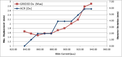 |
Feasibility of Monitoring Geometric Distortion for Radiation Therapy Planning using ACR Weekly QA
Chen Lin1, Stephanie Tensfeldt1, and Christopher Serago2
1Radiology, Mayo Clinic, Jacksonville, FL, United States, 2Radiation Oncology, Mayo Clinic, Jacksonville, FL, United States
To demonstrate the distortion measurements based on automatic analysis of American College of Radiology (ACR) weekly quality assurance (QA) scans is adequate for monitoring the geometric distortion of a MR scanner for radiation therapy planning by comparing with distortion measurements using a 3D grid phantom.
|
|
3396.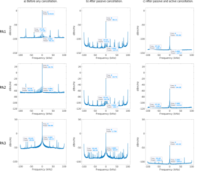 |
Noise Analysis for Simultaneous Transmission and Reception Enabled MRI Scanner
Bilal Tasdelen1,2, Alireza Sadeghi-Tarakameh1,2, Ugur Yilmaz2, and Ergin Atalar1,2
1Department of Electrical and Electronics Engineering, Bilkent University, Ankara, Turkey, 2National Magnetic Resonance Research Center (UMRAM), Ankara, Turkey
In simultaneous transmission and reception (STAR), injection of noise and spurs stemming from transmit chain is a problem as well as the leak power. Investigation of noise and spurs are important in order to determine minimum necessary isolation to achieve similar noise levels compared to conventional imaging. Noise sources during STAR are investigated and compared with experiments. It has been shown that using an active cancellation method where dominant noise source is transmit chain, with combination of passive cancellation can reduce noise and spurs. Necessary isolation is shown to depend on input noise and total gain in transmit chain.
|
|
3397.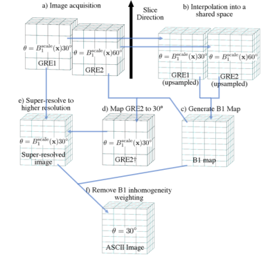 |
ASCII: Acquisition with Self-Correction for Image Inhomogeneity at 7T
Edward M Green1,2, James C Korte1,3, Bahman Tahayori1,4,5, Yasmin Blunck1,2, and Leigh A Johnston1,2
1Department of Biomedical Engineering, University of Melbourne, Parkville, Australia, 2Melbourne Brain Centre Imaging Unit, University of Melbourne, Parkville, Australia, 3Department of Physical Sciences, Peter MacCallum Cancer Centre, Parkville, Australia, 4Department of Medical Physics and Biomedical Engineering, Shiraz University of Medical Sciences, Shiraz, Iran (Islamic Republic of), 5Center for Neuromodulation and Pain, Shiraz, Iran (Islamic Republic of)
Inhomogeneity of the RF transmit field results in undesirable shading and brightening in high field imaging due to range of flip angles across the imaged object. A new method is proposed whereby two image volumes with coarse resolution in the slice direction are acquired at different flip angles and combined to produce an image volume at higher resolution and free of B1 inhomogeneity. Reconstruction quality is comparable to conventional super-resolution imaging, while image inhomogeneity caused by flip angle error is reduced. This is a flexible method applicable to a range of MRI sequences to correct B1 inhomogeneity.
|
|
3398.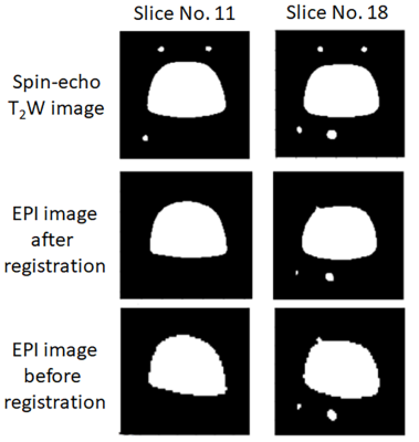 |
Phantom Development for the Standardization of fMRI Data across Multiple Imaging Sites and Scanners
Daisuke Kokuryo1, Chika Sato2, Takashi Itahashi3, Shigeyoshi Saito4, Hiroyuki Ueda1, Etsuko Kumamoto1,5, Toshiya Kaihara1, Nobutada Fujii1, Noriaki Yahata2,6, and Ichio Aoki2,6
1Graduate School of System Informatics, Kobe University, Kobe, Japan, 2National Institute of Radiological Sciences, National Institutes for Quantum and Radiological Science and Technology, Chiba, Japan, 3Medical Institute of Developmental Disabilities Research, Showa University, Tokyo, Japan, 4Graduate School of Medicine, Osaka University, Suita, Japan, 5Information Science and Technology Center, Kobe University, Kobe, Japan, 6Institute for Quantum Life Science, National Institutes for Quantum and Radiological Science and Technology, Chiba, Japan
Establishing data harmonization in fMRI “big data” is necessary to ensure the reliability and reproducibility of fMRI research. Our group recently developed two distinct phantoms: a brain-shaped phantom to correct field heterogeneity and a functional image-specific phantom to calibrate shape distortion and signal irregularity. A preliminary experiment confirmed a shape distortion and signal irregularity caused by field inhomogeneity when using EPI signals, and these were corrected using an affine transformation. Therefore, our developed phantoms have the potential to contribute to achieving fMRI data standardization.
|
3399.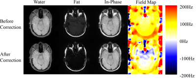 |
A semi-autofocus method to improve two-point Dixon spiral imaging
Tzu-Cheng Chao1, Dinghui Wang1, Guruprasad Krishnamoorthy2, and James G. Pipe1
1Department of Radiology, Mayo Clinic, Rochester, MN, United States, 2Philips Healthcare, Gainesville, FL, United States
Deblurring with water/fat separation plays a critical role in spiral imaging. The reconstruction quality depends on good off-resonant frequency estimation. A field map before each spiral imaging exam is often acquired to approximate the B0 of the scanned subject. However, the estimation errors occur due to nonconcurrent collection of the imaging data. The proposed work introduces an iterative method to update the field map based on the acquired images to improve spiral image reconstruction. The results have shown that the blurring can be reduced along with improved fat/water separation after updating the reference B0 used for reconstruction.
|
|
3400.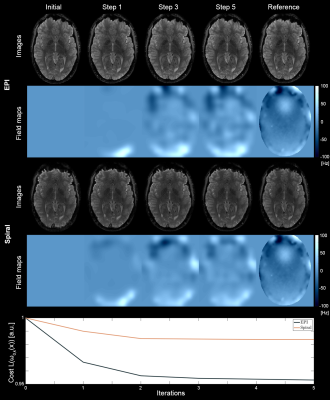 |
Off-Resonance Self-Correction for Single-Shot Imaging
Franz Patzig1, Bertram Wilm1, Simon Gross1, David Brunner1, and Klaas Paul Pruessmann1
1Institute for Biomedical Engineering, ETH Zurich and University of Zurich, Zurich, Switzerland
We present a novel algorithm capable of determining a B0 map and an off-resonance corrected reconstructed image from a single-shot acquisition. The functionality of the algorithm was shown in-vivo for multiple imaging geometries using regular undersampled single-shot EPI and Spiral readouts showing stable convergence. In addition, the method is tested for the common situation of inter-scan subject movement, in which pre-acquired field maps would be rendered invalid.
|
|
3401. |
Optimal Coil Combination with Relatively Aligned Phase (RAP) for Phase-Cycled bSSFP Imaging
Qing-San Xiang1 and Michael Nicholas Hoff2
1Radiology, University of British Columbia, Vancouver, BC, Canada, 2Radiology, University of Washington, Seattle, WA, United States
Phase-Cycled (PC) bSSFP imaging is useful for qualitative and quantitative studies of tissue. But the images generated should be properly combined when they are acquired with multiple receiver coils; in particular, phase information in the complex images must be preserved. We describe a straightforward yet optimal coil combination algorithm for PC-bSSFP applications, where the phase is preserved through relative phase alignment along the measurement dimension. The method is demonstrated with experimental results from phantom and volunteer scans.
|
|
3402.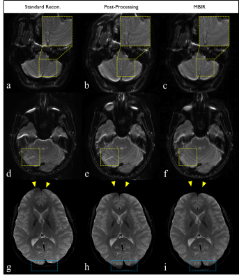 |
Model-Based Iterative Reconstruction for Echo Planar Imaging: A Preliminary Evaluation for Neuro MRI
Uten Yarach1,2, Matthew Bernstein1, John Huston III1, Norbert Campeau1, Petrice Cogswell1, Daehun Kang1, Myung-Ho In1, Yunhong Shu1, Nolan Meyer1, Erin Gray1, and Joshua Trzasko1
1Radiology, Mayo Clinic, Rochester, MN, United States, 2Department of Radiologic Technology, Faculty of Associated Medical Sciences, Chiang Mai University, Chiang Mai, Thailand
A recently proposed model-based iterative reconstruction (MBIR) method enables to prospectively manage B0-inhomogeneity and gradient-nonlinearity in EPI, and has been demonstrated to generate images with high SNR and minimal spatial blurring and geometric distortion. However, the clinical significance and degree of benefit remains undetermined. In this study, using commodity EPI acquisition protocols for whole-brain imaging, MBIR was radiologically compared against standard scanner-generated EPI images and post-processed versions. The results show that significant advantage of MBIR (p<0.05) over both the standard scanner-generated EPI results and post-processed variants was observed in three categories: SNR, geometric accuracy, and overall image quality.
|
|
3403.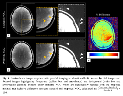 |
Model-Based Nyquist Ghost Correction for Reversed Readout Echo Planar Imaging
Uten Yarach1,2, Matthew Bernstein1, John Huston III1, Myung-Ho In1, Yi Sui1, Daehun Kang1, Yunhong Shu1, Erin Gray1, Nolan Meyer1, and Joshua Trzasko1
1Radiology, Mayo Clinic, Rochester, MN, United States, 2Department of Radiologic Technology, Faculty of Associated Medical Sciences, Chiang Mai University, Chiang Mai, Thailand
Single-polarity reference scan-based paradigms are routinely used to combat Nyquist ghosting artifacts, they often fail to the fully suppress them because of statistical biases in estimated correction coefficients that result from noise and off-resonance effects. Prior work has shown that use of dual-polarity reference scans can mitigate the latter. In this work, we propose that this concept can be generalized to enable robust Nyquist ghost correction (NGC) directly from two reversed-readout EPI acquisitions – without explicit reference scanning. In-vivo results show the similar trends as in phantom examples, with the proposed-NGC mitigating aliasing artifacts representing up to 20% intensity errors.
|
|
3404.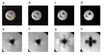 |
Reverse-Encoding MRI Near Metallic Implants Using Missing Pulse Steady-State Free Precession
Michael Mullen1 and Michael Garwood1
1Center for Magnetic Resonance Research, Department of Radiology, University of Minnesota, Minneapolis, MN, United States
Clinical MRI sequences for imaging near metallic implants are mainly multi-spectral approaches, with fast spin-echo acquisitions to achieve clinically relevant scan times. Due to the large bandwidths necessary for refocusing all off-resonance spins near metallic implants, usually the refocusing pulse flip angles must be limited to a sub-optimal value to permit a safe specific absorption rate. Here, a low peak amplitude, radiofrequency refocused sequence is used to limit energy deposition. Reversed encoding is employed, where two acquisitions are collected with opposite frequency-encoding gradient polarity. An estimate of the displacement field is found using a B-spline basis and the magnitude images.
|
|
3405.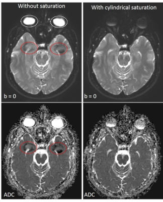 |
Mitigating artifacts from eye-motion in interleaved multi-shot DWI
Naveen Bajaj1, Jaladhar Neelavalli1, Rupesh VK1, Karthik Gopalakrishnan1, and Marc Van Cauteren2
1Philips Healthcare, Bangalore, India, 2Philips Healthcare, Tokyo, Japan
A novel approach for mitigating artifacts from localized motion of the eye, during interleaved multi-shot diffusion weighted imaging, is presented. A 2D cylindrical spatial saturation pulse is used to suppress signal from the orbit region, thereby significantly suppressing the ghost artifacts from eye movement in low b-value images. The saturation being localized to the orbit region does not influence the surrounding tissues. Placement of such a cylindrical pulse on the orbit region is relatively straight forward. In addition, the total scan time is not significantly affected by the cylindrical pulse saturation.
|
|
3406.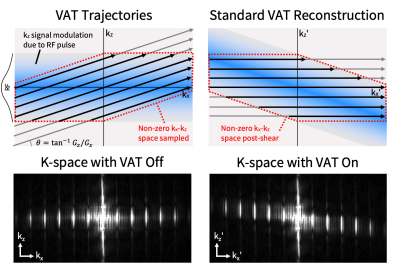 |
Mitigation of Blurring due to View-Angle Tilting in Multispectral Imaging
Alexander R Toews1,2, Philip K Lee1,2, Daehyun Yoon1, and Brian A Hargreaves1,2,3
1Radiology, Stanford University, Stanford, CA, United States, 2Electrical Engineering, Stanford University, Stanford, CA, United States, 3Bioengineering, Stanford University, Stanford, CA, United States
Imaging near metal is challenging due to severe metal-induced field inhomogeneities. Multispectral imaging sequences employ view-angle tilting (VAT) to correct in-plane distortions due to nearby metal at the expense of blurring the entire image. We exploit the additional phase-encoding dimension acquired in multispectral imaging in order to reduce the VAT-induced blur. The method primarily consists of regridding k-space measurements to avoid the VAT-induced shear. Results on a grid phantom demonstrate the ability of this method to reduce readout blur. This work highlights an important limitation of VAT and demonstrates strategies for mitigating this effect.
|
|
3407.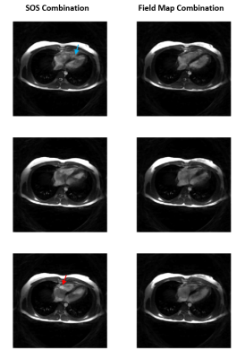 |
BMART-Enabled Field-Map Combination of Phase-Cycled Projection-Reconstruction Cardiac Cine for Banding-Free Balanced SSFP
Anjali Datta1, Dwight Nishimura1, and Corey Baron2
1Electrical Engineering, Stanford University, Stanford, CA, United States, 2Medical Biophysics, Western University, London, ON, Canada
For banding-free cardiac cine bSSFP imaging, we present a non-Cartesian accelerated frequency-modulated sequence that acquires three phase-cycles within a short breath-hold. A projection-reconstruction trajectory is used and data is also acquired on the rewinds, enabling generation of a B0 field map using BMART. This facilitates field-map combination of the phase-cycle images, which results in more homogeneous blood pool and reduced signal contributions from out-of-slice spins than root-sum-of-squares.
|
|
3408.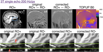 |
Interleaved reversed polarity MPRAGE for in vivo B0 readout distortion correction
Robert Frost1,2, Divya Varadarajan1,2, Jesper Andersson3, Jonathan R Polimeni1,2, Bruce Fischl1,2,4, and André J. W. van der Kouwe1,2
1Athinoula A. Martinos Center for Biomedical Imaging, Massachusetts General Hospital, Charlestown, MA, United States, 2Department of Radiology, Harvard Medical School, Boston, MA, United States, 3Wellcome Centre for Integrative Neuroimaging, University of Oxford, Oxford, United Kingdom, 4Computer Science and Artificial Intelligence Laboratory, Massachusetts Institute of Technology, Cambridge, MA, United States
Readout distortion can affect structural imaging with MPRAGE particularly in temporal regions. Interleaved readout polarity MPRAGE allows for estimation of distortion without motion confound between oppositely distorted images. This approach could be useful for single echo MPRAGE imaging with ~200 Hz/px.
|
|
3409.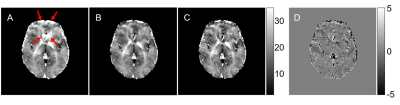 |
Fast library-driven approach for the two-step iterative calculation of the voxel spread function
Jie Wen1, Dmitriy A Yablonskiy2, Alexander L Sukstanskii2, Feiyan Zeng1, and Ying Liu1
1Radiology, The First Affiliated Hospital of USTC, Hefei, China, 2Radiology, Washington University in St. Louis, St. Louis, MO, United States
Macroscopic B0 magnetic field inhomogeneity adversely affects Gradient-Recalled-Echo (GRE) MRI images. A previously introduced voxel spread function (VSF) method has been proposed to correct for these adverse effects if data are acquired using multi-GRE (mGRE) sequences. The method accounts for both, through-slice and in-slice field inhomogeneities and accounts for between-voxel signal propagation effects. The direct voxel-wise VSF calculation requires long computation time. We propose a library-driven approach for the VSF calculation that significantly reduces computation time, thus providing a platform for the applications of mGRE-based quantitative techniques in clinical practices.
|
|
3410. |
Improving 7T Cervical Spine (C-spine) MRI using the Uniform Combined Reconstruction (UNICORN) Algorithm
Venkata Veerendranadh Chebrolu1,2, Stefan Sommer3,4, Constantin von Deuster3,4, and Julien Galley5
1Siemens Medical Solutions USA, Inc., Rochester, MN, United States, 2Department of Radiology, Mayo Clinic, Rochester, MN, United States, 3Siemens Healthcare AG, Zurich, Switzerland, 4SCMI, Swiss Center for Musculoskeletal Imaging, Zurich, Switzerland, 5Department of Radiology, Balgrist University Hospital, University of Zurich, Zurich, Switzerland
Ultra-high field (UHF) 7T MRI provides high spatial resolution with significantly better signal-to-noise ratio compared to 3T and C-spine imaging at 7T is being pursued at a few research sites across the globe. MRI of the C-spine at 7T is challenging due to multiple factors. Recently, an algorithm ‘Uniform Combined Reconstruction’ (UNICORN) was used to correct receive-coil intensity inhomogeneity and improve the uniformity and overall image quality of 7T knee MRI. In this work, we present preliminary results from the application of UNICORN to C-spine imaging and demonstrate the utility of UNICORN in improving uniformity of C-spine MRI at 7T.
|
|
3411. |
Imaging the Internal Acoustic Meatus (IAMs) of patients with cochlear implants in-situ using Slice Encoded Metal Artefact Reduction.
Simon Shah1, Fran Padormo 1, Kristine Knott1, Suki Thomson1, Steve Conner1, Geoff Charles-Edwards1, and Phil Touska1
1Guy's and St Thomas' NHS Foundation Trust, London, United Kingdom
Neurofibromatosis type 2 (NF2) patients commonly have bilateral vestibular schwannomas, which typically require regular monitoring with MRI. However these patients also have cochlear implants which makes MRI challenging. We have used Slice Encoded Metal Artefact Correction (SEMAC) to improve the imaging of IAMs in patients with cochlear implants in-situ. The implementation of SEMAC imaging reduced the geometric distortion and the volume of the signal void around the implants by over half.
|
|
3412.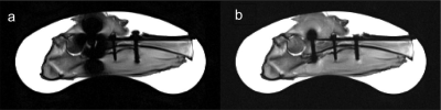 |
MRI Near Metal Using Multi-Spectral 3D Variable Flip-Angle TSE
Sina Tafti1, John P Mugler III2, Kevin Vu1, William J Garrison3, and G Wilson Miller2
1Department of Physics, University of Virginia, Charlottesville, VA, United States, 2Department of Radiology and Medical Imaging, University of Virginia, Charlottesville, VA, United States, 3Department of Biomedical Engineering, University of Virginia, Charlottesville, VA, United States
Multi-spectral imaging techniques such as MAVRIC, SEMAC, and MAVRIC SL based on 3D TSE sequence significantly reduce metal artifacts. MAVRIC and MAVRIC-SL techniques traditionally employ Gaussian RF pulses to achieve an optimal sum of squares composite image while SEMAC uses windowed-sinc RF pulses. In this work, we implement multi-spectral acquisition into the variable flip-angle TSE with hard RF pulses which yields a desirable sum of squares composite image while achieving high turbo-factor and minimal echo spacing in imaging near metallic screws in phantom and an ex vivo lamb leg.
|
|
3413.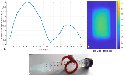 |
Local B1-Enhanced 3D Spin-Echo Imaging for Metal Artifact Reduction
Kilian Stumpf1, Andreas Horneff1, Jan-Bernd Hövener2, and Volker Rasche1
1Department of Internal Medicine II, University Medical Center Ulm, Ulm, Germany, 2Department of Radiology and Neuroradiology, University Medical Center Kiel, Kiel, Germany
Artifacts due to the presence of metallic dental materials often limit the application of MRI. These materials, such as dental implants or fillings, often cause substantial artifacts e.g. in the oral cavity impairing diagnostic accuracy. In this contribution, we present an approach for conducting spin-echo based sequences with significantly reduced flip angles and high excitation bandwidths of up to 17 kHz by using an inductively coupled local coil, which in combination with single-point methods enables almost completely artifact-free local imaging of e.g. dental titanium implants.
|
|
3414. |
Unsupervised Deep Learning Method for EPI Distortion Correction using Dual-Polarity Phase-Encoding Gradients
Jee Won Kim1, Kinam Kwon2, Byungjai Kim1, Sunho Kim1, and Hyunwook Park1
1Korea Advanced Institute of Science and Technology (KAIST), Daejeon, Korea, Republic of, 2Samsung Advanced Institute of Technology (SAIT), Suwon, Korea, Republic of
We propose a new scheme for EPI distortion correction, which implements unsupervised learning, trained with readily available images, such as ImageNet2012 dataset. The distortion-corrected image is obtained by the MR image generation function using the input distorted images and the frequency-shift maps that are the outputs of the network. Two distorted images obtained with dual-polarity phase-encoding gradients are the inputs of the neural network. The neural network estimates the frequency-shift maps from the distorted images. To train the neural network, unsupervised learning was conducted by minimizing the L1 loss between input distorted images and the estimated distorted images.
|
3415.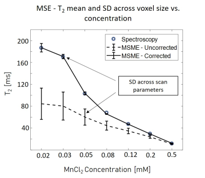 |
Compensating Diffusion Bias of Quantitative T2 mapping on High-Field MRI Scanners
Natalie Bnaiahu1, Ella Wilczynski1, Shir Levy2, Noam Omer1, Tamar Blumenfeld-Katzir1, and Noam Ben-Eliezer1,3,4
1Biomedical Engineering, Tel Aviv University, Tel Aviv, Israel, 2School of Chemistry, Tel Aviv University, Tel Aviv, Israel, 3Sagol school of Neuroscience, Tel Aviv University, Tel Aviv, Israel, 4Center for Advanced Imaging Innovation and Research, New York, NY, United States
On high-field scanners, strong imaging gradients induce spurious diffusion signal decay, which distorts qT2 calculation. By modeling the pulse sequence gradients and coherence pathways, an effective b value was calculated to assess signal attenuation caused by diffusion. ADC values were measured, extensive phantom scans and in-vivo experiments were performed using the MSME protocol with varied voxel sizes to examine the effect and the solution. Before correction T2 values were short with high variability across different voxel sizes, after correction T2 values increased and became more accurate and consistent. Tissue properties also influenced the correction as their ADC varies.
|
|
3416.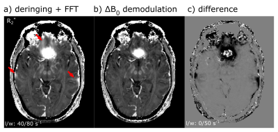 |
Reduction of Static Off-Resonance Artifacts in T2*-Weighted Gradient-Echo MRI
Jakob Meineke1 and Tim Nielsen1
1Philips Research, Hamburg, Germany
A reconstruction method is presented to reduce off-resonance induced artifacts in gradient-echo MRI. The method reconstructs the magnetization without the off-resonance induced phase and in this way reduces the effects of intra-voxel dephasing. The method is applied to simulations and volunteer scans, showing reduced signal voids and improved homogeneity in R2*-maps.
|
|
3417.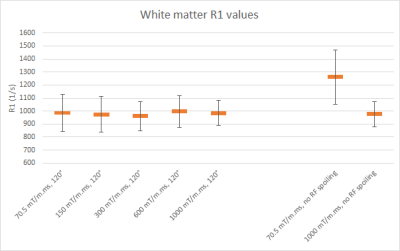 |
Diffusion spoiling for fast steady-state gradient echo imaging of the human brain using a 300 mT/m gradient system
Kerrin J Pine1, Luke J Edwards1, and Nikolaus Weiskopf1
1Department of Neurophysics, Max Planck Institute for Human Cognitive and Brain Sciences, Leipzig, Germany
In gradient echo imaging sequences, transverse coherences persisting across repetitions must be managed to avoid large variations in the steady state signal. Ultra-high amplitudes for gradient spoiling can be effective, however, conventional human MR scanners do not achieve the necessary gradient amplitudes. By using a 3T Connectom MRI scanner with a 300 mT/m gradient amplitude, we demonstrate that by operating in the diffusion spoiling regime, it is now possible to eliminate the contribution of unwanted transverse magnetization to the steady state by gradient spoiling alone. It can serve as a reference standard for validating spoiling schemes and for R1 quantification.
|
|
3418.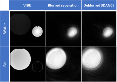 |
3DANCE VDS: Three-points Dixon golden Angle with off-resoNance CorrEction Variable Density Spiral
Marina Manso Jimeno1,2, Sairam Geethanath1,2, and John Thomas Vaughan Jr.1,2
1Columbia University, New York, NY, United States, 2Columbia Magnetic Resonance Research Center (CMRRC), New York, NY, United States
3DANCE is a sequence designed to be a good candidate for a whole-body sequence for multiple applicaitons. Phantom and in vivo experiments demonstrated a good spatial resolution and consistent ability to separate water and fat tissues. Off-resonance correction of the water and fat images have showed good results in vitro but needs more testing for in vivo datasets. Future steps include improving the robustness of the methods and testing for motion tolerance.
|
|
3419.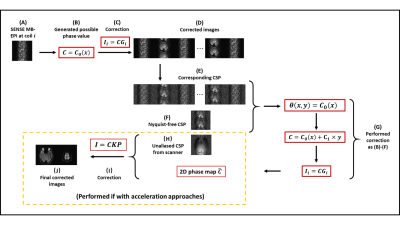 |
Two-Dimensional Coil-signature-based Phase Cycled Reconstruction for Inherent Correction of Echo-Planar Imaging Nyquist Ghost Artifacts
Silu Han1, Mahesh Bharath Keerthivasan2, Chidi Patrick Ugonna1, and Nan-kuei Chen1
1Biomedical Engineering Department, The University of Arizona, Tucson, AZ, United States, 2Medical Imaging Department, The University of Arizona, Tucson, AZ, United States
A coil-signature-based phase cycled correction method has been developed for Nyquist artifact removal in echo planar imaging (EPI). In our study, we investigated a novel coil-signature-based Nyquist artifact correction method that can be applied to EPI integrating various acceleration schemes including through-plane Multi-band Imaging (MB) and in-plane parallel SENSitivity Encoding Imaging (SENSE). Our method uses a coil-signature-based phase-cycled reconstruction without requiring EPI reference scans, and can inherently correct 2D phase errors. Our results show that the developed method can effectively reduce EPI Nyquist artifacts under different acceleration approaches.
|
|
3420.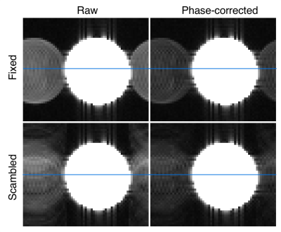 |
Scrambled readout polarities in 3D-encoded EPI markedly reduce the coherence of N/2 ghosts
M. Dylan Tisdall1
1Radiology, Perelman School of Medicine, University of Pennsylvania, Philadelphia, PA, United States
N/2 ghosts in EPI result from consistent discrepancies between readouts with positive and negative polarities. We present 3D EPI with scrambled readout polarities as a strategy to reduce the coherence of N/2 ghosts in 4D x-f space and reduce their impact on quantitative analyses (e.g., motion tracking or resting state fMRI). We demonstrate that the method can produce either individual volumes with N/2 ghost energy spread out in the partition direction, or complete 4D x-f data with ghosts spread in both partition and frequency directions.
|
|
3421.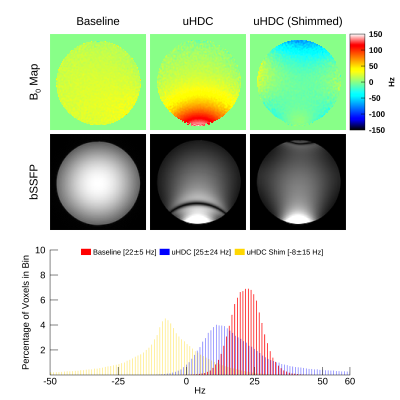 |
Susceptibility effects of uHDC material upon static field homogeneity
Christopher T Sica1, Navid Pourramzan Gandji2, and Qing X Yang2
1Radiology, PennState University College of Medicine, Hershey, PA, United States, 2Neurosurgery, PennState University College of Medicine, Hershey, PA, United States
We performed a study to assess the effects of ultra-high dielectric constant material (uHDC) on the static field homogeneity at 3T. We characterized these effects on both a spherical and body phantom with 3D off-resonance field mapping and balanced SSFP imaging. The static field experienced shifts of up to 100 Hz in the presence of uHDC monolithic blocks. Shimming was able to partially compensate for these shifts. Future studies incorporating these materials should take their susceptibility effects into account.
|
|
3422.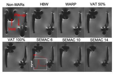 |
Quantitative Assessment of Metal Artefact Reduction Sequence Used In Imaging in Patients with Orthopaedic Total Hip Replacement Implants
Simon Shah1, Danoob Dalili2, Jan Fritz 3, Geoff Charles-Edwards1, and Amanda Isaac1
1Guy's and St Thomas' NHS Foundation Trust, London, United Kingdom, 2King's College London, London, United Kingdom, 3Johns Hopkins University, Baltimore, MD, United States
Musculoskeletal (MSK) imaging of patients with hip implants can be challenging due to metal artefacts. We have quantitatively assessed a number of metal artefact reduction sequences (MARS) on commonly implanted hip implants. The MARS methods used were: high receiver bandwidth, high transmit bandwidth, view angle titling (VAT) and slice encoded metal artefact correction (SEMAC). Signal pile-up and voids were most reduced across implants when SEMAC was employed and also artefacts at the bone metal interface were reduced.
|
|
3423.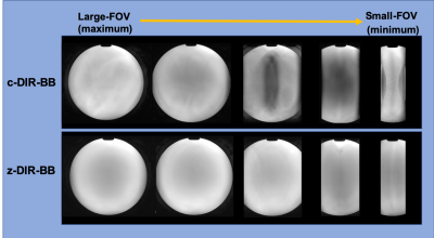 |
Improved dual inversion recovery T2-weighted black-blood image on myocardial MRI using zoomed technique
Kohei Yuda1, Takashige Yoshida1, Masami Yoneyama2, Yuki Furukawa1, and Nobuo kawauchi1
1Tokyo Metropolitan Police Hospital, Nakano, Japan, 2Philips healthcare, Tokyo, Japan, minato, Japan
The conventional Dual Inversion Recovery T2-weighted Black Blood (c-DIR-BB) on myocardial MRI is useful for the clinical diagnosis. One of the problems of c-DIR-BB is aliasing artifact, resulting in degradation of image quality. The zoomed method for improved aliasing artifact, orthogonal selection (i.e. phase / slice direction) excites to suppress tissue signals outside with a smaller FOV in phase encoding direction. Hence the DIR-BB using zoomed methods was improved image quality by orthogonal select excitation RF pulse.
|
|
3424.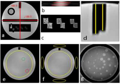 |
Fast, Automatic MRI Quality Assurance: Validation and Comparison with Manual Analysis using the ACR phantom
Hamzeh Ahmad Mohammad Al Masri1,2,3, Tonima Ali1, Katie McMahon4, and Markus Barth1,3,5
1Centre for Advanced Imaging, The University of Queensland, Brisbane, Australia, 2Medical Imaging Department, The Hashemite University, Al-Zarqa, Jordan, 3ARC Training Centre for Innovation in Biomedical Imaging Technology, The University of Queensland, Brisbane, Australia, 4Royal Brisbane & Women's Hospital, Queensland University of Technology, Brisbane, Australia, 5School of Information Technology and Electrical Engineering, The University of Queensland, Brisbane, Australia Poster Permission Withheld
Quality assurance (QA) is mandatory to ensure the stable performance of MR scanners over time. Automated analysis of QA tests can be useful to increase operator efficiency and overcome the manual processing issues such as time constraints and human bias. In this paper, we compare the manual and automated analysis approaches of the QA image datasets that have been collected from 3T MRI scanners using the American College of Radiology (ACR) accreditation phantom. We found that the automated method can significantly reduce the QA analysis time and the results of both methods were in agreement with each other.
|
|
3425.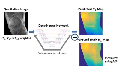 |
Estimation of B1 Map from a Single MR Image Using a Self-Attention Deep Neural Network
Yan Wu1, Yajun Ma2, Jiang Du2, and Lei Xing1
1Radiation Oncology, Stanford University, Stanford, CA, United States, 2Radiology, University of California San Diego, La Jolla, CA, United States
Inhomogeneity of the radiofrequency field (B1) is one of the main problems in quantitative MRI. Leveraging from the unique ability of deep learning, we propose a data driven strategy to derive quantitative B1 map from a single qualitative MR image without specific requirements on the weighting of the input image. B1 estimation is accomplished using a self-attention deep convolutional neural network, which makes efficient use of local and non-local information. Without additional data acquisition, an accurate estimation of B1 map is achieved, which is useful for the compensation of field inhomogeneity in T1 mapping as well as for other applications.
|
|
3426.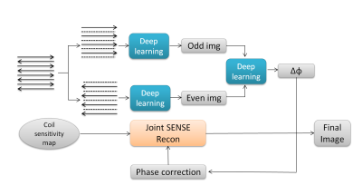 |
EPI Phase correction using model-based deep learning reconstruction
Lili Wang1, Fanwen Wang1, Yinghua Chu2, Xucheng Yu1, Chengyan Wang3, and He Wang1,3
1Institute of Science and Technology for Brain-Inspired Intelligence, Fudan University, Shanghai, China, 2MR Collaboration, Siemens Healthcare Ltd., Shanghai, China, 3Human Phenome Institute, Fudan University, Shanghai, China
The commonly used approach of Nyquist ghost correction in echo planar imaging (EPI) include linear phase correction and model-free 2D phase correction. The recent proposed method termed ‘PEC-SENSE’ incorporates 2D phase error correction with parallel imaging can robustly eliminate Nyquist ghost for EPI data,while does not act well when a distortion mismatch exsisted between the calibration data and image data. The proposed model-based deep learning method can obtain more robust phase maps than PEC-SENSE to remove image ghost and preserve the image SNR in low or high-accelerated EPI data.
|
|
3427. |
The impact of multi-coil combination techniques on multi-baseline temperature imaging methods
John Austin Roberts1, Henrik Odeen1, Sunil Patil2, Waqas Majeed2, Bradley Bolster3, Himanshu Bhat4, and Dennis L Parker1
1Radiology and Imaging Sciences, University of Utah, Salt lake City, UT, United States, 2Siemens Medical Solutions USA, Inc, Baltimore, MD, United States, 3Siemens Medical Solutions USA, Inc, Salt Lake City, UT, United States, 4Siemens Medical Solutions USA, Inc, Boston, MA, United States Poster Permission Withheld
In magnetic resonance thermometry based on proton resonance frequency shift, temperature is computed based on changes in phase as measured: with respect to previously acquired images (baselines); between regions within the same image (referenceless); or some hybrid of the two approaches. In phase reconstruction algorithms, how multi-coil data is combined affects both computational efficiency and temperature measurement precision. We modify a hybrid thermometry phase reconstruction algorithm to test several coil combination methods for computational efficiency and to evaluate their effect on temperature measurement precision. Results suggest applying coil-combination prior to phase extraction both for efficiency and SNR.
|
|
3428.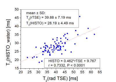 |
Fast radial T2 mapping of the liver
Diana Bencikova1,2, Stephan Kannengiesser3, Gert Reiter4, Ahmed Ba-Ssalamah1, Siegfried Trattnig1,2, and Martin Krššák2,5
1Department of Radiology, Medical University Vienna, Vienna, Austria, 2Christian Doppler Laboratory for Clinical Molecular Imaging, MOLIMA, MUW, Vienna, Austria, 3Siemens Healthcare GmbH, Erlangen, Germany, 4Siemens Siemens Healthcare Diagnostics GmbH, Graz, Austria, 5Division of Endocrinology and Metabolism, Department of Medicine III, Medical University of Vienna, Vienna, Austria
Radial T2 mapping of the liver is fast and motion insensitive, which is ideal for clinical applications. Here we tested a prototype radial turbo-spin-echo sequence for T2 mapping of the liver in phantoms and in-vivo in patients. In the phantoms we compared it to conventional Cartesian multi-SE sequence and tested the effect of fat suppression by comparing fat-suppressed and fat-unsuppressed values. From the patient evaluation, comparing it to single-voxel multi-echo MRS, we proved it to be suitable for clinical applications and a promising tool for characterization of diffuse liver disorders, but there are systematic differences between different methods.
|
|
3429.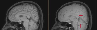 |
Spin-echo sequence with interleaved spiral acquisition avoids pulsation artifact for sagittal T1-weighted brain imaging at 3.0T
Ke Jiang1, Jiazheng Wang1, and Xiaofang Xu1
1Philips Healthcare Greater China, Beijing, China
T1-weighted imaging is a necessary component of clinical brain MR to provide anatomical information at high spatial resolution. T1w brain image in clinical practice is regularly acquired with two-dimensional turbo-spin-echo sequence, which suffers from the pulsation artifacts in the phase-encode direction from superior sagittal sinus, the straight sinus, and the sigmoid sinus. This study exploits the spiral trajectory for the spin-echo sequence to achieve the T1 weighting and to avoid the pulsation artifacts.
|