
Digital Poster Session
Acquisition, Reconstruction & Analysis: ML: Image Reconstruction
Acquisition, Reconstruction & Analysis
3585 -3599 ML: Image Reconstruction - Machine Learning for Image Reconstruction 1
3600 -3614 ML: Image Reconstruction - Machine Learning for Image Reconstruction 2
3615 -3628 ML: Image Reconstruction - Machine Learning for Image Reconstruction 3
3629 -3643 ML: Image Reconstruction - Machine Learning for Image Reconstruction 4
3585.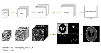 |
Untrained Modified Deep Decoder for Joint Denoising and Parallel Imaging Reconstruction
Sukrit Arora1, Volkert Roeloffs1, and Michael Lustig1
1UC Berkeley, Berkeley, CA, United States
An untrained deep learning model based on a Deep Decoder was used for image denoising and parallel imaging reconstruction. The flexibility of the modified Deep Decoder to output multiple images was exploited to jointly denoise images from adjacent slices and to reconstruct multi-coil data without pre-determed coil sensitivity profiles. Higher PSNR values were achieved compared to the traditional methods of denoising (BM3D) and image reconstruction (Compressed Sensing). This untrained method is particularly attractive in scenarios where access to training data is limited, and provides a possible alternative to conventional sparsity-based image priors.
|
|
3586.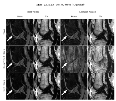 |
Deep Dixon: Deep learning-based chemical-shift corrected water-fat separation with only simulated training data
Frank Zijlstra1 and Peter R Seevinck1
1Image Sciences Institute, UMC Utrecht, Utrecht, Netherlands
Deep learning has been successfully applied to Dixon reconstruction, but requires good training data, which limits clinical applicability. We propose a deep learning-based Dixon method with chemical-shift correction that is trained with only simulated data. Results on three anatomies show that the method produces equivalent or better results than conventional methods for Dixon water-fat separation with chemical-shift correction. This approach is fundamentally different from conventional linear and non-linear solvers and shows promise for extension to more complex problems.
|
|
3587.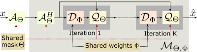 |
Joint Optimization of Sampling Patterns and Deep Priors for Improved Parallel MRI
Hemant Kumar Aggarwal1 and Mathews Jacob1
1Electrical and Computer Engineering, University of Iowa, Iowa City, IA, United States
Model-based deep learning (MoDL) frameworks, which combine deep learned priors with imaging physics, are now emerging as powerful alternatives to compressed sensing in a variety of reconstruction problems. In this work, we investigate the impact of sampling patterns on the image quality. We introduce a scheme to jointly optimize the sampling pattern and the reconstruction network parameters in MoDL scheme. Experimental results demonstrate the significant improvement in reconstruction quality with sampling optimization. The results also show that the decoupling between imaging physics and image properties in MoDL offers improved performance over direct inversion scheme in the joint optimization scheme.
|
|
3588.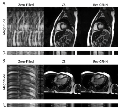 |
Real-Time Cardiac Cine MRI with Residual Convolutional Recurrent Neural Network
Eric Z. Chen1, Xiao Chen1, Jingyuan Lv2, Yuan Zheng2, Terrence Chen1, Jian Xu2, and Shanhui Sun1
1United Imaging Intelligence, Cambridge, MA, United States, 2UIH America, Inc., Houston, TX, United States
Real-time cardiac cine MRI does not require ECG gating in the data acquisition and is more useful for patients who can not hold their breaths or have abnormal heart rhythms. However, to achieve fast image acquisition, real-time cine commonly acquires highly undersampled data, which imposes a significant challenge for MRI image reconstruction. We propose a residual convolutional RNN for real-time cardiac cine reconstruction. To the best of our knowledge, this is the first work applying deep learning approach to Cartesian real-time cardiac cine reconstruction. Based on the evaluation from radiologists, our deep learning model shows superior performance than compressed sensing.
|
|
3589. |
Single-shot T2 mapping improvement through Multi-train Multiple Overlapping-Echo Detachment planar imaging sequence
Xiaoyin Wang1, Qizhi Yang2, Hongjian He1, Congbo Cai2, Yi-Cheng Hsu3, and Jianhui Zhong1,4
1Center for Brain Imaging Science and Technology, Department of Biomedical Engineering, Key Laboratory for Biomedical Engineering of Ministry of Education, Zhejiang University, Hangzhou, China, 2Department of Electronic Science, Fujian Provincial Key Laboratory of Plasma and Magnetic Resonance, Xiamen University, Xiamen, China, 3MR Collaboration, Siemens Healthcare Ltd., Shanghai, China, 4Department of Imaging Sciences, University of Rochester, Rochester, NY, United States
Magnetic Resonance parametric mapping can provide quantitative information to characterize tissue properties. Recently, a single-shot T2 mapping method based on Multiple Overlapping-Echo Detachment (MOLED) planar imaging was proposed. However, limited echo time ranges still affected the reconstruction accuracy of the T2 values, especially when large T2 value ranges were present. In this abstract, MOLED was expanded through multiple-echo-train acquisitions that achieved high accuracy and better texture. The deep convolution neural network was used to reconstruct T2 maps, B1 maps and spin densities in synchrony. The sequence efficiencies were demonstrated in digital-brain, phantom and human-brain experiments.
|
|
3590.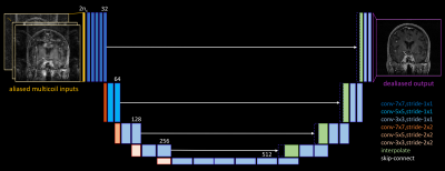 |
Model-Free Deep MRI Reconstruction: A Robustness Study
Gopal Nataraj1 and Ricardo Otazo1,2
1Medical Physics, Memorial Sloan Kettering Cancer Center, New York, NY, United States, 2Radiology, Memorial Sloan Kettering Cancer Center, New York, NY, United States
Speed is often claimed as a key advantage of deep learning (DL) for undersampled parallel MRI reconstruction. However, leading DL methods require repeated application of the MR acquisition model and its adjoint, just as in conventional iterative methods. This work investigates the feasibility and robustness of model-free DL reconstruction, which has the potential to be much faster. Results in varied patient cases of increasing pathological rarity demonstrate that while model-free DL can reasonably reconstruct anatomies similar to those seen in training, performance can degrade drastically in more challenging situations.
|
|
3591.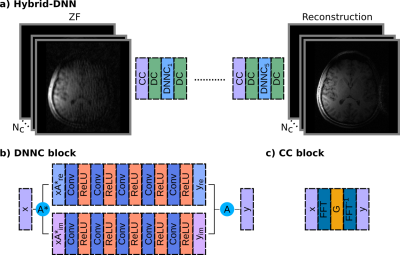 |
Hybrid Deep Neural Network Architectures for Multi-Coil MR Image Reconstruction
Salman Ul Hassan Dar1,2, Mahmut Yurt1,2, Muzaffer Özbey1,2, and Tolga Çukur1,2,3
1Department of Electrical and Electronics Engineering, Bilkent University, Ankara, Turkey, 2National Magnetic Resonance Research Center (UMRAM), Bilkent University, Ankara, Turkey, 3Neuroscience Program, Aysel Sabuncu Brain Research Center, Bilkent University, Ankara, Turkey
Two main frameworks for
|
|
3592.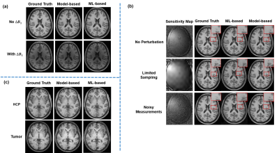 |
A Further Analysis of Deep Instability in Image Reconstruction
Yue Guan1, Yudu Li2,3, Yao Li1, Yiping Du1, and Zhi-Pei Liang2,3
1Institute for Medical Imaging Technology, School of Biomedical Engineering, Shanghai Jiao Tong University, Shanghai, China, 2Department of Electrical and Computer Engineering, University of Illinois at Urbana-Champaign, Urbana, IL, United States, 3Beckman Institute for Advanced Science and Technology, University of Illinois at Urbana-Champaign, Urbana, IL, United States
Deep learning (DL) has emerged as a new tool for solving ill-posed image reconstruction problems and generated a lot of interest in the MRI community. However, image learning is a very high-dimensional problem and deep networks, if not trained properly, would have instability problems. Building upon a recent analysis, we present a further analysis of the instability problems, highlighting: a) the overfitting problem due to limited training data, b) inaccurate density estimation, and c) inadequate sampling from a probability density function. We also present a theoretical analysis of the prediction error based on statistical learning theory.
|
|
3593.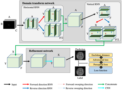 |
A direct MR image reconstruction from k-space via End-To-End reconstruction network using recurrent neural network (ETER-net)
Changheun Oh1, yeji han2, and HyunWook Park1
1Electrical Engineering, Korea Advanced Institute of Science and Technology, Daejeon, Korea, Republic of, 2Biomedical engineering, Gachon University, Incheon, Korea, Republic of
In this work, we propose a novel neural network architecture named ‘ETER-net’ as a unified solution to reconstruct an MR image directly from k-space data. The proposed image reconstruction network can be applied to k-space data that are acquired with various scanning trajectories and multi or single-channel RF coils. It also can be used for semi-supervised domain adaptation. To evaluate the performance of the proposed method, it was applied to brain MR data obtained from a 3T MRI scanner with Cartesian and radial trajectories.
|
|
3594.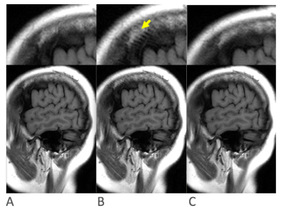 |
A deep network for reconstruction of undersampled fast-spin-echo MR images with suppressed fine-line artifact
Sangtae Ahn1, Anne Menini2, and Christopher J Hardy1
1GE Global Research, Niskayuna, NY, United States, 2GE Healthcare, Menlo Park, CA, United States
Fine-line artifact is suppressed in Fast Spin Echo (FSE) images reconstructed with a deep-learning network. The network is trained using many examples of fully sampled Nex=2 data. In each case the two excitations are combined to generate fully sampled ground-truth images with no fine-line artifact, which are used for comparison with the generated image in the loss function. However only one of the excitations is retrospectively undersampled and fed into the input of the network during training. In this way the network learns to remove both undersampling and fine-line artifacts. At inferencing, only Nex=1 undersampled data are acquired and reconstructed.
|
|
3595.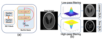 |
Exploiting Coarse-Scale Image Features for Transfer Learning in Accelerated Magnetic Resonance Imaging
Ukash Nakarmi1, Joseph Y. Cheng1, Edgar P. Rios1, Morteza Mardani1, John M. Pauly2, and Shreyas S Vasanawala1
1Department of Radiology, Stanford University, Stanford, CA, United States, 2Department of Electrical Engineering, Stanford University, Stanford, CA, United States
This work investigates coarse-scale image features for transfer learning in accelerated magnetic resonance imaging. The model uses multi-scale unrolled CNN architecture that captures image features at coarse and fine scale to efficiently reduce the training sample size for deep learning model training.
|
|
3596.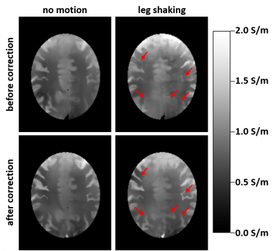 |
Deep-learning based motion correction for brain conductivity reconstruction
Jan Hendrik Wuelbern1, Ulrich Katscher1, Karsten Sommer1, Axel Saalbach1, and Jalal B Andre2
1Philips Research Europe, Hamburg, Germany, 2University of Washington, Seattle, WA, United States
Since tissue conductivity is determined by the numerical second derivative of the phase map, it is particularly susceptible to motion. This abstract investigates the application of deep-learning based methods for retrospective correction of motion artifacts to obtain suitable phase maps as input for conductivity reconstruction. Different types of motion were investigated in the framework of volunteer experiments, revealing that the applied motion correction was indeed capable of improving conductivity reconstruction.
|
|
3597. |
Calgary-Campinas raw k-space dataset: a benchmark for brain magnetic resonance image reconstruction
Roberto Souza1, M Louis Lauzon1, Marina Salluzzi1, Letícia Rittner2, and Richard Frayne1
1University of Calgary, Calgary, AB, Canada, 2University of Campinas, Campinas, Brazil
Machine learning is a new frontier for magnetic resonance (MR) image reconstruction, but progress is hampered by a lack of benchmark datasets. Our datasets provides ~200 GB of brain MR data (both raw and reconstructed data) acquired with different acquisition parameters on different scanners from different vendors and different magnetic field intensities. The fastMRI initiative (https://fastmri.org/), also provides raw data but otherwise is complementary. For instance, fastMRI provides raw k-space data corresponding to 2D acquisitions, while our dataset is composed of 3D acquisitions (i.e., with our data, you can under-sample in two directions).
|
|
3598. |
Attention Based Scale Recurrent Network for Under-Sampled MRI Reconstruction
Gabriel della Maggiora1,2,3, Alberto Di Biase1,2,3, Carlos Castillo-Passi2,3,4, and Pablo Irarrazaval1,2,3,4
1Electrical Engineering Department, Pontificia Universidad Católica de Chile, Santiago, Chile, 2Biomedical Imaging Center, Pontificia Universidad Católica de Chile, Santiago, Chile, 3Millennium Nucleus for Cardiovascular Magnetic Resonance, Santiago, Chile, 4Institute for Biological and Medical Engineering, Pontificia Universidad Católica de Chile, Santiago, Chile
We propose an Attention Based Scale Recurrent Network for reconstructing under-sampled MRI data. This network is a variation of the recently proposed Scale Recurrent Network for blind deblurring1. We treat the reconstruction problem as a deblurring problem. Thus the under-sampling pattern does not need to be known. We trained and tested our network with the NYU knee dataset available for the fastMRI challenge. The proposed model shows promising results for single-coil reconstruction outperforming both baselines.
|
|
3599. |
Uncertainty Quantification for Deep MRI
Vineet Edupuganti1, Morteza Mardani1, Joseph Cheng1, Shreyas Vasanawala2, and John Pauly1
1Electrical Engineering, Stanford University, Stanford, CA, United States, 2Radiology, Stanford University, Stanford, CA, United States
Reliable MRI reconstruction is crucial for accurate diagnosis. However, high resolution imaging leaves substantial uncertainty about the authenticity of the recovered pixels especially when using overparameterized deep learning. Leveraging variational autoencoders (VAEs), this study proposes a Bayesian imaging algorithm that distills the uncertainty in a low-dimensional latent code. One can then simply draw independent samples from the decoder to procure pixel variance maps along with the image. To further quantify the prediction risk of unseen images, we adopt Stein's Unbiased Risk Estimator (SURE), which we find correlates well with the true risk.
|
 View the Poster
 Watch the Video
3600. |
Deep learning-based referenceless distortion correction for single-shot non-Cartesian spatiotemporally encoded MRI
Wei Wang1, Jian Wu1, Qinqin Yang1, Jian Han1, Congbo Cai1, Shuhui Cai1, and Zhong Chen1
1Xiamen University, Xiamen, China
Non-Cartesian spatiotemporally encoded MRI sequence shows obvious advantages in spatial Non-Cartesian spatiotemporally encoded MRI sequence shows obvious advantages in spatial selectivity and sampling efficiency. However, the resulting image is susceptible to severe distortion due to the cumulative effect of the B0 field inhomogeneity. In this work, this issue is addressed by introducing a deep learning-based method that is specifically tailored to inhomogeneous field correction. Simulation and in vivo rat brain experiments show that our method can effectively correct the image distortion and obtain the field map without extra reference scan.
|
|
3601. |
Rapid Region-of-Interest MRI Reconstruction Using Context-Aware Non-Local U-Net
Xinwen Liu1, Jing Wang1,2, Fangfang Tang1, Hongfu Sun1, Feng Liu1, and Stuart Crozier1
1School of Information Technology and Electrical Engineering, the University of Queensland, Brisbane, Australia, 2School of Information and Communication Technology, Griffith University, Brisbane, Australia
In MRI, region-of-interest (ROI) imaging is frequently used in clinical applications. The sub-sampling-based scheme is capable of accelerating the ROI-focused image reconstruction process but degrades the image quality. The degradation could be alleviated by ROI-weighted optimization; however, existing methods mainly focus on the local signal restoration and have no explicit control of the noise from the entire image. In this abstract, we propose to reconstruct the ROI using a non-local U-net method that incorporates contextual information from the whole image. The results show the proposed algorithm improves PSNR and SSIM over conventional methods.
|
|
3602. |
Multi-contrast MR imaging with enhanced denoising autoencoder prior network learning
Xiangshun Liu1,2, Minghui Zhang1, Qiegen Liu1, Leslie Ying3, Xin Liu2, Hairong Zheng2, and Shanshan Wang2
1Department of Electronic Information Engineering, Nanchang University, Nanchang, China, 2Paul C. Lauterbur Research Center for Biomedical Imaging, SIAT, Chinese Academy of Sciences, Shenzhen, China, 3Department of Biomedical Engineering and Electrical Engineering, The State University of New York, Buffalo, New York, NY, United States
This paper proposes an enhanced denoising autoencoder prior (EDAEP) network learning method for multi-contrast MR reconstruction using deep learning. Specifically, a multi-model autoencoder with various noise levels was developed to capture different features from multi-contrast images. A weighted aggregation strategy was also adopted to balance the impact of multiple model outputs. These designs empower the network to explore the correlations and similarities among multi-contrast images, handle different acceleration trajectories and avoid a lot of cumbersome retraining. Experimental results demonstrate that our method can improve the quality of reconstructed images compared to other classical methods.
|
|
3603. |
ODE-based Deep Network for MRI Reconstruction
Ali Pour Yazdanpanah1,2, Onur Afacan1,2, and Simon K. Warfield1,2
1Harvard Medical School, Boston, MA, United States, 2Boston Children's Hospital, Boston, MA, United States
Fast data acquisition in MRI is vastly in demand and scan time depends on the number of acquired k-space samples. The data-driven methods based on deep networks have resulted in promising improvements, compared to the conventional methods. The connection between deep network and Ordinary Differential Equation (ODE) has been studied recently. Here, we propose an ODE-based deep network for MRI reconstruction to enable the rapid acquisition of MR images with improved image quality. Our results with undersampled data demonstrate that our method can deliver higher quality images in comparison to the reconstruction methods based on the UNet and Residual networks.
|
|
3604.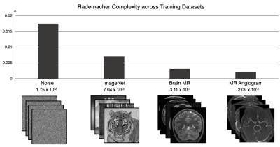 |
Quantitative characterization of image reconstruction training dataset complexity with Rademacher Complexity measures
Bo Zhu1,2,3, Neha Koonjoo1,2,3, Bragi Sveinsson1,2,3, and Matthew S Rosen1,2,3
1Department of Radiology, A.A Martinos Center for Biomedical Imaging/MGH, Charlestown, MA, United States, 2Harvard Medical School, Boston, MA, United States, 3Department of Physics, Harvard University, Cambridge, MA, United States
Here we propose to quantitatively measure the data complexity of training datasets using the Rademacher Complexity metric, and demonstrate its effectiveness in analyzing dataset composition and its effect on neural network training for image reconstruction tasks.
|
|
3605.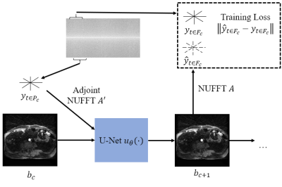 |
Unsupervised Reconstruction of Continuous Dynamic Radial Acquisitions via CNN-NUFFT Self-Consistency
Matthew Muckley*1, Tullie Murrell*2, Suvrat Booshan2, Hersh Chandarana1, Florian Knoll1, and Daniel K. Sodickson1
1Radiology, NYU School of Medicine, New York, NY, United States, 2Facebook AI Research, Menlo Park, CA, United States
The introduction of machine learning for medical image reconstruction has opened up new opportunities for reconstruction speed and subsampling; however, acquiring ground truth data is expensive or impossible in the case of dynamic imaging. Here we investigate a technique for optimizing a CNN on continuous radial data by treating the NUFFT-CNN function as an autoencoding deep image prior. Using this method, we are able to reconstruct images that increment over time frames as short as a single spoke. The technique opens up new possibilities for dynamic image reconstruction.
|
|
3606.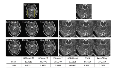 |
ISTA-nets: enhancing the performance of the unrolled deep networks for fast MR imaging
Jing Cheng1, Yiling Liu1, Qiegen Liu2, Ziwen Ke1, Haifeng Wang1, Yanjie Zhu1, Leslie Ying3, Xin Liu1, Hairong Zheng1, and Dong Liang1
1Shenzhen Institutes of Advanced Technology, Chinese Academy of Sciences, Shenzhen, China, 2Nanchang University, Nanchang, China, 3University at Buffalo, The State University of New York, Buffalo, Buffalo, NY, United States
We introduce an effective strategy to maximize the potential of deep learning and model-based reconstruction based on the network of ISTA-net, which is the unrolled version of iterative shrinkage-thresholding algorithm for compressed sensing reconstruction. By relaxing the constraints in the reconstruction model and the algorithm, the reconstruction quality is expected to be better. The prior of the to-be-reconstructed image is obtained by the trained networks and the data consistency is also maintained through updating in k-space for the reconstruction. Brain data shows the effectiveness of the proposed strategy.
|
|
3607.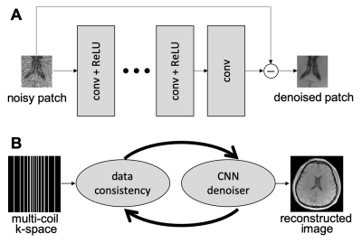 |
A Parameter-free Plug-and-Play Method for Accelerated MRI Reconstruction
Sizhuo Liu1, Ning Jin2, Philip Schniter3, and Rizwan Ahmad1
1Biomedical Engineering, Ohio State University, Columbus, OH, United States, 2Siemens Medical Solutions Inc, Columbus, OH, United States, 3Electrical and Computer Engineering, Ohio State University, Columbus, OH, United States
The recently proposed Plug-and-Play (PnP) methods provide an avenue to combine physics-driven MR models with sophisticated, learned image models instantiated by image denoising subroutines. The performance of PnP methods, however, is sensitive to changes in the measurement signal-to-noise ratio (SNR) and algorithmic parameters that balance the contributions from the data fidelity and denoising terms. We propose a discrepancy-principle-based scheme that mediates the impact of the denoising subroutine, leading to more consistent performance across different measurement SNRs without manual intervention. For validation, the proposed scheme is applied to cine images collected at 3T, 1.5T, and 0.35T scanners.
|
|
3608.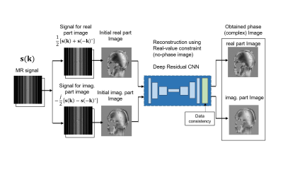 |
Efficient Phase-varied Image Reconstruction using Single Deep Convolutional Neural Network without Estimation of Phase Distribution.
Shohei OUCHI1 and Satoshi ITO1
1Utsunomiya University, Utsunomiya, Japan
A novel single image domain learning CNN based reconstruction method for phase-varied images is proposed in which real and imaginary part of complex image are reconstructed independently. Proposed method uses symmetrical sub-sampling which enable reconstruction for real and imaginary part of complex images independently of each other without estimating phase distribution on the image. Reconstruction experiments showed that higher PSNR images are obtained in proposed method compared to phase estimating CNN or ADMM-CSNet. Proposed method is highly practical since it is robust to phase variation and is easy for training because of its simple CNN structure
|
|
3609. |
Deep Learning to Produce Realistic MR Images through Fréchet Inception Distance Monitoring
Sunghun Seo1, Seung Hong Choi2, and Sung-Hong Park1
1Department of Bio and Brain Engineering, Korea Advanced Institute of Science and Technology, Daejeon, Korea, Republic of, 2Department of Radiology, Seoul National University College of Medicine, Seoul, Korea, Republic of
It is known that optimizing a deep learning model based on best validation loss achieves best quantitative results in image reconstruction, but resulting images are often blurry. In this study we propose an alternative way of optimization in which convolutional neural network (CNN) is trained beyond best validation loss to produce realistic MR images by monitoring Fréchet Inception Distance. The new approach generated sharper and more realistic images than the conventional optimization, providing a new insight into optimization for MR image reconstruction.
|
|
3610. |
Calibrationless SENSE Reconstruction with Deep Coil Sensitivity Learning
Chengyan Wang1, Yan Li2, Jun Lv3, Bo Li4, Fei Dai5, Weibo Chen6, and He Wang1,5
1Human Phenome Institute, Fudan University, Shanghai, China, 2Department of Radiology, Ruijin Hospital, Shanghai Jiao Tong University School of Medicine, Shanghai, China, 3Department of Computer Science, Yantai University, Yantai, China, 4The Third Affiliated Hospital of Nanchang University, Nanchang, China, 5Institute of Science and Technology for Brain-Inspired Intelligence, Fudan University, Shanghai, China, 6Philips Healthcare, Shanghai, China
Conventional SENSE requires accurate estimation of coil sensitivity maps, which remains to be a challenge in practical scenarios. This study aims to apply CNN to extract coil sensitivity information from the undersampled center k-space, and use the estimated sensitivity maps for parallel imaging. Results show that no obvious residual signal can be seen in the reconstructed images for all cases, which indicates the efficacy of the proposed method. Besides, the CNN based SENSE image without ACS appears to be less noisy than conventional SENSE results with ACS, which may benefit from the denoising effect from CNN on the sensitivity maps.
|
|
3611. |
Parallel Imaging with a Combination of SENSE and Generative Adversarial Networks (GAN)
Jun Lyu1, Peng Wang1, and Chengyan Wang2
1Yantai University, Yantai, China, 2Fudan University, Shanghai, China
This study aims to use GAN architecture to remove the g-factor artifacts in SENSE reconstruction. The proposed method outperforms SENSE and ZF+GAN in terms of the measured quality metrics (decreases of NMSE and increases of PSNR and SSIM). Besides, our method performs well in preserving images details with under-sampling factor of up to 6-fold, which is promising to be applied in clinical applications.
|
|
3612. |
An Information Theoretical Framework for Machine Learning Based MR Image Reconstruction
Yudu Li1,2, Yue Guan3, Ziyu Meng2,3, Fanyang Yu2,4, Rong Guo1,2, Yibo Zhao1,2, Tianyao Wang5, Yao Li3, and Zhi-Pei Liang1,2
1Department of Electrical and Computer Engineering, University of Illinois at Urbana-Champaign, Urbana, IL, United States, 2Beckman Institute for Advanced Science and Technology, University of Illinois at Urbana-Champaign, Urbana, IL, United States, 3Institute for Medical Imaging Technology (IMIT), School of Biomedical Engineering, Shanghai Jiao Tong University, Shanghai, China, 4Department of Bioengineering, University of Illinois at Urbana-Champaign, Urbana, IL, United States, 5Department of Radiology, The Fifth People's Hospital of Shanghai, Shanghai, China
Machine learning (ML) based MR image reconstruction leverages the great power and flexibility of deep networks in representing complex image priors. However, ML image priors are often inaccurate due to limited training data and high dimensionality of image functions. Therefore, direct use of ML-based reconstructions or treating them as statistical priors can introduce significant biases. To address this limitation, we treat ML-based reconstruction as an initial estimate and use an information theoretical framework to incorporate it into the final reconstruction, which is optimized to capture novel image features. The proposed method may provide an effective framework for ML-based image reconstruction.
|
|
3613. |
Super-resolution MRI using deep convolutional neural network for adaptive MR-guided radiotherapy: a pilot study
Yihang Zhou1, Hongyu Li2, Jing Yuan1, Leslie Ying2, Kin Yin Cheung1, and Siu Ki Yu1
1Medical Physics & Research Department, Hong Kong Sanatorium & Hospital, Hong Kong, China, 2Department of Biomedical Engineering, Department of Electrical Engineering, The State University of New York at Buffalo, Buffalo, NY, United States
MR-guided radiotherapy (MRgRT) is creating new perspectives towards an individualized precise radiation therapy solution. However, spatial resolution of fractional MRI can be much restricted, in order to shorten scan time, by patient tolerance of immobilization, intra-fractional anatomical motion and complicated MRgRT workflow. We hypothesized that the quality of low-resolution daily MRI could be greatly restored to generate super-resolution MRI, whose quality should be comparable of high-resolution planning MRI, by applying deep learning techniques. In this study, we aimed to investigate the feasibility of deep learning super-resolution MRI generation in the head-and-neck for adaptive MRgRT purpose.
|
|
3614. |
Model-augmented deep learning for VFA-T1 mapping
Lea Bogensperger1,2, Oliver Maier1, and Rudolf Stollberger1,3
1Institute of Medical Engineering, Graz University of Technology, Graz, Austria, 2Institute for Computer Graphics and Vision, Graz University of Technology, Graz, Austria, 3Biotechmed, Graz, Austria
A deep learning approach is proposed to estimate M0 and T1 maps from undersampled variable flip angle (VFA) data to explore the potential of this method for acceleration and rapid reconstruction even without parallel imaging. A U-Net was implemented with a model consistency term containing the signal equation to ensure the physical validity and to include prior knowledge of B1+. Training is performed on numerical brain phantoms and by means of transfer-learning on retrospectively undersampled in-vivo data. Qualitative and quantitative results show the acceleration potential for both numerical and in-vivo data for acceleration factors R=1.89, 3.43, and 5.84.
|
3615. |
Relax-ADMM-Net: A Relaxed ADMM Network for Compressed Sensing MRI
Yiling Liu1, Jing Cheng2, Yanjie Zhu2, Haifeng Wang2, Ziwen Ke1, Qiegen Liu3, Xin Liu2, Hairong Zheng2, Leslie Ying4, and Dong Liang1,2
1Research center for Medical AI, Shenzhen Institutes of Advanced Technology, shenzhen, China, 2Paul C. Lauterbur Research Center for Biomedical Imaging, Shenzhen Institutes of Advanced Technology, shenzhen, China, 3Department of Electronic Information Engineering, Nanchang University, Nanchang, China, 4Departments of Biomedical Engineering and Electrical Engineering, University at Buffalo,the State University of New York, Buffalo, NY, United States
ADMM is a popular algorithm for Compressed sensing (CS) MRI. ADMM-based deep networks have also achieved a great success by unrolling the ADMM algorithm into deep neural networks. Nevertheless, ADMM-Nets only make the components in the regularization term learnable. In this work, we propose a relaxed version of ADMM-Net (i.e. Relax-ADMM-Net) to further improve its performance for fast MRI, where the additional data consistency term and variable combinations in the updating rules are all freely learned by the network. Experiments reveal the effectiveness of the proposed network compared with several competing model-driven networks.
|
|
3616. |
Deep Network Interpolation for Accelerated Parallel MR Image Reconstruction
Chen Qin1, Jo Schlemper1,2, Kerstin Hammernik1, Jinming Duan3, Ronald M Summers4, and Daniel Rueckert1
1Imperial College London, London, United Kingdom, 2Hyperfine Research Inc., Guilford, CT, United States, 3School of Computer Science, University of Birmingham, Birmingham, United Kingdom, 4NIH Clinical Center, Bethesda, MD, United States
We present a deep network interpolation strategy for accelerated parallel MR image reconstruction. In particular, we examine the network interpolation in parameter space between a source model that is formulated in an unrolled scheme with L1 and SSIM losses and its counterpart that is trained with an adversarial loss. We show that by interpolating between the two different models of the same network structure, the new interpolated network can model a trade-off between perceptual quality and fidelity.
|
|
3617.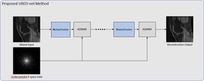 |
Unsupervised Deep Learning Reconstruction Using the MR Imaging Model
Peizhou Huang1, Chaoyi Zhang2, Hongyu Li2, Sunil Kumar Gaire2, Ruiying Liu2, Xiaoliang Zhang1, Xiaojuan Li3, Liang Dong4, and Leslie Ying1,2
1Biomedical Engineering, State University of New York at Buffalo, Buffalo, NY, United States, 2Electrical Engineering, State University of New York at Buffalo, Buffalo, NY, United States, 3Program of Advanced Musculoskeletal Imaging (PAMI), Cleveland Clinic, Cleveland, OH, United States, 4Paul C. Lauterbur Research Center for Biomedical Imaging, SIAT, CAS, Shenzhen, China
Deep learning has been applied to MRI image reconstruction successfully. Most existing works require labeled ground-truth images to learn network parameters for image reconstruction, which is not practical in some MR applications where acquisition of fully sampled images takes too long. In this abstract, we propose a novel unsupervised deep neural network for reconstruction from undersampled data. The proposed network, named URED-net, is built upon conventional ADMM algorithm for compressed sensing reconstruction, but incorporating noise2noise, an unsupervised deep denoising network. The experimental results demonstrate proposed URED-net is superior to the standard noise2noise network with and without ground-truth images for training.
|
|
3618.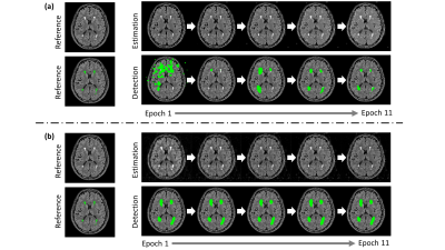 |
Multi-objective Deep Learning for Joint Estimation and Detection Tasks in MRI
Zhiyang Fu1, Maria I Altbach2, Diego R Martin2, and Ali Bilgin1,2,3
1Electrical and Computer Engineering, University of Arizona, Tucson, AZ, United States, 2Department of Medical Imaging, University of Arizona, Tucson, AZ, United States, 3Biomedical Engineering, University of Arizona, Tucson, AZ, United States MR images are often reconstructed first and then used for medical image analysis tasks such as segmentation or classification. This sequential procedure can compromise the performance of the image analysis task. In this work, we propose a multi-task learning framework that jointly reconstructs underlying images and detects multiple sclerosis lesions. This framework outperforms the conventional sequential processing pipeline. We also introduce a multi-objective optimization as an effective and automated approach to balance the trade-off among multi-task losses. Experimental results suggest that taking into account subsequent detection tasks during image reconstruction may lead to enhanced detection performance. |
|
3619.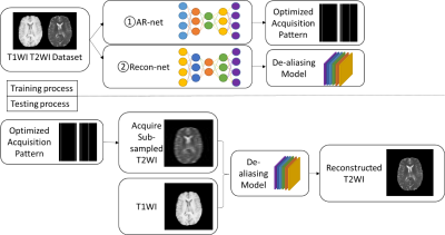 |
Deep Simultaneous Optimization of Sampling and Reconstruction for Multi-contrast MRI
Xinwen Liu1, Jing Wang1,2, Fangfang Tang1, Shekhar S. Chandra1, Feng Liu1, and Stuart Crozier1
1School of Information Technology and Electrical Engineering, the University of Queensland, Brisbane, Australia, 2School of Information and Communication Technology, Griffith University, Brisbane, Australia
MRI images of the same subject in different contrasts contain shared information, such as the anatomical structure. Utilizing the redundant information amongst the contrasts to sub-sample and faithfully reconstruct multi-contrast images could greatly accelerate the imaging speed, improve image quality and shorten scanning protocols. We propose an algorithm that generates the optimized sampling pattern and reconstruction scheme of one contrast (e.g. T2-weighted image) when images with different contrast (e.g. T1-weighted image) have been acquired. The proposed algorithm achieves increased PSNR and SSIM with the resulting optimal sampling pattern compared to other acquisition patterns and single contrast methods.
|
|
3620.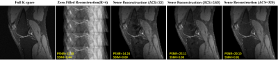 |
A Hybrid SENSE Reconstruction Combined with Deep Convolution Neural Network
Hangfei Liu1, Jingjing Li1, Qing Tang1, and Tao Zhang1,2,3
1School of Life Science and Technology, University of Electronic Science and Technology of China, chengdu, China, 2High Field Magnetic Resonance Brain Imaging Laboratory of Sichuan, Chengdu, China, 3Key Laboratory for Neuro Information, Ministry of Education, Chengdu, China
Recently parallel imaging reconstruction based on deep learning has made lots of progresses, however, there still exist several common challenges, i.e. generalization, transferability and robustness. On the contrary, SENSE reconstruction has been routinely used in clinical scans due to its high robustness and excellent image quality. A high-quality coil sensitivity map (HQCSM) is the key to achieve good SENSE reconstruction. We proposed a hybrid SENSE reconstruction frame, combining the SENSE reconstruction algorithm with a deep convolutional neural network to learn HQCSM from a few automatic calibration lines (ACS), which shows good generalization for different under-sampling ratio and enhanced robustness.
|
|
3621.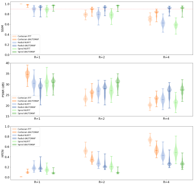 |
Reconstructing non-Cartesian acquisitions using dAUTOMAP
Maarten L Terpstra1,2, Federico d'Agata1,2,3, Bjorn Stemkens1,2, Jan JW Lagendijk1, Cornelis AT van den Berg1,2, and Rob HN Tijssen1,2
1Department of Radiotherapy, Division of Imaging & Oncology, University Medical Center Utrecht, Utrecht, Netherlands, 2Computational Imaging Group for MR diagnostics & therapy, Center for Image Sciences, University Medical Center Utrecht, Utrecht, Netherlands, 3Department of Neurosciences, University of Turin, Turin, Italy
Recently, dAUTOMAP has been presented to perform deep learning-based image reconstruction. dAUTOMAP uses gridded k-space points and so far it has only been used to reconstruct Cartesian acquisitions. In this work, we demonstrate that dAUTOMAP can produce high-quality reconstructions on radial and spiral non-Cartesian acquisitions and can resolve artifacts beyond those introduced by the undersampled acquisition.
|
|
3622. |
End-to-End Deep Learning Reconstruction for Ultra-Short MRF
Mingdong Fan1, Brendan Eck2, Nicole Seiberlich3, Michael Martens1, and Robert Brown1
1Physics, Case Western Reserve University, Cleveland, OH, United States, 2Cardiovascular and Metabolic Sciences, Cleveland Clinic, Cleveland, OH, United States, 3Radiology, University of Michigan, Ann Arbor, Ann Arbor, MI, United States
There are two major challenges in MRF reconstruction, the aliasing artifacts that results from the largely under-sampled k-space, and the very long MRF sequence used in practice to improve the reconstruction accuracy. In this study, we propose an end-to-end deep learning based reconstruction model that aims to address the issue of the spatial aliasing artifacts and provide accurate reconstruction with ultra-short MRF signals.
|
|
3623.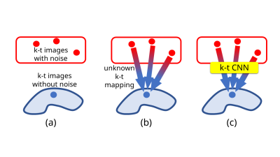 |
k-t CNN for Modeling Spatio-temporal Mappings and an Application to Reconstruction of k-space Data with Stack-of-spirals Trajectory
Hidenori Takeshima1 and Hideaki Kutsuna2
1Advanced Technology Research Department, Research and Development Center, Canon Medical Systems Corporation, Kanagawa, Japan, 2MRI Systems Development Department, MRI Systems Division, Canon Medical Systems Corporation, Kanagawa, Japan
The authors propose a new model using a convolutional neural network (CNN) named k-t CNN for approximating non-linear spatio-temporal mappings used in various applications. Existing studies imply that spatio-temporal mappings are non-linear. Most existing studies developed various methods using linear models for spatio-temporal applications. Meanwhile, the effectiveness of non-linear models was shown for spatial-domain applications.
As an application of k-t CNN, the effectiveness of the proposed method is shown experimentally in the case of reconstruction of stack-of-spirals k-space data. |
|
3624.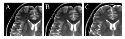 |
Deep Learning-Based Adaptive Noise Reduction for Improving Image Quality of 1.5T MR Images
Shigeru Kiryu1, Yasutaka Sugano2, Tomoyuki Ohta3, and Kuni Ohtomo4
1Radiology, International University of Health, School of medicine, Chiba, Japan, 2Canon Medical Systems Corporation, Kanagawa, Japan, 3International University of Health and Welfare Hospital, Tochigi, Japan, 4International University of Health and Welfare, Tochigi, Japan
We assessed the performance of the Deep Learning-based Reconstruction (dDLR) technique in improving 1.5T MR images. Eleven volunteers underwent MR imaging at 3T and 1.5T on the same day with the same imaging parameters. We applied the dDLR to the 1.5T image data (dDLR-1.5T), and then compared the 1.5T and dDLR-1.5T datasets with reference to the 3T dataset. The structure similarity of dDLR-1.5T was higher than that of 1.5T and dDLR increased SNR at 1.5T. The dDLR technique improves the image quality of MR images obtained at 1.5T.
|
|
3625.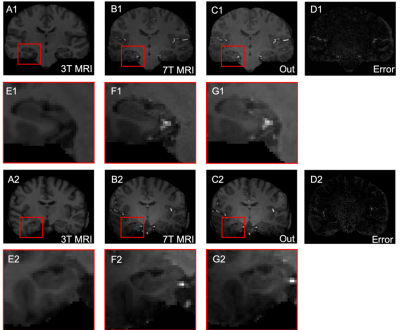 |
Simultaneous Brain Anatomical and Arterial Imaging by 3T MRI: Reconstruction Based on a Generative Adversarial Network
Wei Yu1,2, Lixin Wang1,2, Xianchang Zhang3, Zhentao Zuo4, Rong Xue4,5,6, and Tianyi Qian1,2
1Sinovation Ventures AI Institute, Beijing, China, 2QuantMind, Beijing, China, 3MR Collaboration, Siemens Healthcare, Beijing, China, 4State Key Laboratory of Brain and Cognitive Science, Beijing MRI Center for Brain Research, Institute of Biophysics, Chinese Academy of Sciences, Beijing, China, 5University of Chinese Academy of Sciences, Beijing, China, 6Beijing Institute for Brain Disorders, Beijing, China
This study investigates a reconstruction method designed to generate 7T magnetic resonance images (MRI) from 3T MRI based on a generative adversarial network. Compared with current reconstruction methods, this method can simultaneously reconstruct well-defined anatomical details and salient blood vessels. This reconstruction method may be useful for increasing the efficiency of brain MRI examinations.
|
|
3626.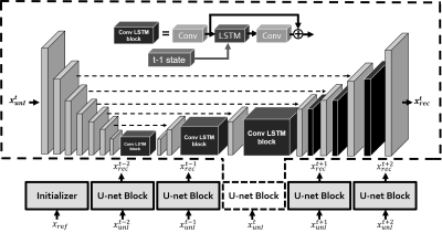 |
A Recurrent Neural Network (RNN) based reconstruction of extremely undersampled neuro-interventional MRI
Ruiyang Zhao1, Tao Wang2, Kang Yan1, Chengcheng Zhang3, Zhipei Liang4, Yiping Du1, Dianyou Li3, Bomin Sun3, and Yuan Feng1
1Institute for Medical Imaging Technology, Shanghai Jiao Tong University, Shanghai, China, 2Functional Neurosurgery,Ruijin Hospital affiliated to Shanghai Jiao Tong University, Shanghai, China, 3Shanghai Jiao Tong University Medical School Affiliated Ruijin Hospital, Shanghai, CHINA, Shanghai, China, 4Beckman Institute for Advanced Science & Technology, Department of Electrical & Computer Engineering,University of Illinois at Urbana-Champaign, Urbana-Champaign, IL, United States
Real –time MR image-guided neurosurgery could greatly improve the surgery accuracy and outcome. However, real-time guidance requires highly accelerated imaging. In this study, we proposed a Convolutional Long Short-term Memory (Conv-LSTM) based U-net to reconstruct consecutive image frames with golden-angle sampling. The Conv-LSTM based architecture was developed to explore time coherence information. Training and test datasets were generated from MR images of patients treated with Deep Brain Stimulation (DBS). Results showed that our model could achieve an acceleration rate ~80x, which provided great potentials for application in MR-guided interventional therapy.
|
|
3627.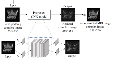 |
Partial Fourier MRI Reconstruction Using Convolutional Neural Networks
Peibei Cao1,2, Linfang Xiao1,2, Yilong Liu1,2, Yujiao Zhao1,2, Yanqiu Feng3, Alex T Leong1,2, and Ed X Wu1,2
1Laboratory of Biomedical Imaging and Signal Processing, The University of Hong Kong, Hong Kong, China, 2Department of Electrical and Electronic Engineering, The University of Hong Kong, Hong Kong, China, 3School of Biomedical Engineering, Southern Medical University, Guangzhou, China
Convolutional neural network (CNN) has emerged as a powerful tool for medical image reconstruction. In this study, we designed and implemented a CNN model for partial Fourier MRI reconstruction, and compared its performance with the existing projection onto convex sets (POCS) method. The results demonstrated that our proposed deep learning approach could effectively recovered the high frequency components and outperformed the POCS method especially when partial Fourier fraction is close to 50%.
|
|
3628.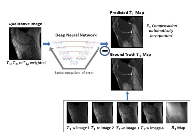 |
Derivation of quantitative T1 map from a single MR image using a self-attention deep neural network
Yan Wu1, Yajun Ma2, Jiang Du2, and Lei Xing1
1Radiation Oncology, Stanford University, Stanford, CA, United States, 2Radiology, University of California San Diego, La Jolla, CA, United States
The application of quantitative MRI is limited by additional data acquisition for variable contrast images. Leveraging from the unique ability of deep learning, we propose a data-driven strategy to derive quantitative T1 map and proton density map from a single qualitative MR image without specific requirements on the weighting of the input image. The quantitative parametric mapping tasks are accomplished using self-attention deep convolutional neural networks, which make efficient use of local and non-local information. In this way, qualitative and quantitative MRI can be attained simultaneously without changing the existing imaging protocol.
|
3629.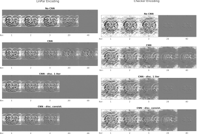 |
Impact of machine learning in iterative motion corrected reconstructions
Rita G. Nunes1, Santiago Sanz-Estébanez2, Joseph V. Hajnal3, Lucilio Cordero-Grande3, and Carlos Alberola-López2
1ISR-Lisbon/LARSyS and Department of Bioengineering, Instituto Superior Técnico – University of Lisbon, Lisbon, Portugal, Lisbon, Portugal, 2Laboratorio de Procesado de Imagen, Universidad de Valladolid, Valladolid, Spain, Valladolid, Spain, 3Centre for the Developing Brain and Department of Biomedical Engineering, School of Biomedical Engineering and Imaging Sciences, King’s College London,London U.K, London, United Kingdom
Incorporation of machine learning (ML) approaches in MR reconstruction is currently a hot topic because of the enormous potential that deep learning solutions have shown in vision and imaging communities. Recently, a procedure known as NAMER has been proposed; this procedure incorporates a ML module into an iterative reconstruction for multishot acquisitions with inter-shot motion estimation and correction (referred to as aligned reconstruction). In this abstract we provide some insight on the benefits and limitations associated to NAMER by analyzing its behavior both with a steady and a discontinued use of the ML artifact cleaning step.
|
|
3630. |
Assessment of the Generalization of Learned Unsupervised Deep Learning Method
Ziwen Ke1,2, Yanjie Zhu3, Jing Cheng2,3, Leslie Ying4, Xin Liu3, Hairong Zheng3, and Dong Liang1,3
1Research Center for Medical AI, Shenzhen Institutes of Advanced Technology,Chinese Academy of Sciences, Shenzhen, China, 2Shenzhen College of Advanced Technology, University of Chinese Academy of Sciences, Shenzhen, China, 3Paul C. Lauterbur Research Center for Biomedical Imaging, Shenzhen Institutes of Advanced Technology,Chinese Academy of Sciences, Shenzhen, China, 4Department of Biomedical Engineering and Department of Electrical Engineering, The State University of New York, Buffalo, NY, United States
In our previous work, we proposed an unsupervised deep learning method for parallel MR cardiac imaging via time interleaved sampling. The comparisons with classical methods on in vivo data have shown that this method can achieve improved reconstruction results. However, the proposed unsupervised framework is based on the time interleaved sampling scheme. Does the model trained with time interleaved undersampling pattern have good generalization to other sampling patterns? In this paper, we will explore the generalization performance of the learned unsupervised deep learning method under different sampling patterns.
|
|
3631. |
Deep-learning reconstruction for 3D Delayed Myocardial Enhancement
Gaspar Delso1, Suryanarayanan Kaushik2, Graeme C McKinnon2, Daniel Lorenzatti3, Julián Vega3, Teresa M. Caralt3, Adelina Doltra3, José T. Ortiz-Pérez3, Rosario J. Perea3, Susanna Prat3, Marta Sitges3, and Martin A. Janich4
1ASL MR, GE Healthcare, Barcelona, Spain, 2GE Healthcare, Waukesha, WI, United States, 3Hospital Clínic de Barcelona, Barcelona, Spain, 4GE Healthcare, Munich, Germany
We present the evaluation results of a novel Deep Learning reconstruction framework, applied to clinical Delayed Myocardial Enhancement datasets acquired with a 3D inversion-recovery T1-weighted gradient echo sequence.
|
|
3632. |
Deep learning for undersampled spiral DENSE reconstruction
Samuel W Fielden1,2, Eric D Carruth1, Christopher D Nevius1, Christopher M Haggerty1, and Brandon K Fornwalt1,3
1Imaging Sciences & Innovation, Geisinger, Danville, PA, United States, 2Medical & Health Physics, Geisinger, Danville, PA, United States, 3Radiology, Geisinger, Danville, PA, United States
Displacement Encoding with Stimulated Echoes (DENSE) is a powerful technique that has found great utility in accurately measuring cardiac tissue displacement. However, DENSE remains time-consuming to acquire, particularly for 3-dimensionally encoded or higher resolution schemes, and so methods to accelerate image acquisition are needed. Here, we apply the Deep Cascade of Convolutional Neural Networks (DCCNN) to the complex-valued, non-Cartesian data of DENSE to show that accelerated imaging via k-space undersampling is feasible using a deep learning-based reconstruction.
|
|
3633.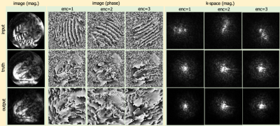 |
Suppression of Artifact-Generating Echoes in Cine DENSE using Deep Learning
Mohammad Abdishektaei1, Xue Feng1, Craig H Meyer1, and Frederick H Epstein1
1Biomedical Engineering, University of Virginia, Charlottesville, VA, United States
Cine displacement encoding with stimulated echoes (DENSE) is an accurate and reproducible method of strain imaging. The stimulated echo (STE), which carries the tissue displacement information in it’s phase, is simultaneously acquired with two artifact-generating echoes. A combination of phase-cycled acquisitions and through-plane dephasing are typically used to suppress the artifact-generating echoes. The limitations of these methods are longer acquisition times, susceptibility to breathing motion and the loss of signal-to-noise ratio due to intravoxel dephasing. To potentially overcome these limitations, the use of a deep convolutional neural network to suppress the undesired echoes from a single acquisition was investigated.
|
|
3634.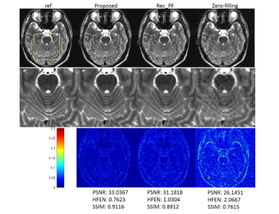 |
Learning reconstruction without ground-truth data: an unsupervised way for fast MR imaging
Jing Cheng1, Ziwen Ke1, Haifeng Wang1, Yanjie Zhu1, Leslie Ying2, Xin Liu1, Hairong Zheng1, and Dong Liang1
1Shenzhen Institutes of Advanced Technology, Chinese Academy of Sciences, Shenzhen, China, 2University at Buffalo, The State University of New York, Buffalo, Buffalo, NY, United States
Most deep learning methods for MR reconstruction heavily rely on the large number of training data pairs to achieve best performance. In this work, we introduce a simple but effective strategy to handle the situation where collecting lots of fully sampled rawdata is impractical. By defining a CS-based loss function, the deep networks can be trained without ground-truth images or full sampled data. In such an unsupervised way, the MR image can be reconstructed through the forward process of deep networks. This approach was evaluated on in vivo MR datasets and achieved superior performance than the conventional CS method.
|
|
3635.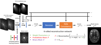 |
Learning-based Optimization of the Under-sampling PattErn with Straight-Through Estimator (LOUPE-ST) for Fast MRI
Jinwei Zhang1,2, Hang Zhang2,3, Cagla Deniz Bahadir4, Alan Wang3, Mert Rory Sabuncu1,2,3, Pascal Spincemaille2, Thanh D. Nguyen2, and Yi Wang1,2
1Biomedical Engineering, Cornell University, Ithaca, NY, United States, 2Weill Cornell Medicine, New York, NY, United States, 3Electrical and Computer Engineering, Cornell University, Ithaca, NY, United States, 4Siemens Healthineers, Princeton, NJ, United States
In this work, we propose LOUPE-ST, which extends the previously introduced optimal k-space sampling pattern learning framework called LOUPE by employing a straight-through estimator to better handle the gradient back-propagation in the binary sampling layer and incorporating an unrolled optimization network (MoDL) to reconstruct T2w images from under-sampled k-space data with high fidelity. Our results indicate that, compared with the variable density under-sampling pattern at the same under-sampling ratio (10%), superior reconstruction performance can be achieved with LOUPE-ST optimized under-sampling pattern. This was observed for all reconstruction methods that we experimented with.
|
|
3636.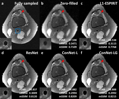 |
Locally and Globally Concatenated Network for MR Image Reconstruction
Zechen Zhou1, Christophe Schülke2, Chun Yuan3, and Peter Börnert2
1Philips Research North America, Cambridge, MA, United States, 2Philips Research Hamburg, Hamburg, Germany, 3Department of Radiology, University of Washington, Seattle, WA, United States
Recently, the convolutional neural network (CNN) based reconstruction concept has emerged as a promising implementation of compressed sensing tailored for specific fast imaging applications. The reconstruction performance of such data-driven models may depend on the CNN structure which determines the feature extraction process for sparse representation. In this study, a locally and globally concatenated network is proposed and compared with the residual network as well as the traditional L1-wavelet ESPIRiT. Preliminary experiments on a public knee imaging database showed that the proposed approach provided improved fine structure (e.g. vessel wall) restoration and background noise reduction.
|
|
3637.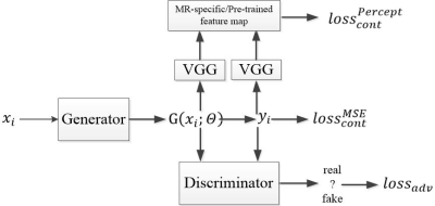 |
Image Reconstruction Using Generative Adversarial Networks with MR-Specific Feature Map
Ruiying Liu1, Hongyu Li1, Dong Liang2, Xiaojuan Li3, Chaoyi Zhang1, Peizhou Zhou1, Leslie Ying1, and Xiaoliang Zhang4
1Department of Biomedical Engineering, Department of Electrical Engineering, The State University of New York at Buffalo, Buffalo, NY, United States, 2Paul C. Lauterbur Research Center for Biomedical Imaging, SIAT, CAS,, Shenzhen, China, 3Program of Advanced Musculoskeletal Imaging (PAMI), Cleveland Clinic, Cleveland, OH, United States, 4Department of Biomedical Engineering, The State University of New York at Buffalo, Buffalo, NY, United States
Deep learning methods have demonstrated great potential in image reconstruction due to its ability to learn the non-linearity relationship between the undersampled k-space data and the corresponding desired image. Among these methods, Generative Adversarial Networks (GANs) is known to reconstruct images that are sharper and more realistic-looking. In this abstract, we study whether an MR-specific feature map that is trained on a large number of MRI images and used in the loss function can improve the GAN-based reconstruction. We demonstrate that the MR-specific feature map is superior to the pre-trained feature map typically used for GAN-based reconstruction.
|
|
3638.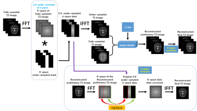 |
A deep-learning based synthesized T2 weighted imaging with multi-modality information and k-space correction
Qing Tang1, Ye Li1, Hangfei Liu1, and Tao Zhang1,2,3
1School of Life Science and Technology, University of Electronic Science and Technology of China, Chengdu, China, 2High Field Magnetic Resonance Brain Imaging Laboratory of Sichuan, Chengdu, China, 3Key Laboratory for Neuro Information, Ministry of Education, Chengdu, China
T2 weighted image (T2WI) usually takes more time and thus is more vulnerable to motion artifacts. With the recent development of applying deep learning to MR imaging, many neural networks are proposed to synthesize high-quality T2 images from under-sampled T2 or other modalities (such as T1). Here we develop a Simple-ResNet network to synthesize high-quality T2 images based on multi-modality information and followed by a k-space correction module. Results show that our model is very easy to train and the synthesized T2 images can achieve comparable image quality as the fully-sampled T2 images.
|
|
3639. |
Quantitative and Volumetric Assessment of a Deep Cascade Network for MR Reconstruction Under Different Acceleration Factors
Wallace Souza Loos1, Roberto Souza1, Mariana Bento1, Robert Marc Lebel1,2, and Richard Frayne1
1University of Calgary, Calgary, AB, Canada, 2General Electric Healthcare, Calgary, AB, Canada
Magnetic resonance (MR) imaging still has a high acquisition time due to inherent sequential procedure required to fill k-space. Deep-cascade networks have been used to reconstruct MR images from an under-sampled k-space in order to reduce acquisition time. In this work we investigate a deep-cascade to reconstruct MR images of the brain. We trained the network with 14 different acceleration factors (R). Relevant brain structures were preserved until R = 7x. For R ≥ 8x, MR images presented noticeable blurring artifact. The quality of the segmentation of the brain structures were similar to the reference MR image until R=9.
|
|
3640.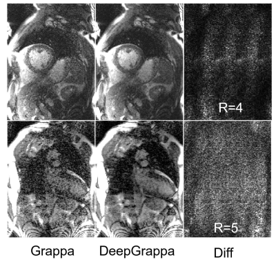 |
Deep Residual Grappa (DeepGrappa): A General Purpose Self-calibrated AI based MR Reconstruction
Hui Xue1, James C Moon2, and Peter Kellman1
1NHLBI, NIH, Bethesda, MD, United States, 2Barts Heart Centre, Barts Health NHS Trust, London, United Kingdom
In this abstract, we proposed a novel self-calibration AI based MR reconstruction algorithm to utilize the power of a deep neural network. Unlike most deep learning MR reconstruction, this algorithm does not require extra training data and only works on the auto-calibration kspace lines. This algorithm is integrated to run on MR scanner via the Gadgetron InlineAI toolbox. We demonstrated this algorithm on cardiac cine imaging, showing improved image quality without introduced unrealistic anatomical structures.
|
|
3641.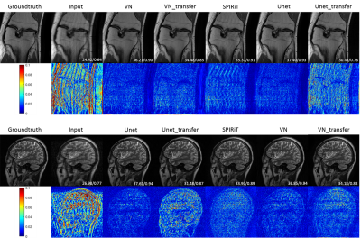 |
Are deep learning MR reconstruction models robust against adversarial attacks?
Taohui Xiao1, Cheng Li1, Haoyun Liang1, Hairong Zheng1, and Shanshan Wang1
1Paul C. Lauterbur Research Center for Biomedical Imaging, SIAT, Chinese Academy of Sciences, Shenzhen, P.R.China, Shenzhen, China Poster Permission Withheld
This paper investigates the robustness of deep learning MR reconstruction models for adversarial attacks like new lesions, different anatomy and noise pollutions. Specifically, three popular MR reconstruction algorithms were selected to investigate this issue. Experimental results show that model-based deep learning MR reconstruction method is relatively more robust than end-to-end data-driven reconstruction networks when transfer to other organs or face new lesions. Data-driven approaches can achieve better results when the testing images follow similar distributions as the training images. Severe noise can be a big issue for both deep learning methods and the traditional method.
|
|
3642. |
Plug-and-Play Deep Learning Module for Faster Parallel MR Imaging
Kamlesh Pawar1, Gary Egan1, and Zhaolin Chen1
1Monash Biomedical Imaging, Monash University, Melbourne, Australia
Deep learning (DL) methods are superior to the conventional method of accelerated imaging such as parallel imaging and compressed sensing but the integration of DL methods into the MR scanners is still in its infancy. The integration of the DL methods into the MR scanner requires the design of new pulse sequences with a modified sampling patterns/trajectories and development of DL reconstruction framework within the MR scanner. In this work, we present an effective plug-and-play approach of integrating the DL reconstruction into the MR scanner that eliminates the need to modify the existing image acquisition and reconstruction pipeline.
|
|
3643.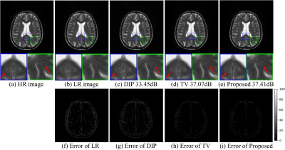 |
Brain MR image super-resolution using regularized deep image prior
Yue Hu1, Peng Li1, and Dong Nan2
1Harbin Institute of Technology, Harbin, China, 2The First Affiliated Hospital of Harbin Medical University, Harbin, China
We propose a novel algorithm for the super-resolution of brain MR images based on feature regularized DIP network, where no prior training pairs are required. We formulate the network by including the total variation (TV) term as the sparsity regularization and the Laplacian as the sharpness regularization. The network is iteratively updated using the image feature regularizations and the measured image. Numerical experiments demonstrate the improved performance offered by the proposed method.
|