
Digital Poster Session
Diffusion: Diffusion Acquisition, Reconstruction and Signal Analysis
Diffusion
4297 -4312 Diffusion Acquisition, Reconstruction and Signal Analysis - Diffusion: Acquisition 1
4313 -4326 Diffusion Acquisition, Reconstruction and Signal Analysis - Diffusion: Acquisition 2
4327 -4342 Diffusion Acquisition, Reconstruction and Signal Analysis - Diffusion: Acquisition & Reconstruction
4343 -4358 Diffusion Acquisition, Reconstruction and Signal Analysis - Diffusion: Reconstruction & Artefact Correction 1
4359 -4373 Diffusion Acquisition, Reconstruction and Signal Analysis - Diffusion: Reconstruction & Artefact Correction 2
4374 -4389 Diffusion Acquisition, Reconstruction and Signal Analysis - Diffusion: Methods
4390 -4404 Diffusion Acquisition, Reconstruction and Signal Analysis - Diffusion Signal Analysis 1
4405 -4517 Diffusion Acquisition, Reconstruction and Signal Analysis - Diffusion Signal Analysis 2
 |
4297.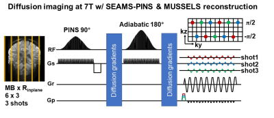 |
SNR efficient diffusion imaging at 7T with B1+ mitigated multi-shot SMS-EPI, using semi adiabatic PINS RF and low-rank completion reconstruction
SoHyun Han1, Rebecca E. Feldman2, Mary Kate Manhard3,4, Congyu Liao3,4, Seong-Gi Kim1, Priti Balchandani5, and Kawin Setsompop3,4
1Center for Neuroscience Imaging Research, Institute for Basic Science (IBS), Suwon, Korea, Republic of, 2Department of Computer Science, Mathematics, Physics, and Statistics, University of British Columbia, Kelowna, BC, Canada, 3Athinoula A. Martinos Center for Biomedical Imaging, Massachusetts General Hospital, Charlestown, MA, United States, 4Department of Radiology, Harvard Medical School, Boston, MA, United States, 5Biomedical Engineering and Imaging Institute, Icahn School of Medicine at Mount Sinai, New York, NY, United States
B1+ inhomogeneity, SAR, and shortened T2-relaxation are the main challenges to leverage the higher SNR at ultra-high-field MRI. Here, we develop a new method by combining navigation-free multi-shot SMS-EPI with low-rank matrix completion reconstruction with semi-adiabatic PINS pulse for B1+ insensitive SMS imaging. Using this combined approach, we demonstrated mitigated B1+ inhomogeneity by comparing with conventional-SE pulse and the feasibility of low-rank completion reconstruction at high b-value. Finally, 1.2 mm isotropic whole-brain diffusion MRI was acquired across 64 diffusion directions with high-SNR in 11 minutes at 7T.
|
4298. |
Noninvasive Detection of Cell Membrane Permeability with Filter-Exchange Imaging
Athanasia Kaika1,2, Mathias Schillmaier1,2, Geoffrey J. Topping1,2, and Franz Schilling1,2
1Technical University of Munich, Munich, Germany, 2Nuclear Medicine, Klinikum rechts der Isar, Munich, Germany
Filter-Exchange Imaging (FEXI) is a noninvasive double-diffusion imaging method, sensitive to transmembrane water exchange, which is strongly connected to cell viability. A FEXI sequence was implemented and tested in vitro with baker’s yeast. Upon permeabilization with ethanol, AXR increased whereas ADC decreased, more so with increasing ethanol concentration. AXR reduced over time, but only minor changes in ADC, intracellular volume and Trypan staining were detected.
|
|
4299.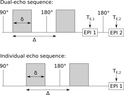 |
Rapid DTI-based free water elimination and mapping with explicit T2 modelling using a dual-echo Stejskal-Tanner EPI sequence
Ezequiel Farrher1, Richard P. Buschbeck1, Kuan-Hung Cho2, Ming-Jye Chen2, Seong Dae Yun1, Zaheer Abbas1, Chang-Hoon Choi1, Li-Wei Kuo2,3, and N. Jon Shah1,4,5,6
1Institute of Neuroscience and Medicine 4, Forschungszentrum Jülich, Jülich, Germany, 2Institute of Biomedical Engineering and Nanomedicine, National Health Research Institutes, Miaoli, Taiwan, 3Institute of Medical Device and Imaging, National Taiwan University College of Medicine, Taipei, Taiwan, 4Department of Neurology, RWTH Aachen University, Aachen, Germany, 5JARA - BRAIN - Translational Medicine, Aachen, Germany, 6Institute of Neuroscience and Medicine 11, JARA, Forschungszentrum Jülich, Jülich, Germany
We propose and investigate a dual-echo (DE) Stejskal-Tanner EPI sequence for rapid DTI-based free water elimination and mapping with explicit T2 modelling (FWET2) in vivo. DTI maps from the DE sequence are artefact-free and similar to the standard, individual echo (IE) approach. Compared to the IE case, an underestimation of T2 values calculated from the DE sequence is observed. The T2 underestimation stems from reduced signal amplitudes in the second echo of the DE sequence, which we demonstrate to correlate with imperfect refocusing RF pulses. A simple correction method is proposed. FWET2 model parameters derived from both sequences are comparable.
|
|
4300. |
Time-dependent and anisotropic diffusion in the heart: linear and spherical tensor encoding with varying degree of motion compensation
Samo Lasic1,2, Henrik Lundell2, Filip Szczepankiewicz3,4,5, Markus Nilsson3, Jürgen E. Schneider6, and Irvin Teh6
1Random Walk Imaging, Lund, Sweden, 2Danish Research Centre for Magnetic Resonance, Centre for Functional and Diagnostic Imaging and Research, Copenhagen University Hospital Hvidovre, Copenhagen, Denmark, 3Clinical Sciences, Lund University, Lund, Sweden, 4Harvard Medical School, Boston, MA, United States, 5Brigham and Women's Hospital, Boston, MA, United States, 6Leeds Institute of Cardiovascular and Metabolic Medicine, University of Leeds, Leeds, United Kingdom
Spherical tensor encoding (STE) can potentially shorten acquisition of mean diffusivity (MD) compared to the traditional linear tensor encoding (LTE). To avoid negative effects of motion, e.g. in the heart, motion compensation is needed. However, motion compensation requires altering diffusion gradient waveforms and their sensitivities to time-dependent diffusion. To exclude motion, we first investigated LTE and STE with different degrees of motion compensation in ex vivo pig hearts. We observed significantly different MD, which can be attributed to time-dependent diffusion and microscopic diffusion anisotropy. Our analysis suggests that time-dependent diffusion is a critical determinant of MD in the myocardium.
|
|
4301.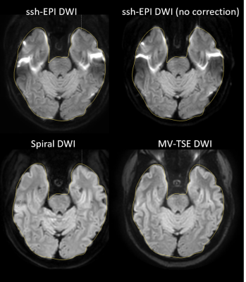 |
SENSE accelerated multishot spiral diffusion: application in brain on a clinical platform
Maarten J. Versluis1, Kim van de Ven1, Velmurugan Gnanaprakasam1, Viswanath Kasireddy2, Suthambhara Nagaraj2, and Silke Hey1
1BIU MR, Philips Healthcare, Best, Netherlands, 2BIU MR, Philips Healthcare, Bangalore, India
In this study we compare SENSE accelerated multi-shot variable density spiral diffusion to the current clinical standards: single shot EPI and MultiVane TSE diffusion. A variable density sampling strategy was employed to correct for the phase of the different shots and iterative SENSE was used to reduce the number of shots and scanning duration. This technique was applied on a clinical platform with clinically acceptable reconstruction times. We showed that spiral diffusion reduces distortions in difficult to shim brain regions compared to ssh-EPI, and spiral diffusion has at a reduced scan duration compared to the TSE-based approach.
|
|
4302.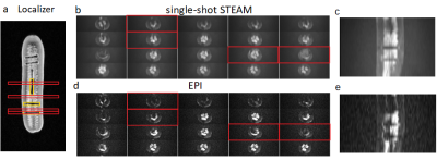 |
Robust Diffusion-Weighted Imaging near Metallic Objects with Inner-FOV Single-Shot STEAM based on 2D-Selective RF Excitations
Caspar Florin1 and Jürgen Finsterbusch1
1Department of Systems Neuroscience, University Medical Center Hamburg-Eppendorf, Hamburg, Germany
The potential of a single-shot stimulated echo acquisition mode (STEAM) sequence based on RF refocused echoes for DW imaging close to metallic objects is evaluated. It is optimized for spinal cord applications by combining it with inner-FOV technique based on 2D-selective RF (2DRF) excitations and half-Fourier sampling which improves its signal-to-noise ratio (SNR) efficiency significantly. Its robustness in the presence of metallic objects is investigated and compared to EPI showing a better performance with smaller regions suffering from signal losses.
|
|
4303.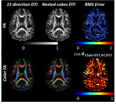 |
Simultaneous Acquisition of Dynamic Diffusion Imaging and Diffusion Tensor Imaging in the Brain
Mihika Gangolli1, Wen-Tung Wang1, Neville Gai2, Dzung L. Pham1, and John Butman1,2
1Center for Neuroscience and Regenerative Medicine, Bethesda, MD, United States, 2National Institutes of Health, Bethesda, MD, United States
We propose a diffusion acquisition scheme, called “nested cubes”, consisting of five triplets of three unique mutually orthogonal directions, providing diffusion weighted data sampled across fifteen noncollinear directions distributed uniformly across a spherical shell. Data acquired using this setup facilitates the simultaneous acquisition of dynamic maps of trace and other diffusion metrics while producing DTI measurements comparable to those from a standard DTI sequence.
|
|
4304.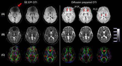 |
Diffusion tensor imaging in human subjects wearing metallic orthodontic braces
Xinyuan Miao1,2, Yuankui Wu1,2,3, Dapeng Liu1,2, Hangyi Jiang1, Qin Qin1,2, Peter C.M van Zijl1,2, Jay J. Pillai4,5, and Jun Hua1,2
1Neurosection, Division of MRI Research, Russell H. Morgan Department of Radiology and Radiological Science, Johns Hopkins University School of Medicine, Baltimore, MD, United States, 2F.M. Kirby Research Center for Functional Brain Imaging, Kennedy Krieger Institute, Baltimore, MD, United States, 3Department of Medical Imaging, Nanfang Hospital, Southern Medical University, Guangzhou, China, 4Johns Hopkins University School of Medicine, Division of Neuroradiology, Russell H. Morgan Department of Radiology and Radiological Science, Baltimore, MD, United States, 5Department of Neurosurgery, Johns Hopkins University School of Medicine, Baltimore, MD, United States
Metallic objects such as dental braces bring substantial susceptibility artifacts in MR images acquired using echo-planar-imaging (EPI) sequences. Here, we demonstrate that diffusion-prepared diffusion tensor imaging (DTI) with three-dimensional fast gradient-echo readout can significantly reduce susceptibility artifacts that are commonly seen in conventional spin-echo (SE) EPI DTI in the presence of metallic orthodontic braces.
|
|
4305.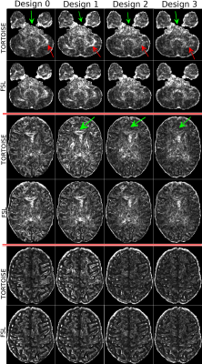 |
Reproducibility of Diffusion MRI Metrics Using 4-way Phase-Encoding Acquisition Design
M. Okan Irfanoglu1, Neda Sadeghi2, Joelle Sarlls3, and Carlo Pierpaoli2
1QMI, NIBIB/NIH, Bethesda, MD, United States, 2NIBIB/NIH, Bethesda, MD, United States, 3NINDS/NIH, Bethesda, MD, United States
In this work, we assessed the reproducibility of diffusion MRI metrics w.r.t different experimental and acquisition designs within the same scan time limits. The design that employed identical diffusion gradients and b-values for blip-up phase-encoding and blip-down phase-encoding provided significant improvements in terms of data reproducibility compared to the design using a single b=0 blip-down image in terms of distortions. The proposed 4-way encoding scheme not only improved upon this design but also consistently reduced the effects of other imaging artifacts; therefore, is suggested to be the acquisition scheme of choice for dMRI studies where biological differences are subtle.
|
|
4306.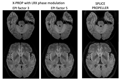 |
Improving X-PROP with a more stable echo train for diffusion weighted MRI
Zhiqiang Li1, Melvyn B Ooi1,2, and John P Karis1
1Neuroradiology, Barrow Neurological Institute, Phoenix, AZ, United States, 2Philips Healthcare, Gainesville, FL, United States
EPI-based DWI is widely used in the clinic but suffers from geometric distortions. DW-PROPELLER, based on FSE, is free from geometric distortions but has low scan efficiency. X-PROP was developed to improve FSE-based DW-PROPELLER scan efficiency by employing a GRASE readout. Although more efficient, XY2 phase modulation used in X-PROP is sensitive to the flip angles of the RF pulse train. This project improves X-PROP image quality by incorporating LRX phase modulation to increase SNR and signal stability. Image quality improvement was illustrated by comparing in vivo images produced with LRX phase modulation, XY2 phase modulation, and SPLICE PROPELLER imaging.
|
|
4307.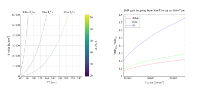 |
Practical considerations of DW-MRS with ultra-strong diffusion gradients
Christopher Jenkins1, Elena Kleban1, Lars Mueller1, John Evans1, Umesh Rudrapatna1, Derek Jones1, Francesca Branzoli2, Itamar Ronen3, and Chantal M.W Tax1
1CUBRIC, School of Psychology, Cardiff University, Cardiff, United Kingdom, 2Centre for NeuroImaging Research - CENIR, Brain and Spine Institute - ICM, Paris, France, 3Department of Radiology, Leiden University Medical Center, Leiden, Netherlands Diffusion-weighted magnetic resonance spectroscopy benefits from the use of ultra-strong gradients. Slow diffusing metabolites necessitate a large range of b-values to accurately model the diffusion properties. Ultra-strong gradients open the possibility of higher b-values and reduced diffusion times, alleviating some of these constraints. We present initial data acquired with DW-PRESS on a 300mT/m gradient Connectom scanner, and introduce the practical considerations associated with ultra-strong gradients. |
|
4308.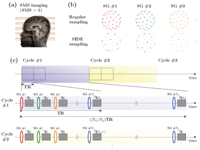 |
50-Fold Acceleration of Diffusion MRI via Slice-Interleaved Diffusion Encoding (SIDE)
Yoonmi Hong1, Wei-Tang Chang1, Geng Chen2, Ye Wu1, Weili Lin1, Dinggang Shen1, and Pew-Thian Yap1
1University of North Carolina at Chapel Hill, Chapel Hill, NC, United States, 2Inception Institute of Artificial Intelligence, Abu Dhabi, United Arab Emirates
We present a sampling and reconstruction scheme that, when combined with multi-band imaging, accelerates dMRI acquisition by as much as 50 folds. In contrast to the conventional approach of acquiring a full diffusion-weighted (DW) volume for each diffusion wavevector, we acquire for each repetition time (TR) a volume consisting of interleaved slice groups, each corresponding to a different diffusion wavevector. This in effect results in a subsample of slices for each diffusion wavevector, based on which we can recover the full volumes for all wavevectors using a graph convolutional neural network (GCNN).
|
|
4309.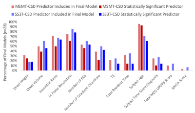 |
Investigating the effect of diffusion MRI acquisition parameters on free water signal fraction estimates from 3-tissue CSD techniques
Benjamin T Newman1,2, Thijs Dhollander3,4, and T. Jason Druzgal1,2
1Department of Radiology & Medical Imaging, Division of Neuroradiology, University of Virginia Health System, University of Virginia, Charlottesville, VA, United States, 2Brain Institute, University of Virginia, Charlottesville, VA, United States, 3The Florey Department of Neuroscience, University of Melbourne, Melbourne, Australia, 4The Florey Institute of Neuroscience and Mental Health, Melbourne, Australia
The CSF-like free water signal fraction is an advanced diffusion MRI metric representing the freely diffusing water in brain tissue. Different methods to calculate the free water signal fraction using constrained spherical deconvolution exist but it is still unknown how variation in data quality and acquisition affect measurements. Using a large clinical dataset with highly variable acquisition schemes, this study shows that the various acquisition parameters significantly affect outcome free water signal fraction, though the multi-shell analysis method is more susceptible than the single-shell method. This highlights the importance of harmonization and quality clinical imaging.
|
|
4310. |
Clinical Microscopic Fractional Anisotropy Imaging in 4 Minutes: an Optimization Approach
Nico J. J. Arezza1,2, Desmond H. Y. Tse2, Aidin Arbabi2, and Corey A. Baron1,2
1Medical Biophysics, Western University, London, ON, Canada, 2Center for Functional and Metabolic Mapping, Robarts Research Institute, London, ON, Canada
Microscopic diffusion anisotropy ($$$\mu A$$$) and microscopic fractional anisotropy ($$$\mu FA$$$) quantify water diffusion anisotropy in tissue with no influence from neuron fiber orientation. Here, we characterized $$$\mu A^2$$$ signal-to-noise ratio using standard error propagation to determine the b-value and ratio of linear to isotropic encodings needed to maximize image quality. This optimization enabled an MRI protocol that utilizes efficient isotropic diffusion encoding to acquire high-quality full-brain $$$\mu A^2$$$ and $$$\mu FA$$$ maps in 2.4 and 4 minutes, respectively, which are demonstrated in two healthy volunteers at 3T.
|
|
4311. |
Novel practical SNR determination method for MRI using combined largest b-value and echo time (COLBET)
Hirotaka Oyabu1, Tosiaki Miyati1, Naoki Ohno1, Toshifumi Gabata1, and Satoshi Kobayashi1
1Graduate School of Medical Sciences, Kanazawa University, Kanazawa, Japan Poster Permission Withheld
We developed a novel practical SNR measurement method using combined the largest b-value and echo time (COLBET). The COLBET method makes it possible to simply and practically perform image SNR quantitation including the long T2 region in human with parallel imaging.
|
|
4312. |
Investigating the reproducibility of 4th order Spherical Harmonics dMRI Rotation Invariant Features in White Matter
Mauro Zucchelli1, Samuel Deslauriers-Gauthier1, and Rachid Deriche1
1Athena Project-Team, Inria Sophia Antipolis - Méditerranée, Université Côte d'Azur, Sophia Antipolis - Méditerranée, France
Rotation invariant features can potentially be used as biomarkers for diffusion MRI. One of the most important characteristics of biomarkers is their reproducibility. In the case of diffusion MRI, reproducibility means that if we acquire data from the same subject twice with a short time gap between the two acquisitions we should obtain the same values for the biomarkers. In this work, we investigate the reproducibility of 12 new rotation invariant features that we obtained from 4th order spherical harmonics. Our results suggest that the new invariants are reproducible and can be selected as biomarker-candidates for white matter.
|
 View the Poster
 Watch the Video
4313.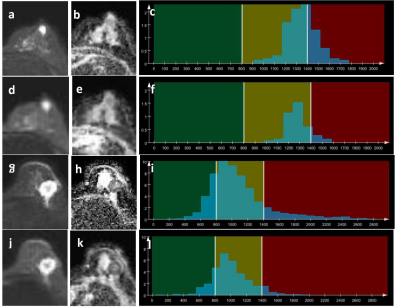 |
Whole-Tumor Histogram Analysis of Breast Lesions Based on Simultaneous Multi-slice Readout-segmented Echo-planar Imaging
Xue Li1, Kun Sun1, Wei Liu2, Robert Grimm3, Caixia Fu2, and Fuhua Yan1
1Department of Radiology, Ruijin Hospital, Shanghai Jiao Tong University School of Medicine, Shanghai, China, 2Application Development, Siemens Shenzhen Magnetic Resonance Ltd., Shenzhen, China, 3Application Predevelopment, Siemens Healthcare, Erlangen, Germany
This study compared the diagnostic performance of the ADC derived from the diffusion-weighted readout-segmented EPI DWI accelerated with SMS technique (SMS rs-EPI DWI) with that derived from the conventional rs-EPI DWI on breast lesions using whole-tumor histogram analysis. Our results showed that the SMS technique can increase the spatial resolution of the rs-EPI DWI sequence without prolonging the scan time. It can also improve the diagnostic performance of the derived ADC map based on whole-tumor histogram analysis in distinguishing benign and malignant breast lesions.
|
|
4314. |
Determination of the optimal set of b-values for Intravoxel Incoherent Motion (IVIM) parameter mapping in liver Diffusion-Weighted MRI
Óscar Peña-Nogales1, Rodrigo de Luis-Garcia1, and Santiago Aja-Fernández1
1Laboratorio de Procesado de Imagen, Universidad de Valladolid, Valladolid, Spain
Estimation of Intravoxel Incoherent Motion (IVIM) parameter maps from a set of diffusion-weighted (DW) images acquired at multiple b-values usually suffers from low SNR, which may increase the variance of the estimated maps. Unfortunately, there is no consensus on the optimal b-values to maximize the noise performance of IVIM parameters. In this work, we determine the optimal b-values to maximize the performance of IVIM parameter mapping by using a Cramér-Rao Lower Bound approach under realistic noise assumptions. The reduction of the estimation variance on the IVIM parameters compared to state-of-the-art b-values suggests the utility of this approach to optimize DW-MRI.
|
|
4315.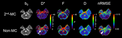 |
Intravoxel incoherent motion analysis of the brain with second-order motion-compensated diffusion encoding
Naoki Ohno1, Tosiaki Miyati1, Tetsuo Ogino2, Yu Ueda2, Yuki Koshino1,3, Yudai Shogan1,3, Toshifumi Gabata4, and Satoshi Kobayashi1
1Faculty of Health Sciences, Institute of Medical, Pharmaceutical and Health Sciences, Kanazawa University, Kanazawa, Japan, 2Philips Japan, Tokyo, Japan, 3Radiology Division, Kanazawa University Hospital, Kanazawa, Japan, 4Department of Radiology, Kanazawa University Graduate School of Medical Sciences, Kanazawa, Japan Poster Permission Withheld
In this study, we compared diffusion parameters with intravoxel incoherent motion (IVIM) analysis of the brain between second-order motion-compensated (2nd-MC) and conventional (non-MC) diffusion encoding schemes. Perfusion-related diffusion coefficient with non-MC was strongly affected by bulk motion in the pons which has the largest motion in the brain. By contrast, the 2nd-MC diffusion gradients compensated the bulk motion-induced signal loss and improved the fitting accuracy of biexponential model. The 2nd-MC diffusion encoding reduces the bulk motion effect on IVIM analysis of the brain, thereby improving the measurement accuracy.
|
|
4316.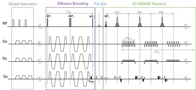 |
Oscillating Gradient (OG) Prepared 3D-GRASE Sequence for Improved OG-Diffusion MRI
Dan Wu1, Dapeng Liu2, Yi-Cheng Hsu3, Haotian Li1, Yi Sun3, Qin Qin2, and Yi Zhang1
1Key Laboratory for Biomedical Engineering of Ministry of Education, Department of Biomedical Engineering, College of Biomedical Engineering & Instrument Science, Zhejiang University, Hangzhou, China, 2Johns Hopkins University School of Medicine, BALTIMORE, MD, United States, 3MR Collaboration, Siemens Healthcare Ltd., Shanghai, China
Oscillating gradient enables access of short diffusion times for time-dependent diffusion MRI (dMRI), but poses challenges for clinical use, including limited oscillating frequencies and b-values, low SNR, and relatively long scan times. This study proposes a 3D oscillating gradient prepared gradient spin-echo sequence (OGprep-GRASE) to improve the SNR and shorten the acquisition time for OG-dMRI. The proposed sequence reduced the scan time by a factor of 1.38 and increased the SNR by 1.74 times, compared with the existing 2D echo-planar imaging (EPI) approach, leading to improved diffusion tensor reconstruction. Diffusivity measurements showed similar time-dependency using the GRASE and EPI sequences.
|
|
4317. |
TGSE diffusion-weighted pulse sequence in the evaluation of optic neuritis: A comprehensive comparison of image quality with RESOLVE DWI
Ting Yuan1, Yan Sha2, Zhongshuai Zhang3, Xilan Liu4, Xinpei Ye5, Yaru Sheng5, Kun Zhou6, and Caixia Fu6
1Shanghai Insititute of Medical Imaging, Shanghai, China, 2Eye & ENT Hospital of Shanghai Medical School, Fudan University, Shanghai, China, 3Siemens Healthcare Ltd, Shanghai, China, 4Department of Radiology, Shanghai Ninth People’s Hospital, Shanghai JiaoTong University, Shanghai, China, 5Department of Radiology, Eye & ENT Hospital of Shanghai Medical School, Fudan University, Shanghai, China, 6Department of Digitalization, Siemens Shenzhen Magnetic Resonance Ltd., Shenzhen, China
This study investigated the role of RESOLVE and TGSE DWI sequences in the evaluation of optic neuritis and compared their image qualities qualitatively and quantitatively. We found that TGSE significantly improved the image quality for the evaluation of optic neuritis by reducing the susceptibility induced image distortion compared with RESOLVE. However, it appeared lower SNR and CNR than that of RESOLVE images.
|
|
4318. |
Automatic no-reference image quality evaluation of DWI in uterine malignancy at 3T with iShim, RESOLVE, and ss-EPI sequences – a feasibility study
Qi Zhang1, Xiaoduo Yu1, Jieying Zhang1, Xinming Zhao1, Han Ouyang1, Hongmei Zhang1, Qinglei Shi2, Xiang Feng2, and Xiaoye Wang2
1Department of Imaging Diagnosis, National Cancer Center/Cancer Hospital, Chinese Academy of Medical Science and Peking Union Medical College, Beijing, China, 2MR Scientific Marketing, Diagnostic Imaging, Siemens Healthcare Ltd, Beijing, China
This study proposed an automatic image quality evaluation method using no-reference image quality metrics of structural similarity index (SSIM), blind/referenceless image spatial quality evaluator (BRISQUE), perception based Image quality evaluator (PIQE), SNR Wavelet and Contrast. The SNR Wavelet was calculated by the quotient of the image before wavelet filtering and the difference between the image before wavelet filtering and the image after wavelet filtering. The contrast was calculated using the average signal difference of three to five gray bars in the middle position. This study showed the automatic no-reference image quality metrics have potentials in future application of evaluating the image quality of uterine malignancy DWI at 3T with a higher efficiency.
|
|
4319. |
Performance comparison of three b-value sampling schemes in multiple diffusion models, including DTI, DKI, NODDI and MAP-MRI
Huiting Zhang1, Ankang Gao2, Shaoyu Wang1, Yang Song3, Jingliang Cheng2, Guang Yang3, and Xu Yan1
1MR Scientific Marketing, Siemens Healthcare, Shanghai, China, 2The First Affiliated Hospital of Zhengzhou University, Zhengzhou, China, 3Shanghai Key Laboratory of Magnetic Resonance, East China Normal Univeristy, Shanghai, China
This study aimed to evaluate the performance of three b-value sampling schemes in calculating multiple diffusion models, including the DTI, DKI, NODDI and the newly proposed mean apparent propagator (MAP)-MRI models. The three schemes includes the conventional diffusion spectrum imaging (DSI) acquisition scheme based on Cartesian grid sampling in q-space, multi-shell sampling with the same (MDDW) or different (FREE) gradient directions in each shell. Each scheme supports the estimation of all the four models. The results showed that generally three schemes generated very similar parameters, and could be all used in future studies.
|
|
4320.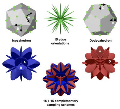 |
Isotropic sampling for skewed encoding: novel rotation schemes for non-axisymmetric encoding objects in diffusion MRI
Carl-Fredrik Westin1,2 and Filip Szczepankiewicz1,2
1Harvard Medical School, Boston, MA, United States, 2Brigham and Women's Hospital, Boston, MA, United States
In this work we propose novel sampling schemes for diffusion MRI required for encoding with objects with more than one directional axis. The presented solution is general and suitable for both axisymmetric and non-axisymmetric encoding schemes. An important feature of the presented sampling schemes is that they can be interleaved and be complimentary. This means that any combination of them can be used to define an isotropic sampling scheme with number of samples: (15, 30, 45, 60, 75, 90).
|
|
4321. |
Optimal experimental design for multi-tissue spherical deconvolution of diffusion MRI
Jan Morez1, Jan Sijbers1, and Ben Jeurissen1
1imec-Vision Lab, Dept. Physics, University of Antwerp, Antwerp, Belgium
Multi-tissue constrained spherical deconvolution of multi-shell diffusion weighted MRI data estimates the white matter fiber orientation distribution function, together with the densities of gray matter and cerebrospinal fluid. In this work, we propose a 5-minute scanning protocol that allows a more precise estimation of WM and GM densities, while maintaining a high angular resolution.
|
|
4322.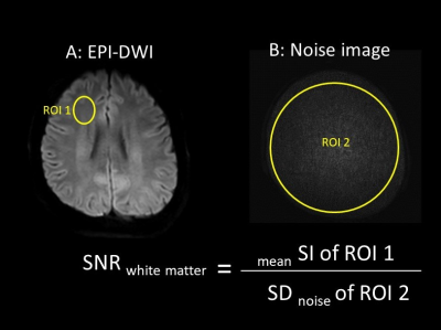 |
Ultra-high b-value single-shot echo planar diffusion-weighted imaging with Compressed SENSE
Kayoko Abe1, Kazufumi Suzuki1, Masami Yoneyama2, and Shuji Sakai1
1Diagnostic Imaging and Nuclear Medicine, Tokyo Women's Medical University, Tokyo, Japan, 2Philips Japan, Tokyo, Japan
High b-value single-shot echo planar diffusion-weighted imaging (EPI-DWI) has been expected to provide more detail information about brain structure and diseases. However, higher b-value causes lower image quality due to an increase in noise-like artifacts. Compressed SENSE (C-SENSE), which is a combination of compressed sensing and parallel imaging technique: SENSE is an accelerating scan technique, which includes noise reduction methods. In this study, we revealed that EPI-DWI images with C-SENSE using high b-values (b:1000, 2000, 3000, 4000, 5000 s/mm2) showed higher SNR and ADC values than EPI-DWI images with SENSE.
|
|
4323.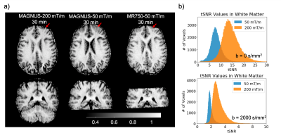 |
Q-Space Trajectory Imaging Using the MAGNUS High-Performance Head Gradient
Grant Kaijuin Yang1,2, Ek Tsoon Tan3, Eric Fiveland4, Thomas Foo4, and Jennifer McNab2
1Electrical Engineering, Stanford University, Stanford, CA, United States, 2Radiology, Stanford University, Stanford, CA, United States, 3Hospital for Special Surgery in Manhattan, New York, NY, United States, 4GE Global Research, Niskayuna, NY, United States
In this work, q-space trajectory imaging was implemented on a whole-body system equipped with a high-performance head-only gradient system capable of 200 mT/m maximum gradient amplitude and 500 T/m/s slew rate. The improved gradient performance enabled the acquisition of q-space trajectory imaging with sub-millimeter in-plane resolution and reduced voxel volume compared to previously published work, while simultaneously improving head coverage, and reducing susceptibility induced image distortions.
|
|
4324. |
Implementation of a Diffusion-Weighted Echo Planar Imaging sequence using the Open Source Hardware-Independent PyPulseq Tool
Rita G. Nunes1, Keerthi Sravan Ravi2, Sairam Geethanath2, and J. Thomas Vaughan Jr2
1Instituto Superior Técnico, Lisbon, Portugal, 2Columbia University Magnetic Resonance Research Center, New York, NY, New York, NY, United States
Diffusion-weighted imaging is an essential sequence for many clinical applications. While the post-processing tools for diffusion are widely available, vendor-neutral, open-source acquisition implementations have not been shared for research purposes. We develop a cross-vendor, open source package of a multi-slice single-shot spin echo-planar imaging based diffusion pulse sequence, capable of multiple b values and directions. We demonstrate this on an in vitro phantom, measuring plausible Apparent Diffusion Coefficient values and in vivo human brain data, obtaining good quality Fractional Anisotropy and Mean Diffusivity maps. We process our data with freely available post-processing tools to generate quantitative diffusion maps.
|
|
4325.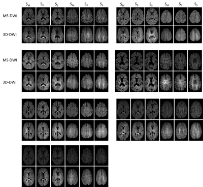 |
Feasibility and evaluation of whole brain single-slab 3D DWI and comparison to 2D multi-slice DWI
Neville D Gai1 and John A Butman1
1National Institutes of Health, Bethesda, MD, United States
While most brain imaging sequences now favor their 3D counterparts, diffusion imaging is an exception. This is due to large diffusion gradients resulting in increased sensitivity to motion exhibited by 3D acquisition. Prior schemes have used limited brain coverage and/or triggering or acquired multiple 3D slabs along with modified reconstruction schemes. The modified sequence used here employs first-order motion compensated diffusion gradients in addition to real-time alignment to acquire whole brain 3D-DWI images as a single slab. Relatively shorter TE (using enhanced gradients) and TR along with other modifications result in faster, reduced artifact diffusion images while providing higher SNR.
|
|
4326.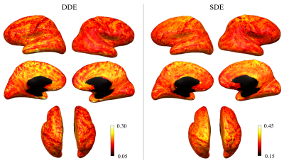 |
Investigating restricted diffusion within different cortical regions using double-diffusion encoding
Qiuyun Fan1, Thomas Witzel1, Slimane Tounekti1, Qiyuan Tian1, Chanon Ngamsombat1, Maya Polackal1, Aapo Nummenmaa1, and Susie Huang1
1Athinoula A. Martinos Center for Biomedical Imaging, Massachusetts General Hospital, Harvard Medical School, Boston, MA, United States
We report the acquisition of whole brain, 2-mm isotropic resolution DDE data in a healthy volunteer using an orientationally invariant sampling scheme and quantify the mean DDE signal intensity across cortical regions as a measure of diffusion restriction within different cortices. Higher mean signal intensities were observed in the cerebellum and limbic cortices, which are thought to reflect a higher degree of restriction in the tissue microstructural environment and may correspond to densely packed, small granule and pyramidal cells known to be present in these regions.
|
4327.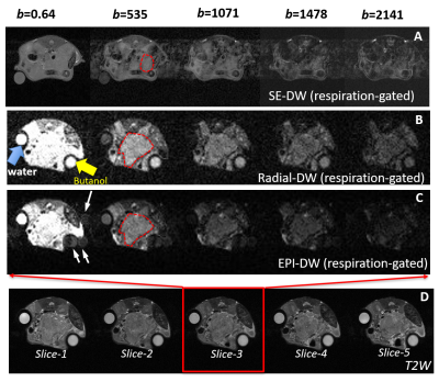 |
Radial diffusion-weighted MRI enables motion-robustness and reproducibility for orthotopic pancreatic cancer in mouse
Jianbo Cao1, Stephen Pickup1, Hanwen Yang1, Victor Castillo1, Cynthia Clendenin2,3, Peter O’Dwyer2,3, Mark Rosen1,3, and Rong Zhou1,3
1Department of Radiology, University of Pennsylvania, Philadelphia, PA, United States, 2Pancreatic Cancer Research Center, University of Pennsylvania, Philadelphia, PA, United States, 3Abramson Cancer Center, University of Pennsylvania, Philadelphia, PA, United States
Diffusion weighted (DW)-MRI is sensitive to tumor microenvironment (TME) thus useful for assessing pancreatic cancer responses to stroma-directed drugs, as they change TME by degradation or reduction of extracellular matrix. Motion-sensitive location of pancreatic tumor and fast respiration rate of mice impose a big challenge for quantitative DW-MRI. We compared radial k-space and echo-planar imaging based DW protocol for their accuracy and test-retest reproducibility. EPI-DW consistently underestimates water ADC value at 37C (reference to literature) where radial-DW does not. Better test-retest producibility measured by within-subject CV is obtained with radial-DW compared to EPI-DW.
|
|
4328.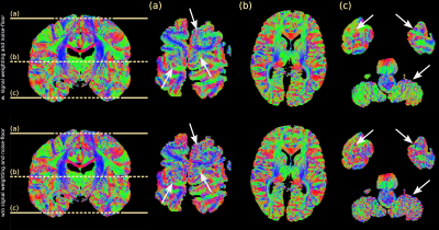 |
Correction for the influence of transmit-inhomogeneity in DW-SSFP on signal and ADC estimates in whole post-mortem brains at 7T
Benjamin C Tendler1, Sean Foxley2, Moises Hernandez-Fernandez3, Michiel Cottaar1, Olaf Ansorge4, Saad Jbabdi1, and Karla Miller1
1Wellcome Centre for Integrative Neuroimaging, Nuffield Department of Clinical Neurosciences, University of Oxford, Oxford, United Kingdom, 2Department of Radiology, University of Chicago, Chicago, IL, United States, 3Centre for Biomedical Image Computing and Analytics, University of Pennsylvania, Philadelphia, PA, United States, 4Nuffield Department of Clinical Neurosciences, University of Oxford, Oxford, United Kingdom
Diffusion-weighted steady-state free precession (DW-SSFP) generates high SNR diffusivity estimates in whole, post-mortem human brains. Improved estimates at 7T has motivated its use at ultra-high field. However, the DW-SSFP signal has a strong dependence on flip angle. This translates into both variable signal amplitude and diffusion contrast. At 7T, transmit-($$$B_{1}^{+}$$$) inhomogeneity leads to $$$B_{1}^{+}$$$-dependent SNR and ADC estimates. Previous work corrected for $$$B_{1}^{+}$$$-inhomogeneity by acquiring DW-SSFP datasets at two flip angles. Here, this approach is extended, utilising the full Buxton model of DW-SSFP to model non-Gaussian diffusion. A noise-floor correction and signal weighting are also incorporated to improve diffusivity estimates.
|
|
4329.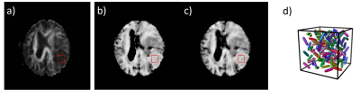 |
Acceleration of multidimensional diffusion MRI data acquisition and post-processing using convolutional neural networks
Yuan Zheng1, Tao Feng1, Sirui Li2, Wenbo Sun2, Qing Wei3, Samo Lasic4, Danielle van Westen5, Karin Bryskhe4, Daniel Topgaard4,5, and Haibo Xu2
1UIH America, Houston, TX, United States, 2Zhongnan Hospital of Wuhan University, Wuhan, China, 3United Imaging Healthcare, Shanghai, China, 4Random Walk Imaging, Lund, Sweden, 5Lund University, Lund, Sweden
Multidimensional diffusion MRI (dMRI) is a powerful tool that even in its simplest form provides more detailed microstructural information than conventional dMRI, such as microscopic anisotropy (µFA) unconfounded by orientation dispersion. However, it requires multiple diffusion encoding modes (usually directional and isotropic encodings) and, for the more advanced versions, prolonged scan and post-processing times. We proposed using convolutional neural networks (CNN) to accelerate multidimensional dMRI data acquisition and analysis, and have demonstrated that satisfactory µFA maps can be generated in real-time with only 50% of the encodings, which might help to better adapt multidimensional dMRI to clinical practices.
|
|
4330. |
Accelerating myelin-water imaging by extracting myelin content from anatomical and diffusion images through machine learning
Gerhard S Drenthen1,2, Walter H Backes1, and Jacobus FA Jansen1,2
1Department of Radiology and Nuclear Medicine, Maastricht University Medical Center, Maastricht, Netherlands, 2Department of Electrical Engineering, Eindhoven University of Technology, Eindhoven, Netherlands
In this study we aim to accelerate the acquisition time of myelin-water imaging by acquiring fewer slices and applying machine learning to extract myelin-specific information from anatomical (T1w and T2w) and diffusion-weighted imaging (DWI), which are commonly available in many clinical research studies. It is shown that with a 6-fold acceleration (from 7:30min to 1:15min) the myelin content can be reconstructed using neural networks with an agreement to the ground-truth that is comparable to the reproducibility of the scan itself.
|
|
4331.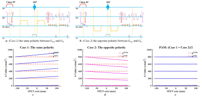 |
Single-Shot Diffusion-Weighted Spatiotemporal Encoding (SPEN) using Polarities Average Mode (PAM) to Correct Spatial-Dependent b-Values
Lisha Yuan1, Yi-Cheng Hsu2, Dan Wu1, Hongjian He1, and Jianhui Zhong1,3
1Center for Brain Imaging Science and Technology, Department of Biomedical Engineering, Key Laboratory for Biomedical Engineering of Ministry of Education, Zhejiang University, Hangzhou, China, 2MR Collaboration, Siemens Healthcare Ltd., Shanghai, China, 3Department of Imaging Sciences, University of Rochester, Rochester, NY, United States
Compared to traditional echo-planar imaging (EPI)-based schemes, spatiotemporal encoding (SPEN) is largely insensitive to magnetic field and chemical shift heterogeneities. However, excitation gradient has different effects for each position, thus the interaction between imaging and diffusion gradients introduces spatial-dependent diffusion weightings along the SPEN axis. A new method named polarities average mode (PAM) was proposed to obtain accurate apparent diffusion coefficient (ADC) map, with two acquisitions of different polarities between excitation and diffusion gradients. Simulation, phantom, and human experiments were designed to assess method performance. The proposed method enables SPEN to obtain ADC maps easily and accurately.
|
|
4332.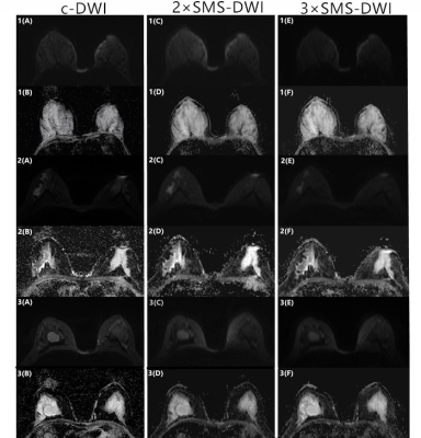 |
Feasibility Study of applying Simultaneous Multi-slice technique in Diffusion Weighted Imaging of Breast Lesions
Fei Wang1, Mengxiao Liu2, and Juan Zhu1
1Department of MRI,AnQing Municipal Hospital, Anqing, China, 2MR scientific Marketing, Diagnostic Imaging, Siemens Healthcare Ltd, Shanghai, China
To evaluate the feasibility of applying simultaneous multi-slice (SMS) single-shot echo planar imaging (EPI) to accelerate MR diffusion imaging for breast carcinoma, fibroadenoma of breast and normal breast. SMS sequences with double and triple acceleration were compared with conventional DWI sequences, respectively. These SMS DWI sequences were compared to conventional DWI in terms of image quality parameters (5-point Likert scale) and SNR, ADC measurements. Comparing with conventional EPI-DWI, the SMS markedly reduces the diffusion scan time and the image SNR still shows a good quality. Thus, SMS technique is recommended for DWI of the MR breast examinations.
|
|
4333. |
The Use of Stimulated-Echo EPI to Obtain High b-Value DTI Data at Short TEs on a Clinical Scanner
R. Allen Waggoner1, Thorsten Feiweier2, and Keiji Tanaka1
1Laboratory for Cognitive Brain Mapping, RIKEN - Center for Brain Science, Wako-shi, Japan, 2Siemens Healthcare GmbH, Erlangen, Germany
On clinical scanners, high b-value diffusion studies using SE-EPI suffer from the need for long TEs, which leads to signal loss due to T2 decay. Stimulated-Echo EPI permits high b-values together with short TEs on clinical scanners. We demonstrate that tractograms obtained from high b-value STE-EPI images are clean even in regions where tractograms from SE-EPI images with the same b-values break down.
|
|
4334. |
Evaluating Diffusion Kurtosis Imaging Precision at Varying Gradient Strength in High Spatial Resolution 3T MRI
Loxlan W Kasa1, Terry Peters2, Roy AM Haast3, and Ali R Khan4
1School of Biomedical Engineering, Imaging Research Laboratories, Robarts Research Institute, Western University, LONDON, ON, Canada, 2Imaging Research Laboratories, Robarts Research Institute, School of Biomedical Engineering,,Department of Medical Biophysics,Departments of Medical Imaging, Western University, London, ON, Canada, 3Imaging Research Laboratories, Robarts Research Institute, Western University, London, ON, Canada, 4Imaging Research Laboratories, Robarts Research Institute, School of Biomedical Engineering, Department of Medical Biophysics, Western University, London, ON, Canada
Diffusion kurtosis imaging (DKI), an extension to diffusion tensor imaging (DTI), aims to improve quantification of the hindered/restricted diffusion pattern due to microstructural complexity in the brain. But in order to capture the non-Gaussian diffusion behaviour of water molecules in biological tissues, stronger gradients larger than those employed in standard diffusion weighted imaging (DWI) are required. Here, we explored the test-retest reliability of DKI derived metrics with respect to different gradient strength in a high spatial resolution dataset. It was observed that DKI precision was comparable between b-value=1000, 2000, 3000 s/mm2 and b-value=1000 & 3000 s/mm2 dataset.
|
|
4335. |
Comparison of iShim, RESOLVE, and ss-EPI diffusion-weighted MR imaging with high b value at 3T MR in the evaluation of uterine malignancy
Qi Zhang1, Jieying Zhang1, Xiaoduo Yu1, Han Ouyang1, Xinming Zhao1, Hongmei Zhang1, Qinglei Shi2, Xiang Feng2, and Xiaoye Wang2
1Department of Imaging Diagnosis, National Cancer Center/Cancer Hospital, Chinese Academy of Medical Science and Peking Union Medical College, Beijing, China, 2MR Scientific Marketing, Diagnostic Imaging, Siemens Healthcare Ltd, Beijing, China
In the evaluation of uterine malignancy, conventional DWI based on single-shot echo-planar imaging (ss-EPI) is prone to imaging artifacts, including susceptibility artifacts from gas, imaging blurring, which limit its diagnostic value, especially in detecting and staging uterine malignancy. The purpose of this study is to compare the detection of uterine malignancy and image quality among DWI based on integrated slice-specific dynamic shimming (iShim), readout segmentation of long variable echo trains (RESOLVE) and ss-EPI sequence. Our results indicated that iShim DWI showed better image quality than ss-EPI and RESOLVE DWI in the terms of subjective image scores and objective quantitative metrics.
|
|
4336. |
Evaluation of simple acceleration strategy for advanced neural diffusion models based on half q-space under-sampling
Min-xiong Zhou1, Huiting Zhang2, Yang Song3, Guang Yang3, and Xu Yan2
1Shanghai University of Medicine & Health Sciences, Shanghai, China, 2MR Scientific Marketing, Siemens Healthcare, Shanghai, China, 3Shanghai Key Laboratory of Magnetic Resonance, East China Normal Univeristy, Shanghai, China
Advanced diffusion models such as NODDI, MAP-MRI are of high interests in brain research, but suffer from long acquisition time. Advanced under-sampling scheme were reported in previous studies for acceleration but are not commercially available. This study evaluates a simple and commercial available under-sampling scheme using the symmetric property of q-space, which could accelerate the acquisition by 2 fold. Results showed that it did not significant sacrifice the accuracy of quantitative maps. In addition, a symmetrically data copy step is needed to improve the estimation accuracy for both MAP-MRI and NODDI models.
|
|
4337.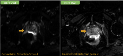 |
Readout-Segment Echo-Planar Imaging of Prostate, a Strategy to Reduce Geometrical Distortion in Prostate Diffusion Weighted Imaging
Melina Hosseiny1, KyungHyun Sung1, Teeravut Tubtawee1, Voraparee Suvannarerg1, Shabnam Mortazavi1, Soheil Kooraki1, Saurab Gupta1, Afshin Azadikhah1, Justin Ching1, Ely R Felker1, David Lu1, and Steven S Raman1
1Abdominal Radiology, David Geffen School of Medicine at UCLA, Los Angeles, CA, United States
Single-Shot Echo-Planar Imaging (ssEPI)is highly susceptible to T2* blurring and geometrical distortion. This study aimed to compare the image quality between ssEPI and Readout-Segment Echo-Planar Imaging (rsEPI) for acquiring prostate DWI on 162 patients. Geometrical distortion was ranked on ssEPI and rsEPI using a five-point scale. The geometrical distortion was significantly less observed in rsEPI compared to ssEPI (P<0.01). Geometrical distortion scores of three and higher were observed in 30 individuals in ssEPI, with all having scores < 3 on rsEPI. In conclusion, using rsEPI for DWI acquisition may augment or replace ssEPI on 3T prostate mpMRI.
|
|
4338.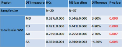 |
DTI in early RRMS patients with correlation to clinical parameters and comparison to Healthy Controls
Abdulaziz Alshehri1,2, Oun Al-iedani1,2, Jameen Arm1,2, Neda Gholizadeh1, Rodney Lea3, Jeannette Lechner-Scott3,4,5, and Saadallah Ramadan1,2
1School of Health Sciences, University of Newcastle, Newcastle, Australia, 2Imaging center, Hunter Medical Research Institute, Newcastle, Australia, 3Hunter Medical Research Institute, Newcastle, Australia, 4School of Medicine and Public Health, University of Newcastle, Newcastle, Australia, 5Department of Neurology, John Hunter Hospital, Newcastle, Australia
This study aims to evaluate and compare DTI parameters in relapsing-remitting MS patients with age and sex-matched healthy controls, and to correlate these DTI metrics with clinical symptoms and brain volumetric measures. As a result, There was a statistically significant increase in most of DTI parameters for RRMS patients compared with healthy controls. FA correlated positively with clinical parameters like EDSS and cognitive assessment. Both MD and RD correlated negatively with cognition parameters and positively with EDSS. Quantitative DTI parameters not only differentiate between RRMS patients and HCs, but are also associated with disability and mental health of RRMS.
|
|
4339.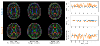 |
SENSE-based Multi-shot DWI Reconstruction with Extra-navigated Rigid Motion and Contrast Correction for Brain EPI
Malte Steinhoff1, Alfred Mertins1, and Peter Börnert2,3
1Institute for Signal Processing, University of Luebeck, Luebeck, Germany, 2Philips Research Europe, Hamburg, Germany, 3Department of Radiology, LUMC, Leiden, Netherlands
We propose an extra-navigated SENSE-based multi-shot DWI reconstruction algorithm that comprises navigator-based phase and rigid in-plane motion corrections at fast reconstruction times. Furthermore, this approach exploits the low-resolution navigator signal to perform diffusion contrast corrections explicitly within the model. The extra-navigated method is compared in-vivo to a self-navigated reference algorithm. The extra-navigated motion estimation from low-resolution navigator data yields decent reconstructions which perfectly coincide with self-navigated results. Moreover, extra-navigation allows for fast reconstruction at the cost of lower scan efficiency and appears to be more robust for strong motion corruption and high segmentations.
|
|
4340.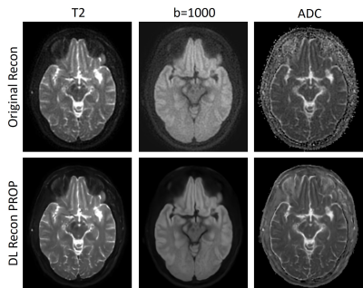 |
Diffusion Weighted Imaging using PROPELLER Acquisition and a Deep Learning based Reconstruction
Xinzeng Wang1, Daniel Litwiller2, Ali Ersoz3, Marc Lebel4, Sagar Mandava5, Lloyd Estkowski3, Arnaud Guidon6, Ann Shimakawa7, and Ersin Bayram1
1Global MR Applications & Workflow, GE Healthcare, Houston, TX, United States, 2Global MR Applications & Workflow, GE Healthcare, New York, NY, United States, 3Global MR Applications & Workflow, GE Healthcare, Waukesha, WI, United States, 4Global MR Applications & Workflow, GE Healthcare, Calgary, AB, Canada, 5Global MR Applications & Workflow, GE Healthcare, Tucson, AZ, United States, 6Global MR Applications & Workflow, GE Healthcare, Boston, MA, United States, 7Global MR Applications & Workflow, GE Healthcare, Menlo Park, CA, United States
PROPELLER DWI, a FSE based DWI method, is increasingly used to reduce susceptibility artifacts and motion artifacts. Multi-shot Echo-Planar diffusion method also can reduce susceptibility artifacts, but PROPELLER DWI shows better image quality where susceptibility artifacts are most problematic, such as in skull base, head-neck and pelvis. However, the acquisition time is often longer compared to ms-DW-EPI, therefore SNR is usually compromised to reduce acquisition time. In this work, we evaluated a deep-learning based reconstruction method (DL Recon PROP) intended to improve image quality and ADC measurements by reducing the noise and artifacts without increasing acquisition time.
|
|
4341.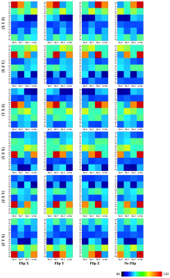 |
Model-Free, Fast, and Automated Correction of Diffusion Gradient Orientations
Ye Wu1, Yoonmi Hong1, Weili Lin1, Pew-Thian Yap1, and the UNC/UMN Baby Connectome Project Consortium1
1Department of Radiology and BRIC, University of North Carolina, Chapel Hill, Chapel Hill, NC, United States
We propose a rapid and automated method to rectify incorrect gradient orientations resulting from inconsistencies in coordinate frame conventions across scanners, file formats, and processing tools. Using these incorrect gradient orientations will invalidate subsequent derived quantities that are dependent on local orientation information, particularly tractography. Our approach to correcting the gradient orientations is based on maximizing an orientation continuity index that is computed directly from the diffusion-weighted images without the need for model fitting.
|
|
4342.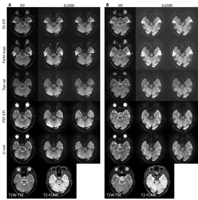 |
High-resolution distortion-free single-shot EPI enabled by deep-learning
Zhangxuan Hu1, Zhe Zhang2, Yishi Wang3, Yajing Zhang4, and Hua Guo1
1Center for Biomedical Imaging Research, Department of Biomedical Engineering, School of Medicine, Tsinghua University, Beijing, China, 2China National Research Center for Neurological Diseases, Beijing Tiantan Hospital, Capital Medical University, Beijing, China, 3Philips Healthcare, Beijing, China, 4MR Clinical Science, Philips Healthcare (Suzhou), Suzhou, China
Single-shot EPI (SS-EPI) is widely used for diffusion-weighted imaging (DWI), but suffers from susceptibility-induced distortion and T2* blurring, which limit its resolution and ability to detect detailed structures. Parallel imaging and multi-shot techniques can be used to improve the resolution and reduce image distortion. However, these techniques have their own drawbacks, such as limited achievable acceleration factors or prolonged acquisition time. In this study, a deep-learning based method is proposed to achieve high-resolution distortion-free DWI using SS-EPI thus to improve the acquisition efficiency and clinical applicability.
|
4343.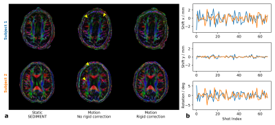 |
Segmented Diffusion Imaging with Iterative Motion Corrected Reconstruction for Self-navigated Brain Echo-planar Imaging at 7T
Malte Steinhoff1, Itamar Ronen2, Andrew Webb2, Alfred Mertins1, and Peter Börnert2,3
1Institute for Signal Processing, University of Luebeck, Luebeck, Germany, 2Department of Radiology, LUMC, Leiden, Netherlands, 3Philips Research Europe, Hamburg, Germany
Segmented diffusion imaging with iterative motion corrected reconstruction (SEDIMENT) is studied at 7T for self-navigated multi-shot DWI reconstruction including rigid in-plane motion correction. Motion-corrupted datasets contain intra-shot motion corrupted data with imperfect diffusion-sensitizing gradient reversal, which have to be identified and removed. The iterative SEDIMENT framework is evaluated in-vivo in conjunction with tailored data rejection strategies to detect corrupted shot datasets and generally improve convergence. The proposed algorithm provides high-quality multi-shot DWI and DTI reconstructions in the presence of gross motion allowing for efficient navigator-free DWI acquisition schemes.
|
|
4344.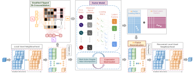 |
NoiseFactors: Blind Denoising of dMRI via Randomized Factor Models
Shreyas Fadnavis1, Hu Cheng2, and Eleftherios Garyfallidis1
1Intelligent Systems Engineering, Indiana University Bloomington, Bloomington, IN, United States, 2Psychological and Brain Sciences, Indiana University Bloomington, Bloomington, IN, United States
NoiseFactors is a probabilistic graphical model to suppress and remove additive noise in a single DWI image. It mitigates the issues caused by noise by preserving correlations in the signal components and suppressing the uncorrelated noise within local neighbourhoods. We solve the low-rank approximation problem by learning a best m-component approximation of a factor model. To do so we also introduce a novel flipped bi-crossvalidation to estimate the factor model. It outperforms the state-of-the-art PCA based methods such as Marchenko-Pastur PCA and Local PCA. The proposed method for denoising will be made available with an open-source implementation in DIPY.
|
|
4345.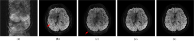 |
Phase-Constrained Reconstruction of High-Resolution Multi-shot Diffusion Weighted Image
Yiman Huang1, Xinlin Zhang1, Hua Guo2, Huijun Chen2, Di Guo3, and Xiaobo Qu1
1Department of Electronic Science, Xiamen University, Xiamen, China, 2School of Medicine, Tsinghua University, Beijing, China, 3School of Computer and Information Engineering, Xiamen University of Technology, Xiamen, China
Multi-shot DWI improves the image resolution, while it induces phase variation at the same time. We introduce a smooth phase constraint of each shot image into multi-shot DWI reconstruction procedures by imposing the low-rankness of Hankel matrix constructed from the k-space data. The image is further improved with a partial sum of singular values in low-rank matrix reconstruction. Results on brain imaging data show that the proposed method outperforms the state-of-the-art methods in terms of artifacts removal and is compatible to partial Fourier sampling in accelerated DWI.
|
|
4346. |
Joint estimation of phase and diffusion tensor parameters from multi-shot k-q-space data: a proof of concept
Banafshe Shafieizargar1, Ben Jeurissen1, Arnold Jan den Dekker1, and Jan Sijbers1
1imec-Vision Lab, Department of Physics, University of Antwerp, Antwerp, Belgium
To address the issue of phase induced artifacts in multi-shot diffusion weighted imaging, we propose a model-based framework which enables the joint estimation of diffusion and phase parameters directly from the multi-shot k-q-space. In a simulation study, we show that using this framework, diffusion parameters can be estimated more accurately and precisely than with the conventional method (image reconstruction followed by voxel-wise model fitting) that ignores phase differences.
|
|
4347. |
Distortion Correction for Isotropic High-Resolution Diffusion Imaging Using 3D Simultaneous Multi-Slab (SMSlab) Acquisition
Simin Liu1, Yuhui Xiong1,2, Erpeng Dai1,3, Jieying Zhang1, and Hua Guo1
1Center for Biomedical Imaging Research, Department of Biomedical Engineering, School of Medicine, Tsinghua University, Beijing, China, 2Neusoft Medical Systems Co., Ltd., Shanghai, China, 3Department of Radiology, Stanford University, Stanford, CA, United States
In EPI-based diffusion imaging, the geometric distortions become more severe with increased image resolution. Various post-processing methods have been proposed to correct for distortions, such as the top-up method and the field-mapping method. Nonetheless, for 3D isotropic high-resolution diffusion imaging, distortion correction becomes more challenging due to decreased SNR. In this study, we applied a modified distortion correction method, which was previously proposed for 2D imaging, to isotropic high-resolution diffusion imaging using 3D simultaneous multi-slab (SMSlab) acquisition. The modified distortion correction method performed well in both phantom and in vivo experiments. It also outperformed the conventional top-up and field-mapping methods.
|
|
4348. |
Rapid, structure-preserving denoising of DTI data via tight framelet thresholding.
Gregory R. Lee1,2
1Radiology, Cincinnati Children's Hospital Medical Center, Cincinnati, OH, United States, 2Radiology, University of Cincinnati College of Medicine, Cincinnati, OH, United States
In this work it is demonstrated that computationally efficient deniosing can be done using thresholding of wavelet coefficients without sacrificing image quality relative to state-of-the-art patch-based methods. This is achieved by combining use of a redundant, directional tight wavelet frame with the Karhunen-Loeve transform along the "directions" dimension. An efficient GPU implementation of the algorithm required less than 30 seconds to process even relatively large DTI datasets (e.g. 96x96x60x203). The proposed approach should find use in SNR-challenged acquisitions such high resolution DTI, DKI and DSI.
|
|
4349.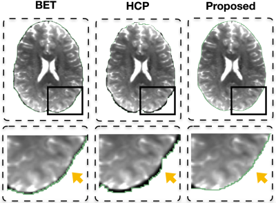 |
Automated Identification of Non-Brain Voxels for Clean Brain Extraction Using Diffusion MRI
Ye Wu1, Yoonmi Hong1, Weili Lin1, Pew-Thian Yap1, and the UNC/UMN Baby Connectome Project Consortium1
1Department of Radiology and BRIC, University of North Carolina, Chapel Hill, Chapel Hill, NC, United States
Automated brain extraction using diffusion MRI is challenging owing to the low spatial resolution. Unremoved residual non-brain voxels characteristically manifest as voxels with high fractional anisotropy (FA). In this abstract, we introduce a fast and robust method to identify non-brain voxels for clean extraction of the brain using diffusion MRI. We show that our method is effective for both adult and infant data.
|
|
4350.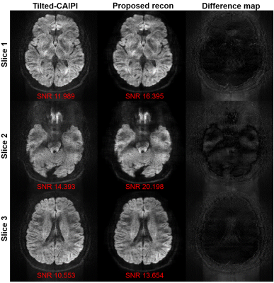 |
A low-rank based reconstruction method for diffusion-weighted point-spread-function encoded EPI (PSF-EPI)
Xinyu Ye1, Guangqi Li1, Yuan Lian1, Yishi Wang2, and Hua Guo1
1Center for Biomedical Imaging Research, Department of Biomedical Engineering, School of Medicine, Tsinghua University, Beijing, China, 2Philips Healthcare, Beijing, China
Recently, a distortion- and blurring-free acquisition method called PSF-EPI has been used in DWI. However, when field inhomogeneity is severe, DW images may become noisy and more shots are needed to get the reliable results. In this work, we introduce a low-rank based reconstruction method using signal correlation along the ky-encoding dimension in PSF-EPI to improve image quality and reduce needed shot number to shorten scan time. High-resolution in-vivo data were used to test the performance of the proposed method. The results show that the quality of the images is improved.
|
|
4351.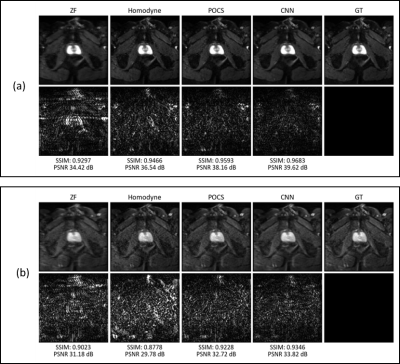 |
Deep learning-based partial Fourier reconstruction for improved prostate DWI
Fasil Gadjimuradov1,2, Seung Su Yoon1,2, Thomas Benkert2, Marcel Dominik Nickel2, Karl Engelhard3, and Andreas Maier1
1Pattern Recognition Lab, Friedrich-Alexander-Universität Erlangen-Nürnberg, Erlangen, Germany, 2Magnetic Resonance, Siemens Healthcare GmbH, Erlangen, Germany, 3Department of Radiology, Martha-Maria Hospital, Nürnberg, Germany
Partial Fourier (PF) acquisition schemes are a common way to increase the inherently low signal-to-noise ratio in diffusion-weighted (DW) images. The naïve solution of zero-filling k-space results in visible blurring and Gibbs ringing. Based on the circumstance that traditional methods such as homodyne reconstruction or POCS often fail to remove blurring and ringing without introducing new artifacts, this work aims to use a Convolutional Neural Network for robust PF reconstruction in prostate DWI. We show that our data-driven approach, which efficiently uses correlations across different b-values, outperforms traditional methods in terms of quantitative measures and visual impression of the images.
|
|
4352. |
Acceleration of diffusion ADC mapping with phase-corrected SUPER
Xin Tang1, Jun Xie2, Guobin Li2, Meng Jiang3, and Chenxi Hu4
1Department of Medical Information Engineering, Wuyuzhang honors college, Sichuan University, Chengdu, China, 2United Imaging Healthcare Co., Ltd, Shanghai, China, 3Department of Cardiology, Renji Hospital, School of Medicine, Shanghai Jiao Tong University, Shanghai, China, 4Institute of Medical Imaging Technology, School of Biomedical Engineering, Shanghai Jiao Tong University, Shanghai, China
A novel method was proposed to accelerate apparent-diffusion-coefficient(ADC) mapping to shorten the EPI echo-train and/or improve resolution. The method was based on SUPER--a Cartesian k-space undersampling strategy for parametric mapping acceleration—and adapted to account for the nonlinear phase variation in diffusion-weighted imaging at different b-values. In healthy subjects, the phase-corrected SUPER(R=2) and SUPER-SENSE(combining SUPER and parallel imaging, R=4) demonstrated similar image quality, reasonable noise amplification, and similar reconstruction time compared with the non-acceleration gold standard, despite a 2-fold and 4-fold reduction of reconstruction data. This suggests that SUPER is a practical and accurate approach for accelerating ADC mapping.
|
|
4353.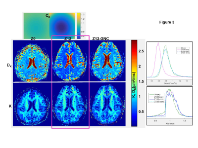 |
Practical correction of gradient nonlinearity bias for mean diffusion kurtosis model parameters
Dariya Malyarenko1 and Thomas L Chenevert1
1University of Michigan, Ann Arbor, MI, United States
Quantitative tissue diffusion parameters derived from diffusion weighted imaging (DWI) models hold promise for diagnostic and prognostic clinical oncology applications. System-dependent spatial DW bias due to gradient nonlinearity (GNL) is known confounding factor for quantitative DWI metrics. Improved accuracy and multiplatform reproducibility was previously demonstrated for mono-exponential apparent diffusion coefficient with correction for platform-dependent GNL bias (GNC). Complex tumor microenvironment often exhibits multi-exponential diffusion described by isotropic kurtosis model. This study proposes analytical extension and demonstrates empirical confirmation for GNC of parametric maps derived from diffusion kurtosis model.
|
|
4354. |
Interleaved Block-Segmented Echo-Planar Imaging (iblocks-EPI) based Diffusion-Tensor Imaging with Retrospective Motion Correction
Liyuan Liang1, Mei-Lan Chu2, Nan-Kuei Chen3,4, Shihui Chen1, and Hing-Chiu Chang1
1Department of Diagnostic Radiology, The University of Hong Kong, Hong Kong, China, 2Graduate Institute of Biomedical Electronics and Bioinformatics, National Taiwan University, Taipei, Taiwan, 3Department of Biomedical Engineering, University of Arizona, Tucson, AZ, United States, 4Brain Imaging and Analysis Center, Duke University Medical Center, Durham, NC, United States
Recently, a self-navigated interleaved block-segmented EPI (iblocks-EPI) has been proposed to acquire DTI data with high spatial resolution and less geometric diction. In addition, the oversampling of central k-space of iblock-EPI can benefit the SNR performance. However, same as the other multi-shot EPI techniques, iblocks-EPI is highly susceptible to minuscule and macroscopic motions during data acquisition. In this study, we developed a self-calibrated and collaborative iblocks-DTI reconstruction framework that can correct image artifacts and diffusion-encoding contrast change caused by minuscule and macroscopic motions.
|
|
4355. |
Use of 2D image registration parameters for correction of eddy-current magnetization-density ADC errors in presence of gradient nonlinearity
Thomas L. Chenevert1 and Dariya I. Malyarenko1
1University of Michigan Hospitals, Ann Arbor, MI, United States Systematic errors confound wide-spread clinical use of apparent diffusion coefficient (ADC) for diagnostic and prognostic applications. Standard clinical diffusion sequences using single-spin-echo echo-planner-imaging are susceptible to gradient channel-specific eddy-currents for b>0 inducing distortions of voxel magnetization-density (MD), as well as geometric distortion. Unlike geometric distortion that are largely correctable by image registration to b=0, persisting signal amplitude distortions lead to systematic spatially-dependent errors mimicking, but physically distinct from, non-uniform diffusion weighting induced by gradient nonlinearity (GNL). This study proposes the use of geometric distortion parameters derived from in-plane image registration for MD correction of ADC in presence of GNL. |
|
4356. |
Accelerating Navigator-free Multi-shot Spiral DTI via Joint Calibrationless Reconstruction with Low-Rank Tensor Completion
Xiaodong Ma1,2, Yilong Liu1,2, Zheyuan Yi1,2,3, Alex T. Leong1,2, Hua Guo4, and Ed X. Wu1,2
1Laboratory of Biomedical Imaging and Signal Processing, The University of Hong Kong, Hong Kong, China, 2Electrical and Electronic Engineering, The University of Hong Kong, Hong Kong, China, 3Electrical and Electronic Engineering, Southern University of Science and Technology, Shenzhen, China, 4Center for Biomedical Imaging Research, Department of Biomedical Engineering, School of Medicine, Tsinghua University, Beijing, China
We propose a novel joint calibrationless reconstruction for accelerating multi-shot navigator-free DTI, using a low-rank completion approach. The redundant information across different directions is utilized to facilitate the reconstruction, including sharable coil sensitivities and anatomical structures. A 3D Hankel tensor was constructed and its concatenated Hankel matrices were used for low-rank approximation. In vivo human brain DTI experiment shows that the proposed joint reconstruction can reduce artifacts in diffusion-weighted images, and yield more accurate DTI metrics, when compared with separate reconstruction for different directions. This method also presents a new potential reconstruction strategy for fast high-resolution DTI.
|
|
4357.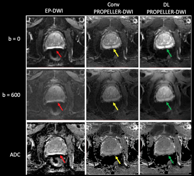 |
PROPELLER Diffusion-Weighted Imaging of the Prostate with Deep-Learning Reconstruction
Xinzeng Wang1, Ersin Bayram1, Daniel Litwiller2, Tetsuya Wakayama3, Alan B McMillan4, Lloyd Estowski5, Ty A Cashen5, and Ali Pirasteh4
1Global MR Applications & Workflow, GE Healthcare, Houston, TX, United States, 2Global MR Applications & Workflow, GE Healthcare, New York, NY, United States, 3Global MR Applications & Workflow, GE Healthcare, Hino, Japan, 4Radiology, University of Wisconsin Madison, Madison, WI, United States, 5Global MR Applications & Workflow, GE Healthcare, Waukesha, WI, United States
While echo-planar diffusion-weighted imaging (EP-DWI) is the main sequence for cancer detection in the prostate peripheral zone, it is susceptible to signal loss and distortion due to B0-field inhomogeneities secondary to a variety of causes, including rectal gas or metal hardware in the pelvis. We were able to demonstrate that a spin-echo based DWI sequence with radial k-space sampling (PROPELLER) can overcome such artifacts and the addition of a deep-learning reconstruction algorithm can overcome the poor signal-to-noise (SNR) profile of the PROPELLER-DWI, overall generating images with minimal-to-no appreciable artifact and favorable SNR.
|
|
4358.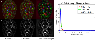 |
Towards individual direction-based deep learning of diffusion weighted images for standard diffusion model analysis.
Peidong He1, Zifei Liang1, Marco Muccio1, Florian Knoll1, Jiangyang Zhang1, and Yulin Ge1
1Department of Radiology, New York University School of Medicine, New York, NY, United States
In diffusion MRI, a higher number of gradient directions benefit to SNR and robustness of fiber rotational invariant estimation for tensor computation, however, it makes the acquisition time to lengthy to be clinically viable. This study was to apply a deep learning approach to generate new individual direction diffusion weighted (DW) source images (e.g., 60 more directions) from original 30 direction DW images based on high-angular-resolution (90 directions) dataset. Such an approach not only significantly reduce the scan time using one-third original DW images, but also be able to compute dMRI-derived parametric maps using a standard tensor model.
|
4359.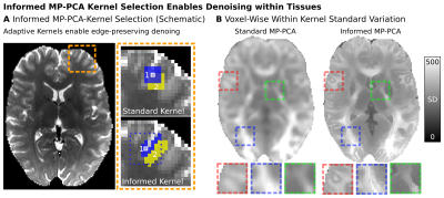 |
Mitigating Impacts of Tissue-Heterogeneity and Noise Bias on MP-PCA Denoising for High-Quality Diffusion MRI
Cornelius Eichner1, Michael Paquette1, Angela D Friederici1, and Alfred Anwander1
1Department of Neuropsychology, Max Planck Institute for Human Cognitive and Brain Sciences, Leipzig, Germany
Advanced diffusion MRI (dMRI) data with high resolution and strong diffusion contrast typically suffer from low SNR levels. Therefore, denoising algorithms such as MP-PCA became an essential part of current dMRI processing pipelines. To overcome challenges related to violations of MP-PCAs assumption of tissue homogeneity in typical dMRI data, we here introduce an informed-MP-PCA (iMP-PCA) algorithm taking local differences in tissue composition into account. Denoising-performance of iMP-PCA was compared to conventional MP-PCA and evaluated on both magnitude and real-valued dMRI data. iMP-PCA was shown to significantly improve denoising-performance, especially at tissue boundaries and in regions of low SNR.
|
|
4360. |
Denoise magnitude diffusion magnetic resonance images via variance-stabilizing transformation and optimal singular-value manipulation
Xiaoping Wu1 and Kamil Ugurbil1
1Center for Magnetic Resonance Research, Radiology, Medical School, University of Minnesota, Minneapolis, MN, United States
We introduce a new denoising framework for denoising magnitude diffusion MRI. The framework synergistically combines the variance stabilizing transform with optimal singular-value manipulation. The usefulness of the proposed framework is demonstrated using both simulation and real-data experiments. Our results show that the proposed denoising framework can significantly improve signal-to-noise ratios across the entire brain, leading to substantially enhanced performances for estimating diffusion-tensor-related indices and for resolving crossing fibers when compared to another competing method. As such, the proposed denoising method is expected to have great utility for high-quality, high-resolution whole-brain diffusion MRI, desirable for many neuroscience and clinical applications.
|
|
4361.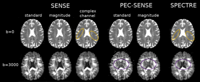 |
SENSE reconstruction with simultaneous 2D phase correction and channel-wise noise removal (SPECTRE)
Elizabeth Powell1,2, Torben Schneider3, Marco Battiston2, Francesco Grussu2,4, Ahmed Toosy2, Jonathan D Clayden5, and Claudia A. M. Gandini Wheeler-Kingshott2,6,7
1Medical Physics and Biomedical Engineering, University College London, London, United Kingdom, 2NMR Research Unit, Queen Square MS Centre, Department of Neuroinflammation, UCL Queen Square Institute of Neurology, Faculty of Brain Sciences, University College London, London, United Kingdom, 3Philips Healthcare, Guildford, United Kingdom, 4Centre for Medical Image Computing, Department of Computer Science, University College London, London, United Kingdom, 5Developmental Imaging and Biophysics Section, Great Ormond Street Institute of Child Health, University College London, London, United Kingdom, 6Department of Brain and Behavioural Sciences, University of Pavia, Pavia, Italy, 7Brain MRI 3T Center, IRCCS Mondino Foundation, Pavia, Italy
Nyquist sampling errors in echo planar imaging (EPI) often require 2D phase correction during reconstruction to remove unwanted ghost artefacts; however, phase corrections can be challenging to translate to high b-value diffusion weighted imaging (DWI) owing to associated noise amplification. We introduce SPECTRE (SENSE with 2D PhasE CorrecTion and channel-wise noise REmoval), and demonstrate that the SNR gains achieved by denoising complex channel data enable robust ghost correction without biasing diffusion parameter estimates.
|
|
4362.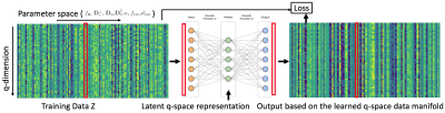 |
Model-Based Deep Learning for Reconstruction of Joint k-q Under-Sampled Diffusion MRI
Merry P. Mani1, Hemant Kumar Aggarwal2, Sanjay Ghosh1, and Mathews Jacob2
1Department of Radiology, University of Iowa, Iowa City, IA, United States, 2Department of Electrical and Computer Engineering, University of Iowa, Iowa City, IA, United States
We propose a model-based deep learning architecture for the reconstruction of highly accelerated diffusion MRI. We introduce the use of a pre-trained denoiser as the regularizer in a model-based recovery for diffusion weighted data from k-q under-sampled acquisition in a parallel MRI setting. The denoiser is designed based on a general tissue microstructure diffusion signal model with multi-compartmental modeling. A neural network was trained in an unsupervised manner using a convolutional auto-encoder to learn the diffusion MRI signal subspace. To demonstrate the acceleration capabilities of the proposed method, we perform MRI reconstruction experiments on a simulated brain dataset.
|
|
4363.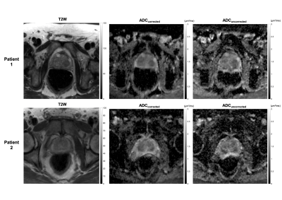 |
Towards the clinical application of model‐based reconstruction framework for distortion correction in prostate diffusion weighted MRI.
Lebina Shrestha Kakkar1, Muhammad Usman1, Alex Kirkham2, Simon Arridge1, and David Atkinson1
1Centre for Medical Imaging, University College London, London, United Kingdom, 2Department of Radiology, University College Hospital, London, United Kingdom
Prostate diffusion-weighted (DW) MRI based on single-shot echo planar imaging (EPI) can suffer from geometric distortions caused by off-resonance magnetic field at the prostate-rectal air interface. Although techniques exist for effective distortion correction in brain imaging, such approaches may struggle with the severe distortions observed in prostate imaging. Here we focus on applying a recently developed model-based reconstruction technique to correct distortions in typical clinically used prostate DW-MRI. The distortion correction is feasible and may improve prostatic zonal anatomy in DW-MRI. Additionally, corrected averaged high b-value datasets show higher SNR than uncorrected dataset and may further help to identify tumours.
|
|
4364. |
Highly Accelerated and High-Quality Intra-voxel Incoherent Motion DWI Enabled by Parametric POCS based multiplexed sensitivity-encoding
Shihui Chen1, Mei-Lan Chu2, Chun-Jung Juan3, Liyuan Liang1, and Hing-Chiu Chang1
1The University of Hong Kong, Hong Kong, Hong Kong, 2Graduate Institute of Biomedical Electronics and Bioinformatics, National Taiwan University, Taipei, Taiwan, Taipei, Taiwan, 3Department of Medical Imaging, China Medical University Hsinchu Hospital, Taiwan, Taipei, Taiwan
Low SNR and long acquisition time hinder the clinical feasibility of intra-voxel incoherent motion (IVIM) diffusion-weighted imaging. In this work, we used a joint reconstruction framework based on POCSMUSE algorithm to simultaneously reconstruct under-sampled multi-b multi-shot DW-EPI data without undesired noise amplification. A proposed parametric correction scheme (PCS) was incorporated into POCSMUSE algorithm for robust reconstruction based on either conventional bi-exponential or simplified IVIM model. The proposed method demonstrated that the high-quality brain IVIM images could be reconstructed from highly accelerated (R=4) data acquired with multi-b multi-shot DW-EPI.
|
|
4365. |
Fast Simultaneous Multi-Slice Multi-Shell Diffusion Tensor Imaging with Model-based Reconstruction
Oliver Maier1, Stefan M Spann1, Lea Bogensperger1,2, and Rudolf Stollberger1,3
1Institute of Medical Engineering, Technical University Graz, Graz, Austria, 2Institute for Computer Graphics and Vision, Technical University Graz, Graz, Austria, 3Biotechmed, Graz, Austria
Multi-Shell DTI suffers from low SNR for high b-value data and prolonged scan time. The Gaussian noise assumption is typically violated due to multi-coil imaging and magnitude forming thus requiring special treatment to avoid biases in the DTI estimates. To this end, we propose a model-based reconstruction technique to exploit the Gaussian noise in the raw k-space data and enable acceleration of the DTI measurement. We show the acceleration potential and quantitative accuracy of the proposed method for mono- and bi-exponential fitting approaches on freely available DTI data and full brain DTI measurements of one healthy volunteer.
|
|
4366.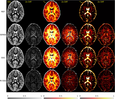 |
Regularized Image Domain Split Slice-GRAPPA for Simultaneous Multi-Slice Diffusion MR Imaging
SeyyedKazem HashemizadehKolowri1, Rong-Rong Chen1, Ganesh Adluru2,3, and Edward V. R. DiBella1,2,3
1Electrical and Computer Engineering, University of Utah, SALT LAKE CITY, UT, United States, 2Radiology and Imaging Science, University of Utah, SALT LAKE CITY, UT, United States, 3Biomedical Engineering, University of Utah, SALT LAKE CITY, UT, United States
Simultaneous multi-slice (SMS) acquisition combined with blipped controlled aliasing in parallel imaging is commonly used to accelerate diffusion imaging with single-shot EPI sequences. In this work, we propose a new method, termed regularized image domain split slice-GRAPPA (RI-SSG), which allows an efficient image domain implementation of SSG coupled with total variation regularization to improve the quality of SMS reconstruction. We process two single-shot EPI datasets acquired using diffusion protocol of Human Connectome Project in Aging to evaluate performance of SMS reconstructions. The RI-SSG yields less noisy results than SENSE and SSG in estimating diffusion-weighted images and parametric maps of diffusion.
|
|
4367. |
Magnitude-regularized Phase Estimation (MAPE) with U-Net Support for Self-navigated Multi-shot Echo-planar DWI in the Brain
Malte Steinhoff1, Alfred Mertins1, and Peter Börnert2,3
1Institute for Signal Processing, University of Luebeck, Luebeck, Germany, 2Philips Research Europe, Hamburg, Germany, 3Department of Radiology, LUMC, Leiden, Netherlands
We propose a self-navigated iterative reconstruction algorithm for multi-shot DWI which effectively performs the shot phase updates with a fixed joint image prior. This framework further nicely incorporates deep learning generated image priors into the shot phase estimation while keeping the joint image production isolated. A U-Net is trained on extra-navigated data to mitigate phase cancellation artifacts. The algorithm with and without U-Net support is compared to self- and extra-navigated reference algorithms. The U-Net approach effectively mitigates phase-related signal cancellation artifacts. The improved multi-shot image prior regularizes the shot phase estimation enabling highly segmented self-navigated diffusion echo-planar imaging.
|
|
4368.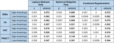 |
An Evaluation of q-Space Regularization Strategies for gSlider with Interlaced Subsampling
Yunsong Liu1, Congyu Liao2, Kawin Setsompop2, and Justin P. Haldar1
1Signal and Image Processing Institute, University of Southern California, Los Angeles, CA, United States, 2Martinos Center for Biomedical Imaging, Charlestown, MA, United States
gSlider is a diffusion MRI method that achieves fast high-resolution data acquisition using a novel slab-selective RF-encoding strategy. Recent work has proposed subsampling of the multidimensional gSlider encoding space (diffusion-encoding/RF-encoding) for further improved scan efficiency. Two different q-space regularization approaches (i.e., Laplace-Beltrami smoothness and spherical ridgelet sparsity) have been proposed to compensate for missing data, but there have been no systematic comparisons between the two. We compare and evaluate the potential synergies of these regularization approaches. Results suggest that there can be small advantages to combining both regularization strategies together, although Laplace-Beltrami regularization alone is simpler and not much worse.
|
|
4369.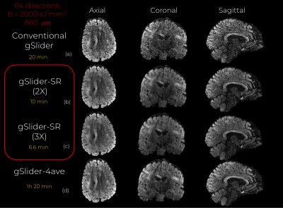 |
Whole-brain in-vivo submillimeter diffusion MRI in 10 minutes with combined gSlider-Spherical Ridgelets reconstruction
Gabriel Ramos-Llordén1, Lipeng Ning1, Congyu Liao2, Rinat Mukhometzianov3, Oleg Michailovich3, Kawin Setsompop2, and Yogesh Rathi1
1Brigham and Women's Hospital, Harvard Medical School, Boston, MA, United States, 2Massachusetts General Hospital, Harvard Medical School, Charlestown, MA, United States, 3University of Waterloo, Waterloo, ON, Canada
gSlider is an efficient, super-resolution, technique to achieve submillimeter diffusion MRI data circumventing the trade-off between image resolution and SNR. Yet, the long acquisition time is still an issue. In this work, we extend gSlider by allowing under-sampling both in q-space and Radio-Frequency (RF)-encoded data, achieving then shorter acquisition time that gSlider. Our method, gSlider-SR, uses a basis of Spherical-Ridgelets to exploit the redundancy of the dMRI data, while at the same time enhancing SNR. We demonstrate that only ten minutes are needed to reconstruct 64 diffusion directions (b=2000s/mm2) at 860 μm data with reliably signal preservation.
|
|
4370.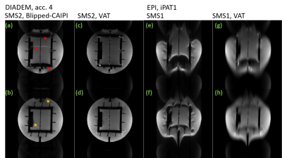 |
Simultaneously multi-slice VAT-DIADEM at ultra-high field
Yi-Hang Tung1, Frank Godenschweger1, Myung-Ho In2, Alessandro Sciarra3, and Oliver Speck1
1Biomedical Magnetic Resonance, Otto-von-Guericke University Magdeburg, Magdeburg, Germany, 2Department of Radiology, Mayo Clinic, Rochester, MN, United States, 3Medicine and Digitalization, Otto-von-Guericke University Magdeburg, Magdeburg, Germany VAT-DIADEM is able to accelerate the distortion-free T2 weighting and diffusion weighting imaging sequence DIADEM at ultra-high field. The VAT gradient is turned on during imaging read-out in the slice selection direction. To further increase the acquisition efficiency, we developed a blipped version of the VAT gradient and applied simultaneous multi-slice imaging, based on blipped-CAIPI. The result is distortion reduction in EPI or acceleration in DIADEM and the increased volume coverage without increasing TR. |
|
4371.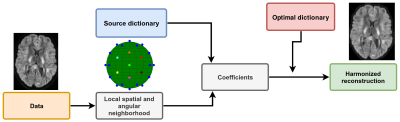 |
Automated feature extraction across scanners for harmonization of diffusion MRI datasets
Samuel St-Jean1, Max A. Viergever1, and Alexander Leemans1
1Image sciences institute, University Medical Center Utrecht, Utrecht, Netherlands
Small variations in diffusion MRI metrics between subjects are ubiquitous due to differences in scanner hardware and are entangled in the genuine biological variability between subjects, including abnormality due to disease. In this work, we propose a new harmonization algorithm based on adaptive dictionary learning to mitigate the unwanted variability caused by different scanner hardware while preserving the biological variability of the data. Results show that unpaired datasets from multiple scanners can be mapped to a scanner agnostic space while preserving genuine anatomical variability, reducing scanner effects and preserving simulated edema added to test datasets only.
|
|
4372.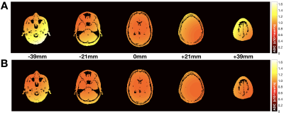 |
Concomitant Gradient Field Corrections for Asymmetric Diffusion Encoding Waveforms
Matthew J. Middione1, Michael Loecher1, Kévin Moulin1,2, and Daniel B. Ennis1,2,3
1Department of Radiology, Stanford University, Palo Alto, CA, United States, 2Cardiovascular Institute, Stanford University, Palo Alto, CA, United States, 3Veterans Administration Health Care System, Division of Radiology, Palo Alto, CA, United States
In DWI, applied diffusion gradients are assumed to be linear in space, but in practice are accompanied by additional undesired concomitant gradient (CG) fields that arise as a consequence of Maxwell’s equations. These CG fields contribute a residual gradient moment for asymmetric diffusion encoding strategies, which may impact the quantitative accuracy of the measured ADC. In this work, a CG correction method that does not increase the minimum achievable TE was implemented for clinically relevant asymmetric diffusion encoding DWI protocols and the mean ADC error reduction was characterized.
|
|
4373.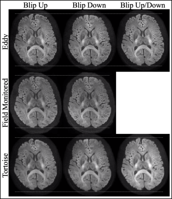 |
A Comparison of Image-based and Field-monitoring-based Correction for Eddy Current and Bo Field Distortions in Diffusion Imaging Data
Yoojin Lee1, Klaas P Pruessmann2, and Zoltan Nagy1
1Laboratory for Social and Neural Systems Research (SNS Lab), University of Zurich, Zurich, Switzerland, 2Institute of Biomedical Engineering, ETH, Zurich, Switzerland Diffusion MRI commonly proceeds with single-shot echo planar imaging (EPI) readout, which renders this modality sensitive to Eddy current and susceptibility-induced distortions. Both image-based and k-space-based correction methods have been suggested previously. The aim of this project was to use a standard dataset and compare output images of two image-based correction methods (topup/Eddy and Tortoise) and the extended signal model method. The latter relies on separate magnetic field monitoring data and a Bo field map. Qualitatively the output of three pipelines were similar. Future work will make the diffusion acquisition more challenging for a more complete systematic evaluation. |
4374. |
Introduction of matrix analysis for hybrid multidimensional prostate diffusion-weighted and T2-weighted imaging
Xiaobing Fan1, Aritrick Chatterjee1, Milica Medved1, Aytekin Oto1, and Gregory S. Karczmar1
1Radiology, The University of Chicago, Chicago, IL, United States
We present a new concept based on matrix analysis for analyzing hybrid multi-dimensional T2-weighted and diffusion-weighted MRI of the prostate. This study evaluates whether matrix analysis is useful in diagnosis of prostate cancer. The hybrid data were linearized first by taking natural logarithms. Then the hybrid symmetric matrix was formed by multiplying by its own transpose matrix for each pixel. The eigenvalues and eigenvectors are calculated for this symmetric matrix to generate color maps. The preliminary results suggest that the combined color eigenvalue map provides new information that could help to identify and stage prostate cancer.
|
|
4375. |
Automatic Segmentation in Ischemic Stroke Rodent Brain with Pattern-recognition of Directional Intravoxel Incoherent Motion MRI
MinJung Jang1, Seokha Jin1, MungSoo Kang1, SoHyun Han2, and HyungJoon Cho1
1Biomedical Engineering, Ulsan national institute of science and technology, Ulsan, Republic of Korea, 2Center for Neuroscience Imaging Research, Institute of Basic Science, Suwon, Republic of Korea
The purpose of this study is to demonstrate automatic segmentation for reduced diffusion and perfusion areas in ischemic stroke brains with pattern-recognition (constrained non-negative matrix factorization (cNMF)) to directional intravoxel incoherent motion MRI (IVIM-MRI). The robustness of region segmentation with pattern-recognition was observed in both simulations and in vivo experiments. Using pattern-recognition analysis of IVIM signal, white matter in which flow may have been aligned, and lesion regions are automatically segmented in ischemic stroke brain. In this study, we successfully implemented pattern-recognition to directional IVIM signals.
|
|
4376. |
Free-Water Diffusion Tensor Imaging (DTI) improves the accuracy and sensitivity of white matter analysis in Alzheimer’s disease
Maurizio Bergamino1, Ryan R Walsh2, and Ashley M Stokes1
1Division of Neuroimaging Research, Barrow Neurological Institute, Phoenix, AZ, United States, 2The Muhammad Ali Parkinson Center, Barrow Neurological Institute, Phoenix, AZ, United States
The objective of this study is to investigate differences in white matter (WM) integrity between Alzheimer’s disease (AD) and healthy subjects (HC) using diffusion tensor imaging (DTI) metrics from standard DTI and free-water (FW)-DTI. Regional changes in DTI metrics were found with both standard and FW-DTI, while FW-DTI improves the reliability and inter-parameter consistency of DTI metrics in the presence of atrophy. We hypothesize that the implementation of FW correction algorithm for DTI may provide more sensitive and specific insight into AD-related pathological changes in WM.
|
|
4377.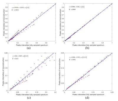 |
Joint Sparsity and Low Rankness-based Spectroscopy Reconstruction for Magnetic Resonance Diffusion-Ordered NMR
Di Guo1, Zhangren Tu1,2, Zifei Zhang3, Tianyu Qiu3, Xiaofeng Du1, Min Xiao2, Zhong Chen3, and Xiaobo Qu3
1School of Computer and Information Engineering, Xiamen University of Technology, Xiamen, China, 2School of Opto-Electronic and Communication Engineering, Xiamen University of Technology, Xiamen, China, 3Department of Electronic Science, Xiamen University, Xiamen, China
The magnetic resonance diffusion ordering spectrum has been widely used in the separation of mixture due to the mobility of molecular self-diffusion reaction molecules. We propose a hybrid time and exponential decay signal recovery method based on low rank Hankel matrix1 to accelerate the acquisition. The experimental results show that this method enables to reduce the error between the recovery diffusion spectrum and the fully sampled signal, and enhance the peak intensity.
|
|
4378.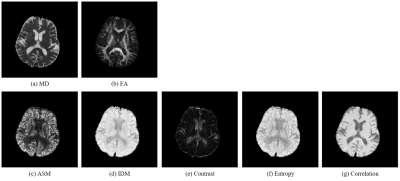 |
Characterizing intravoxel spatial distribution of diffusion based on texture analysis
Suguru Yokosawa1,2, Toru Shirai1, Hisaaki Ochi1, and Yoshitaka Bito3
1Research & Development Group, Hitachi, Ltd., Tokyo, Japan, 2Graduate School of Engineering, Chiba University, Chiba, Japan, 3Healthcare Business Unit, Hitachi, Ltd., Tokyo, Japan
In this study, we proposed a method for characterizing intravoxel spatial distribution of diffusion by using texture analysis with GLCM (gray level cooccurrence matrix) from single-shell diffusion MRI data, which can be acquired in practical scan time. Since the method does not assume a diffusion distribution model, unstable calculation such as fitting process is not required. The method could provide novel diagnostic indices of diffusion MRI without additional acquisition.
|
|
4379.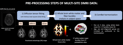 |
Exploring the reliability of ComBat for multi-site diffusion MRI harmonization
Suheyla Cetin Karayumak1,2, Lily O’Sullivan2, Monica Gabriella Lyons2, Marek Kubicki1,2, and Yogesh Rathi1,2
1Harvard Medical School, Boston, MA, United States, 2Brigham and Women's Hospital, Boston, MA, United States
This study thoroughly explores the reliability of a statistical multi-site diffusion-MRI harmonization method, ComBat, using a large diffusion-MRI dataset involving 5 sites. Additionally, we demonstrate the effects of using different software packages to estimate the diffusion tensor on the harmonization performance of ComBat. Even when the processing steps (and software) were identical for all sites, ComBat altered the inter-group variability at each site and in some regions flipped the direction of effect-sizes. Finally, when different software packages were used to estimate the diffusion tensor at different sites, ComBat introduced much drastic effects, thereby reducing the reliability of the results.
|
|
4380.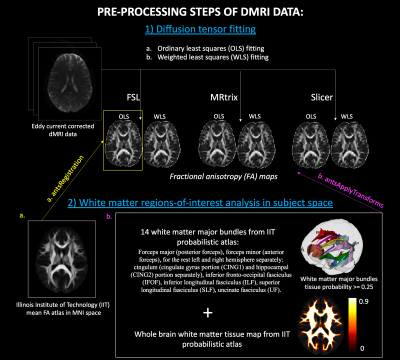 |
Reproducibility crisis in diffusion MRI: Contribution of software processing pipelines
Suheyla Cetin Karayumak1,2, Lily O’Sullivan2, Monica Gabriella Lyons2, Tashrif Billah2, Ofer Pasternak1,2, Sylvain Bouix1,2, Marek Kubicki1,2, and Yogesh Rathi1,2
1Harvard Medical School, Boston, MA, United States, 2Brigham and Women's Hospital, Boston, MA, United States
This study attempts to highlight the reproducibility problem in clinical diffusion MRI studies by comparing different software in a large diffusion MRI data, collected from multiple sites with 291 schizophrenia patients and 251 healthy controls. We fit diffusion tensor using FSL, MRtrix and Slicer with ordinary-least-squares (OLS) and weighted-least-squares (WLS) methods. To assess biological differences (measured by FA) between controls and patients, we computed effect-sizes (Cohen’s d) at each site using each software. We observed significant software effects with each software package producing significantly different results, when either OLS or WLS, was used.
|
|
4381.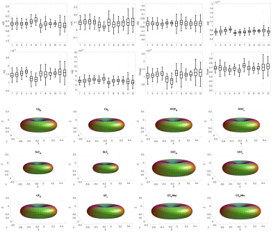 |
Diffusion MRI response function estimates vary more across pathways than across subjects
Kurt G Schilling1, Bennett A Landman1,2,3,4, Alexander Leemans5, Adam W Anderson1,2, and Chantal M.W. Tax6
1Vanderbilt University Institute of Imaging Science, Nashville, TN, United States, 2Biomedical Engineering, Vanderbilt University, Nashville, TN, United States, 3Radiology and Radiological Sciences, Vanderbilt University Medical Center, Nashville, TN, United States, 4Electrical Engineering, Vanderbilt University, Nashville, TN, United States, 5Image Sciences Institute, University Medical Center Utrecht, Utrecht, Netherlands, 6CUBRIC, School of Physics, Cardiff University, Cardiff, United Kingdom
We investigated the diffusion fiber response function by characterizing the modelled diffusivity, the shape, and the size of the diffusion signal in regions of single fiber populations. We show that the various descriptors of the diffusion signal vary significantly across white matter pathways within a subject, and across subjects for a given pathway. Analysis of variance suggests that variability between pathways is greater than across subjects. Understanding the response function and its variation are necessary for accurate fiber orientation characterization, as well as characterizing microstructural effects from geometrical effects within a voxel.
|
|
4382.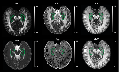 |
High-resolution microscopic diffusion anisotropy imaging in the human hippocampus using multidimensional diffusion encoding
Jiyoon Yoo1, Leevi Kerkelä1, Patrick W. Hales1, Kiran K. Seunarine1, Noemi G. Gyori1,2, Enrico Kaden2, and Christopher A. Clark1
1UCL Great Ormond Street Institute of Child Health, London, United Kingdom, 2UCL Centre for Medical Image Computing, London, United Kingdom
Several neurological diseases are associated with microstructural changes in the hippocampus which can be observed using diffusion MRI. However, DTI derived microstructural metrics such as fractional anisotropy (FA) are not able to differentiate between orientation dispersion and microscopic anisotropy. In this study, we applied an optimised multidimensional diffusion encoding sequence to measure microscopic fractional anisotropy (µFA) and normalised size variance (CMD) in the human hippocampus of healthy subjects. We also defined a clinically feasible acquisition protocol by subsampling the full data set.
|
|
4383.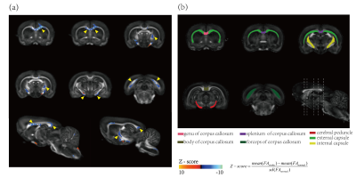 |
Quantifying whole-brain microstructural changes in rat model of focal ischemia on unilateral motor cortex using diffusion MRI at 14T
Zhaoqing Li1, Huan Gao2, Kedi Xu2, and Ruiliang Bai1,3
1Interdisciplinary Institute of Neuroscience and Technology (ZIINT), College of Biomedical Engineering and Instrument Science, Zhejiang University, HangZhou, China, 2Qiushi Academy for Advanced Studies (QAAS), Key Laboratory of Biomedical Engineering of Ministry of Education, College of Biomedical Engineering and Instrument Science, Zhejiang University, HangZhou, China, 3School of Medicine, Zhejiang Univerisity, HangZhou, China
Focal cortical ischemia animal model has been widely used to study neuroanatomical reorganization of cortex with intracortical microstimulation. However, the whole-brain structural changes following focal ischemia on motor cortex is unknown. In this study, high-resolution DTI is performed on focal unilateral motor cortex ischemia rat model at 14T. Voxel-wise based analysis (VBA) and VBA-guided connectivity analysis are performed to explore potential whole-brain microstructure and global structural connectivity changes. Our results show that large-scale white matter microstructural changes mainly distribute in corpus callosum, external capsule, cerebral peduncle. Significant reorganization of sensorimotor network associated with these regions were also found after ischemia.
|
|
4384.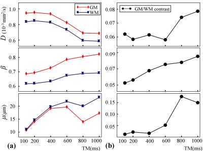 |
A Study on Time Dependency of the Fractional Order Calculus Diffusion Model
Guangyu Dan1,2, Yuxin Zhang3,4, Zheng Zhong1,2, Kaibao Sun1, M. Muge Karaman1,2, Diego Hernando3,4, and Xiaohong Joe Zhou1,2,5
1Center for Magnetic Resonance Research, University of Illinois at Chicago, Chicago, IL, United States, 2Department of Bioengineering, University of Illinois at Chicago, Chicago, IL, United States, 3Department of Medical Physics, University of Wisconsin-Madison, Madison, WI, United States, 4Department of Radiology, School of Medicine and Public Health, University of Wisconsin-Madison, Madison, WI, United States, 5Department of Radiology and Neurosurgery, University of Illinois at Chicago, Chicago, IL, United States
Parameters in many diffusion models depend on diffusion time. However, time-dependent diffusion behaviors in the long diffusion time regime have not been well studied because a longer diffusion time would lead to a longer TE, substantially reducing signal-to-noise ratio in conventional spin-echo diffusion pulse sequences. In this study, we employed a STEAM diffusion sequence to investigate the diffusion-time dependence of parameters in a fractional order calculus diffusion model. Our results showed substantial dependence of all diffusion parameters on diffusion times in the range of 100-1000 ms.
|
|
4385.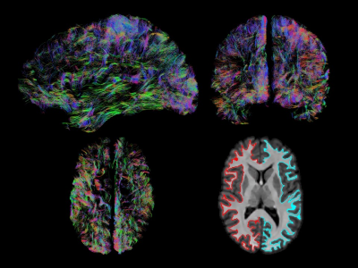 |
Delineating the grey matter-white matter interface directly from diffusion MRI data
Dmitri Shastin1,2, Maxime Chamberland1, Greg Parker1, Chantal M. W. Tax1, Kristin Koller1, Khalid Hamandi1,2, William Gray2,3, and Derek Jones1
1School of Psychology, Cardiff University Brain Research Imaging Centre, Cardiff, United Kingdom, 2School of Medicine, Cardiff University Brain Research Imaging Centre, Cardiff, United Kingdom, 3BRAIN Biomedical Research Unit, Cardiff, United Kingdom
Anatomically constrained tractography aims to reduce the number of false positive streamlines by applying anatomically realistic priors. Grey matter-white matter interface is currently extracted based on T1 information requiring the availability of a T1 volume and its accurate co-registration with diffusion MRI data. Here we describe an alternative method based on multi-shell multi-tissue constrained spherical deconvolution that produces the interface without the need for T1. We go ahead and compare our method with the existing one and show a potential application.
|
|
4386.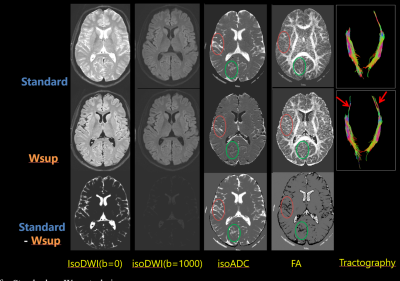 |
Synthetic-DWI with T2-based Water Suppression
Tokunori Kimura1, Kousuke Yamashita1, and Kouta Fukatsu1
1Department of Radiological Science, Shizuoka College of Medicare Science, Hamamatsu-Shi, Japan
We proposed a new Diffusion Weighted Imaging (DWI) with T2-based water suppression technique (Wsup-DWI) and additionally combined with synthetic-MRI technique. Our water suppression was achieved by subtracting heavy T2-weighted (long-TE) image from the standard DWI images after correcting signal intensities due to b-value. We demonstrated that hyperintense artifacts due to CSF partial volume effects (PVE) were dramatically suppressed even at lower b-values (b); and quantitative maps of ADC and FA became close to the tissue values due to CSF reduction. Furthermore, fiber tracts became longer for Wsup-DWI data than for standard-DWI data in a tractography with the same condition.
|
|
4387.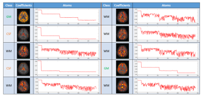 |
Sparse Representation of DWI Images for Fully Automated Brain Tissue Segmentation
Hu Cheng1, Jian Wang1,2, and Sharlene Newman1
1Psychological and Brain Sciences, Indiana University, Bloomington, IN, United States, 2School of Information Science and Engineering, Shandong Normal University, Jinan, China
We propose a fully automated brain tissue segmentation method based on sparse representation of diffusion weighted imaging (DWI) signal. Learning a dictionary from DWI signals, brain voxels are classified into gray matter, white matter, and CSF according to their sparse representation of clustered dictionary atoms. The proposed method was tested on three subjects of the HCP DWI datasets and achieved good agreement with the segmentation on T1-weighted images using SPM12. The method is very fast and robust for a wide range of sparse coding parameter selection and works well on DWI data with less number of shells or gradient directions.
|
|
4388.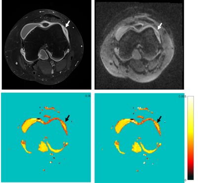 |
Improved parameter maps calculation in IVIM-diffusion-weighted MRI using a noise compensation fitting model
Anna-Katinka Bracher1, Meinrad Beer1, Volker Rasche2, and Neubauer Henning1
1Department of Diagnostic and Interventional Radiology, Ulm University Medical Center, Ulm, Germany, 2Department of Internal Medicine II, Ulm University Medical Center, Ulm, Germany
In standard diffusion-weighted imaging, the parameter maps are calculated based on a set of magnitude images. The standard fitting model of the parameter maps does not take a noise-induced bias into account. A noise offset model for Intravoxel Incoherent Motion (IVIM) in Diffusion Weighted Imaging (DWI) is introduced and the impact on diffusion parameter maps is evaluated for muscle and synovia of the knee joint. Parameter fitting using the noise-offset model results in faster signal decay of the fitted curve and therefore in increased diffusion coefficients compared to standard IVIM mapping.
|
|
4389.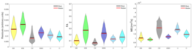 |
Diffusion tensor residuals as a potential biomarker for brain tissue damage in memory clinic patients
Anouk van Rijn1, Alexander Leemans1, Geert Jan Biessels2, and Alberto de Luca 1
1Image Sciences Institute, University Medical Center Utrecht, Utrecht, Netherlands, 2Department of Neurology, University Medical Center Utrecht Brain Center, Utrecht, Netherlands
DTI metrics are often used without assessing the goodness of fit of the estimation model. We investigated local changes in fit residuals induced by pathology. To this end, a template of expected normalized residuals was created with 10 healthy controls, then voxel-wise comparisons were performed against the residuals of subjects affected by dementia. Results show that the residuals of infarcted regions are significantly different as compared to healthy tissue, whereas no differences were observed in hyperintensities. The fit residuals of the DTI model can be used to complement the information of DTI metrics at detecting microstructural changes in brain lesions.
|
4390. |
The efficacy of ADC value of DWI in differentiating extrahepatic cholangiocarcinoma from mass-forming cholangitis
Yaxin Niu1, Ailian Liu1, Ye Li1, and Lizhi Lizhi Xie2
1The First Affiliated Hospital of Dalian Medical University, Dalian Medical University, Da Lian, China, 2GE Healthcare, MR Research China, BeiJing, China, Beijing, China
Diffusion-weighted imaging reflects the micro-movements of water molecules.
|
|
4391. |
Quasi-Diffusion Magnetic Resonance Imaging (QDI): optimisation of acquisition protocol
Catherine A Spilling1, Franklyn A Howe1, and Thomas R Barrick1
1Neurosciences Research Centre, Molecular and Clinical Sciences Research Institute, St George's University of London, London, United Kingdom
Quasi-diffusion image (QDI) is a new ultra-high b-value diffusion magnetic resonance imaging technique which provides standard and non-Gaussian diffusion images. We use permutation analysis to optimise a clinical QDI tensor acquisition from a gold standard multi b-value protocol (28 non-zero b-values in 6 diffusion directions) to identify a 2 minute acquisition protocol with excellent tissue contrast. We achieve this by comparing different b-value combinations (2 non-zero b-values) to the gold standard using χ2 difference in parameterised signal decay curves. We obtain an optimal acquisition protocol of b=0, 1080, 5000 s mm-2 that may be acquired in a clinically acceptable time.
|
|
4392. |
Diffusion tensor imaging in identificationof stageIendometrial carcinoma and endometrial poly
Xuedong Wang1, Ailian Liu1, ShiFeng Tian1, Lizhi Xie2, and Qingwei Song1
1The first affiliated hospital of dalian medical university, DaLian, China, 2GE Healthcare, MR Research China, Beijing, Beijing, China
Diffusion tensor imaging (DTI), a type of MR functional imaging, can reflect the direction of the diffusion motion of water moleculesas well as describe the free diffusion rate of water molecules. The characteristic suggested the DTI were helpful in distinguishing endometrial carcinoma (EC) from endometrial polyp (EP).
|
|
4393.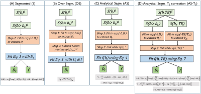 |
An analytical segmented approach for extracting TE independent perfusion fraction in intravoxel incoherent motion (IVIM) MRI
Erick O Buko1, Afis Ajala1, Jiming Zhang2, Pei Herng Hor1, and Raja Muthupillai2
1Physics, Texas Center for Superconductivity at University of Houston, Houston, TX, United States, 2Diagnostic and Interventional Radiology, Baylor St. Luke's Medical Center, Houston, TX, United States
Determination of perfusion fraction (f) from IVIM-MRI data is challenging. Recent studies have shown that estimation of f is influenced by echo time (TE). Eliminating thisTE dependency on f requires acquiring data sets at multiple TEs. Here, we propose an analytical segmented approach to correct for the TE dependency on f arising from T2 differences between the tissue and fluid compartments (AS-T2). Numerical simulations predict, and phantom experiments confirm that, -compared to commonly used segmented approaches to estimate f, AS-T2 approach permits the determination of perfusion fraction without TE dependence, and with fewer measurements.
|
|
4394.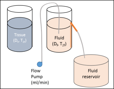 |
A simple phantom for extracting Intravoxel Incoherent Motion (IVIM) model parameters
Erick O Buko1, Afis Ajala1, Jiming Zhang2, and Pei Herng Hor1
1Physics, Texas Center for Superconductivity at University of Houston, Houston, TX, United States, 2Diagnostic and Interventional Radiology, Baylor St. Luke's Medical Center, Houston, TX, United States
We present a simple phantom set-up to model diffusion weighted imaging signal arising from intra-voxel incoherent motion (IVIM). The model provides means to independently control both the perfusion fraction (f) as well as the pseudodiffusion coefficient (Df).
|
|
4395.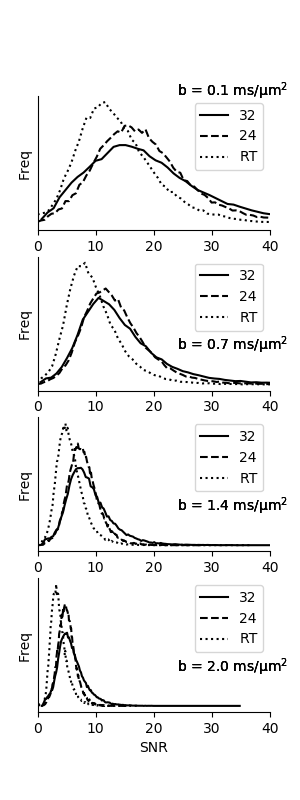 |
Impact of brain and head and neck radiotherapy fixation and coil configuration on tensor-valued dMRI
Patrik Brynolfsson1, Markus Nilsson2, Christian Jamtheim Gustafsson1,3, Tim Sprenger4, Pia C Sundgren5,6, Lars E Olsson1, and Filip Szczepankiewicz7,8
1Dept. of Translational Medicine, Division of Medical Radiation Physics, Lund University, Lund, Sweden, 2Dept. of Clinical Sciences, Division of Radiology, Lund University, Lund, Sweden, 3Dept. of Hematology, Oncology and Radiation Sciences, Lund University, Lund, Sweden, 4MR Applied Science Laboratory Europe, GE Healthcare, Stockholm, Sweden, 5Diagnostic Radiology, Clinical Sciences, Lund University, Lund, Sweden, 6Lund University Bioimaging Center, Lund University, Lund, Sweden, 7Lund University, Lund, Sweden, 8Brigham and Women's Hospital, Boston, MA, United States
Brain and head and neck cancer patients need fixation while undergoing radiotherapy, which prevents the use of a dedicated head coil when imaging patients using MRI in radiotherapy setup. We investigate the impact of coil setup on the technical feasibility of tensor-valued diffusion MRI in a radiation therapy setting. Three coil configurations were evaluated with respect to SNR and parameter bias in the resulting parametric maps using Bland-Altman plots. As expected, the coils configured for radiation therapy imposed a penalty on the SNR, but accuracy was not negatively impacted.
|
|
4396.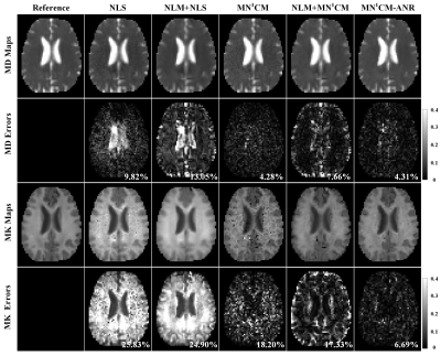 |
Improved estimation of diffusion tensors by noise-corrected curve fitting with adaptive neighborhood regularization
Li Guo1,2,3, Xinyuan Zhang2,3, Changqing Wang4, Jian Lyu2,3, Yingjie Mei5, Ruiliang Lu1, Mingyong Gao1, and Yanqiu Feng2,3
1Department of MRI, The First People’s Hospital of Foshan (Affiliated Foshan Hospital of Sun Yat-sen University), Foshan, China, 2School of Biomedical Engineering, Southern Medical University, Guangzhou, China, 3Guangdong Provincial Key Laboratory of Medical Image Processing, Southern Medical University, Guangzhou, China, 4School of Biomedical Engineering, Anhui Medical University, Hefei, China, 5Philips Healthcare, Guangzhou, China
The noncentral Chi noise in magnitude image may significantly affect the reliability of quantitative analysis in diffusion-weighted (DW) magnetic resonance imaging (MRI), especially at high b-value and/or higher order modeling of diffusion signal such as diffusion kurtosis imaging (DKI). We developed a novel first-moment noise-corrected curve fitting model with adaptive neighborhood regularization (MN1CM-ANR) algorithm for DKI. By fitting the signal to its first-moment (i.e. the expectation of the signal), MN1CM-ANR can effectively compensate the bias due to the noncentral Chi noise. In addition, by exploiting the neighboring pixels to regularize the curve fitting, MN1CM-ANR can reduce the measurement variance.
|
|
4397.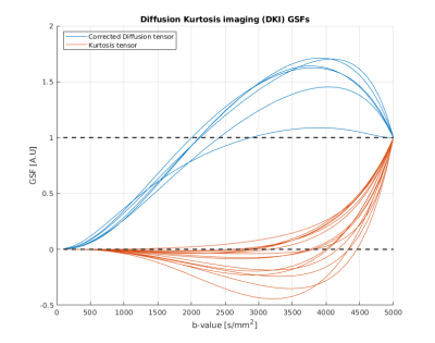 |
Generalized sensitivity functions for optimal b-value selection in diffusion-based MRI modeling
Umberto Villani1,2, Simona Schiavi3, and Alessandra Bertoldo1,2
1Padova Neuroscience Center, University of Padova, Padova, Italy, 2Department of Information Engineering, University of Padova, Padova, Italy, 3Department of Computer Science, University of Verona, Verona, Italy
Generalized sensitivity functions provide insights about which temporal observations are most informative for the estimation of biological model parameters. We formulate the same concept in the dMRI field to investigate how biophysical models/data representations react to HARDI acquisitions of different b-values. This approach handily shows how different parameters feature enhanced estimation precision at different b-values and exposes potential correlations between them, shedding light on possible a posteriori identifiability issues. Requiring only byproducts of standard optimization routines, generalized sensitivity functions can easily be integrated in standard analyses when proposing either a new model or a modification of existing ones.
|
|
4398.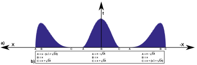 |
Anomalous Diffusion estimation through the solution of the Fractional Time order Bloch-Torrey equation
Óscar Peña-Nogales1, Carlos Castillo2,3,4, Carlos Lizama5, Rodrigo de Luis-Garcia1, Santiago Aja-Fernández1, and Pablo Irarrazaval2,3,4,6
1Laboratorio de Procesado de Imagen, Universidad de Valladolid, Valladolid, Spain, 2Instituto de Ingeniería Biológica y Médica, Pontificia Universidad Catolica de Chile, Santiago, Chile, 3Millennium Nucleus for Cardiovascular Magnetic Resonance, Santiago, Chile, 4Biomedical Imaging Center, Pontificia Universidad Católica de Chile, Santiago, Chile, 5Departamento de Matemáticas y Ciencia de la Computación, Universidad de Santiago de Chile, Santiago, Chile, 6Electrical Engineering Department, Pontificia Universidad Católica de Chile, Santiago, Chile
Diffusion-Weighted (DW) imaging has a monoexponential signal decay at low b-values related to the statistical mechanics of Brownian motion. However, at high b-values the DW signal is no longer monoexponential. Various models have been proposed to better fit the data; nevertheless, none of them assumes that the DW signal is governed by the fractional-time order dynamics of the generalized Brownian motion (a.k.a., anomalous diffusion). In this work, by assuming anomalous diffusion we solve the fractional-time order Bloch-Torrey equation for smooth diffusion-weighting gradients and compare the obtained model to existing ones. The proposed model outperforms state-of-the-art on synthetic anomalous phantom experiments.
|
|
4399.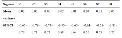 |
Using Diffusional Kurtosis Imaging to Quantify non-Gaussian Water Diffusion in Normal Human Liver
Junying Wang1, Hansen Schie2, Weiqiang Dou3, and Xiaoyi He1
1Medical Imaging, Shandong First Medical University, Jinan, Shandong, China, China, 2Medical Imaging, Shandong Provincial Qianfoshan Hospital,the First Hospital Affiliated with Shandong First Medical University, Jinan City, Shandong Province, China, China, 3GE Healthcare, MR Research China,Bejing, Bejing,China, China This study aimed to test the reproducibility of DKI technique in normal liver and to explore the microscopic and diffusion differences between the left and right hepatic lobes. 32 healthy volunteers were scanned in DKI twice and the interval time was two weeks. All DKI-derived parametrical maps were reconstructed and mean values in eight liver segments were calculated. We demonstrated that DKI in the liver showed excellent reproducibility between two measurements and found interesting regional distributions of the parameters in liver. Additionally, significant difference was found in DKI-derived parameters between the left and right hepatic lobes |
|
4400.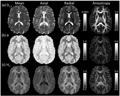 |
Quasi-Diffusion Magnetic Resonance Imaging (QDI): a fast, high b-value diffusion imaging technique
Thomas R Barrick1, Catherine A Spilling1, Carson Ingo2, Jeremy Madigan3, Jeremy D Isaacs3, Philip Rich3, Timothy L Jones3, Richard L Magin4, Matt G Hall5,6, and Franklyn A Howe1
1Neurosciences, St George's, University of London, London, United Kingdom, 2Northwerstern University, Chicago, IL, United States, 3St George's University Hospitals NHS Foundation Trust, London, United Kingdom, 4University of Illinois at Chicago, Chicago, IL, United States, 5National Physical Laboratory, Teddington, United Kingdom, 6University College London, London, United Kingdom
We present a new ultra-high b-value diffusion magnetic resonance imaging (dMRI) methodology, Quasi-Diffusion Imaging (QDI). The QDI technique includes a tensor representation of the dMRI data. We show that high contrast to noise images representing standard and non-Gaussian diffusion measures are obtainable in 1 to 4 minutes. QDI entropy maps show pathological contrast similar to Diffusional Kurtosis Imaging (DKI) in stroke and brain tumours. In addition, QDI overcomes the b-value limitations of DKI.
|
|
4401.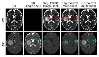 |
Lesion detection using Free-Water elimination DTI - is this technique really specific to free water?
Marc Golub1, R. N. Henriques2, and R. G. Nunes1
1ISR-Lisboa/LARSyS and Department of Bioengineering, Instituto Superior Técnico – Universidade de Lisboa, Lisbon, Portugal, Lisbon, Portugal, 2Champalimaud Neuroscience Programme, Champalimaud Centre for the Unknown, Lisbon, Portugal, Lisbon, Portugal
Free water elimination (FWE) for diffusion tensor imaging (DTI) aims to decouple the free water contribution from tissue's diffusion. Recent studies suggest that FWE-DTI is essential to characterize degenerative processes since it suppresses free water partial volume effects from DTI analysis and provides an index sensitive to edema and tissue degeneration. In this work, the state-of-the-art of FWE-DTI estimation is explored and several fitting procedures are tested on in vivo data to which simulated lesions were added. We found that, unlike the multi-shell approach, the single-shell implementation was unable to discriminate between changes in tissue diffusion and free water increases.
|
|
4402.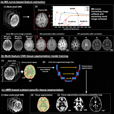 |
Deep Learning Based Brain Tissue Segmentation from Novel Diffusion Kurtosis Imaging Features
Fan Zhang1, Anna Breger2, Lipeng Ning1, Carl-Fredrik Westin1, Lauren J O'Donnell1, and Ofer Pasternak1
1Harvard Medical School, Boston, MA, United States, 2University of Vienna, Wien, Austria
Brain tissue segmentation is important in many diffusion MRI (dMRI) visualization and quantification tasks. We propose a deep learning tissue segmentation method that relies only on dMRI data. We leverage diffusion kurtosis imaging (DKI) and a recently proposed mean-kurtosis-curve (MK-curve) method to create a feature set that is highly discriminative between different types of tissues. We train a Unet model with a recently developed augmented target loss function on dMRI data from the Human Connectome Project. We show improved segmentation performance compared to several other methods and reliable segmentation results when applied on data with a different acquisition.
|
|
4403.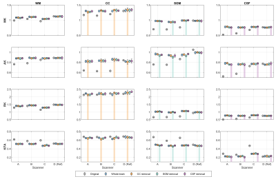 |
Self-validation for deep learning–based diffusion kurtosis imaging harmonization
Qiqi Tong1, Ting Gong1, Hongjian He1, Yi-Cheng Hsu2, and Jianhui Zhong1,3
1Center for Brain Imaging Science and Technology, Department of Biomedical Engineering, Zhejiang University, Hangzhou, China, 2MR Collaboration, Siemens Healthcare, Shanghai, China, 3Department of Imaging Sciences, University of Rochester, Rochester, NY, United States
Deep learning–based harmonization for diffusion imaging data with high efficiency and low cost is gaining popularity. However, the performance of the training-required network depends on the training data, which lack the diversity of the large sets of data in more substantial multicenter projects. We proposed a leave-one-tissue-out training strategy to evaluate the validity and reliability across scanners of a deep learning–based diffusion kurtosis imaging harmonization method. The results confirm that the deep learning–based network can still reconstruct the untrained tissue with validity, although the reliability would be higher when the tissue is trained.
|
|
4404.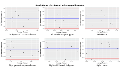 |
Test-retest reliability of Diffusion Kurtosis Imaging (DKI) of the brain in healthy volunteers.
Ernst Christiaanse1,2, Alexander Leemans2, Patrik Wyss1, and Alberto de Luca2
1Department of Radiology, Swiss Paraplegic Centre, Nottwil, Switzerland, 2Image Sciences Institute, University Medical Center Utrecht, Utrecht, Netherlands
It is essential for longitudinal studies to validate the reproducibility of the applied methods. Therefore, we assessed the test-retest reliability of diffusion kurtosis imaging of the brain in 10 healthy participants and showed a good reproducibility with some variation in different white matter regions.
|
4405.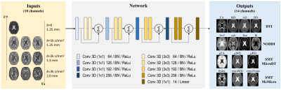 |
DeepHIBRID: How to condense the sampling in the k-q joint space for microstructural diffusion metric estimation empowered by deep learning
Qiuyun Fan1, Qiyuan Tian1, Chanon Ngamsombat1, and Susie Y. Huang1
1Athinoula A. Martinos Center for Biomedical Imaging, Massachusetts General Hospital, Boston, MA, United States
Conventional diffusion imaging protocols may require tens or hundreds of samples in the q-space to generate reliable maps. Knowing that the k-q joint space is highly redundant and given the tradeoffs between k, q and SNR, we trained a deep convolutional neural network using a HIgh B-value and high Resolution Integrated Diffusion (HIBRID) sampling scheme, dubbed DeepHIBRID. We show DeepHIBRID outperforms conventional sampling schemes, and is capable of outputting 14 synthesized diffusion metric maps simultaneously with only 10 input images, without sacrificing the quality of the output maps, using 30x angular downsampling.
|
|
4406.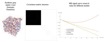 |
A Theoretical Framework for Representing and Estimating a Normal Diffusion Tensor Distribution
Magdoom Kulam Najmudeen1, Dario Gasbarra2, and Peter J Basser1
Video Permission Withheld
1SQITS/NICHD, National Institute of Health, Bethesda, MD, United States, 2University of Helsinki, Helsinki, Finland
A new signal model is introduced for diffusion tensor distribution imaging which is monotonically decreasing for all b-values unlike the cumulant and kurtosis models. A constrained multi-normal distribution is used as the tensor distribution which is fully characterized by the 2nd order mean and 4th order covariance tensors. A theoretical framework is presented showing the richness of covariance tensor, using synthetic gray and white matter voxels, and the ability to estimate the mean and covariance tensor from noisy MR signal.
|
|
4407.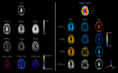 |
Estimating distributions of diffusion tensors and longitudinal relaxation rates in the brain via Monte-Carlo inversion: a proof of principle
Alexis Reymbaut1,2, Jeffrey Critchley3, Giuliana Durighel3, Tim Sprenger4,5, Michael Sughrue6, and Daniel Topgaard1,2
1Physical Chemistry, Lund University, Lund, Sweden, 2Random Walk Imaging AB, Lund, Sweden, 3Spectrum Medical Imaging, Sydney, Australia, 4Karolinska Institute, Stockholm, Sweden, 5GE Healthcare, Stockholm, Sweden, 6Charlie Teo Foundation, Sydney, Australia Conventional $$$T_1$$$-mapping techniques are only sensitive to voxel-averaged $$$T_1$$$ values, which hinders the study of fiber-specific myelination changes in the developing, aging or diseased brain. While recent works have focused on combining diffusion- and $$$T_1$$$- weightings to access orientation-resolved $$$T_1$$$ values, they rely on assumptions regarding the voxel content. This work combines a prototype diffusion-relaxation MR acquisition and a Monte-Carlo inversion method to extract intra-voxel nonparametric 5D distributions of diffusion tensors and longitudinal relaxation rates $$$R_1=1/T_1$$$ without the use of limiting assumptions. Estimated $$$R_1$$$ values are then mapped onto nonparametric orientation distribution functions, thereby yielding fiber-specific longitudinal relaxation rates. |
|
4408.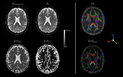 |
General tools for diffusion tensor distributions, and matrix-variate Gamma approximation for multidimensional diffusion MRI data
Alexis Reymbaut1,2
1Physical Chemistry, Lund University, Lund, Sweden, 2Random Walk Imaging AB, Lund, Sweden
Either on the voxel scale or within sub-voxel diffusion compartments, tissue microstructure can be described using a diffusion tensor distribution $$$\mathcal{P}(\mathbf{D})$$$. One way to resolve microstructural heterogeneity relies on choosing a plausible parametric functional form to approximate $$$\mathcal{P}(\mathbf{D})$$$. However, such a high-dimensional mathematical object is usually intractable. Here, we define matrix moments enabling the computation of diffusion metrics for any arbitrary functional choice approximating $$$\mathcal{P}(\mathbf{D})$$$. Applying these general tools to the matrix-variate Gamma distribution on the voxel scale, we obtain a new signal representation, the matrix-variate Gamma approximation, that we validate in vivo and in silico.
|
|
4409.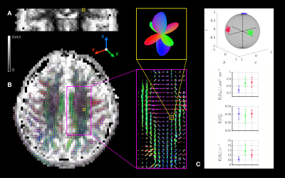 |
Resolving orientation-specific diffusivities and transverse relaxation rates in heterogenous brain tissue
Alexis Reymbaut1,2, João Pedro de Almeida Martins1,2, Chantal M. W. Tax3, Filip Szczepankiewicz2,4,5, Derek K. Jones3, and Daniel Topgaard1,2
1Physical Chemistry, Lund University, Lund, Sweden, 2Random Walk Imaging AB, Lund, Sweden, 3Cardiff University Brain Research Imaging Centre (CUBRIC), School of Psychology, Cardiff University, Cardiff, United Kingdom, 4Harvard Medical School, Boston, MA, United States, 5Radiology, Brigham and Women’s Hospital, Boston, MA, United States
Due to their cubic-millimeter scale, white-matter diffusion MRI voxels are often heterogeneous, comprising not only multiple fibre bundles but also grey matter, cerebrospinal fluid, or pathological tissue. To tackle this problem, conventional approaches rely on assumptions regarding tissue properties. This work combines state-of-the-art diffusion-relaxation MR acquisition and processing methods to extract intra-voxel nonparametric 5D distributions of diffusion tensors and transverse relaxation rates $$$R_2$$$ without the use of limiting assumptions. Orientation-resolved (fibre-specific) means of isotropic diffusivities, diffusion anisotropies and $$$R_2$$$ values are then obtained via clustering within the orientation subspace of these 5D distributions.
|
|
4410.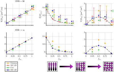 |
Trueness and precision of statistical descriptors obtained from multidimensional diffusion signal inversion algorithms
Alexis Reymbaut1,2, Paolo Mezzani1,3, João Pedro de Almeida Martins1,2, and Daniel Topgaard1,2
1Physical Chemistry, Lund University, Lund, Sweden, 2Random Walk Imaging AB, Lund, Sweden, 3Physics, Università degli Studi di Milano, Milan, Italy
In first approximation, the diffusion signal writes as the Laplace transform of an intra-voxel diffusion tensor distribution (DTD). Several algorithms have been introduced to estimate the DTD’s statistical descriptors (mean diffusivity, variance of isotropic diffusivities, mean squared diffusion anisotropy, etc.) by inverting data obtained from tensor-valued diffusion encoding schemes. However, the trueness and precision of these estimations have not been systematically assessed and compared across methods. Here, we compare such estimations in silico for a 1D Gamma fit, a generalized two-term cumulant approach, and 2D and 4D Monte-Carlo inversion techniques, using a common and clinically feasible tensor-valued acquisition scheme.
|
|
4411.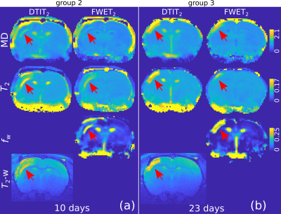 |
Spatiotemporal evolution of ischemic lesions in stroke animal models using free-water elimination and mapping with explicit T2 modelling
Ezequiel Farrher1, Chia-Wen Chiang2, Kuan-Hung Cho2, Richard Buschbeck1, Ming-Jye Chen2, Zaheer Abbas1, Kuo-Jen Wu3, Yun Wang3, Farida Grinberg1, Chang-Hoon Choi1, N. Jon Shah1,4,5,6, and Li-Wei Kuo2,7
1INM-4, Forschungszentrum Jülich, Jülich, Germany, 2Institute of Biomedical Engineering and Nanomedicine, National Health Research Institutes, Miaoli, Taiwan, 3Center for Neuropsychiatric Research, National Health Research Institutes, Miaoli, Taiwan, 4Department of Neurology, RWTH Aachen University, Aachen, Germany, 5JARA – BRAIN – Translational Medicine, RWTH Aachen University, Aachen, Germany, 6Institute of Neuroscience and Medicine 11, JARA, Forschungszentrum Jülich, Jülich, Germany, 7Institute of Medical Device and Imaging, National Taiwan University College of Medicine, Taipei, Taiwan
It is known that excess fluid as a result of vasogenic oedema formation following stroke onset obscures the microstructural characterisation of ischemic tissue by diffusion MRI. DTI-based free water elimination and mapping (FWE) has been proposed as a technique to potentially reduce the partial-volume effect. However, FWE estimation is ill-conditioned, leading to inaccurate results. More recently, it has been shown that the addition of a second dimension spanned by transverse relaxation weighting, mitigates the ill-conditioned problem. We aim here to investigate the latter model in a longitudinal study of MCAo stroke animal models.
|
|
4412.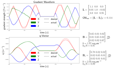 |
Impact of gradient non-linearities on B-tensor diffusion encoding
Michael Paquette1, Chantal M.W. Tax2, Cornelius Eichner1, and Alfred Anwander1
1Neuropsychology, Max Planck Institute for Human Cognitive and Brain Sciences, Leipzig, Germany, 2CUBRIC, School of Physics, Cardiff University, Cardiff, United Kingdom
We investigate the effect of gradient non-linearities (GNL) on free gradient waveform used for B-tensor diffusion encoding. We show the magnitude of the GNL-bias for strong gradients of $$$300 m\text{T}/\text{m}$$$. We derive a closed-form formula of the voxelwise B-tensor under GNL, independent of the choice of gradient waveform used to encode the B-tensor.
|
|
4413.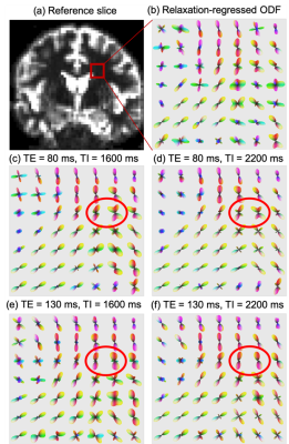 |
Multi-dimensional Moment Imaging (MMI) for probing tissue microstructure
Lipeng Ning1,2, Filip Szczepankiewicz1,2, Borjan Gagoski2,3, Carl-Fredrik Westin1,2, and Yogesh Rathi1,2
1Brigham and Women's Hospital, Boston, MA, United States, 2Harvard Medical School, Boston, MA, United States, 3Boston Children's Hospital, Boston, MA, United States
Multi-dimensional imaging techniques can provide complementary information to investigate the microscopic organizations of biological tissue. Deriving meaningful indices from these multi-dimensional imaging data is important to make these techniques relevant to clinical research. In this work, we introduce a general framework to estimate the moments of the joint probability distribution of T1, T2 relaxation and diffusion coefficients. This framework is an extension of our previously introduced REDIM approach which only focused on joint moments of T2 relaxation and diffusion. We show the cross coupling between different parameters using three datasets acquired using different protocols.
|
|
4414.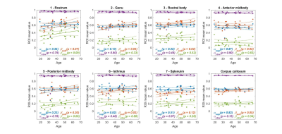 |
Anomalous diffusion MRI model parameters vary in the human corpus callosum sub-regions with age
Qianqian Yang1 and Viktor Vegh2
1School of Mathematical Sciences, Queensland University of Technology, Brisbane, Australia, 2Centre for Advanced Imaging, University of Queensland, Brisbane, Australia Poster Permission Withheld
Diffusion MRI measures of the human brain provide key insight into microstructural variations across individuals and into the impact of central nervous system diseases and disorders. One approach to extract information from diffusion signals has been to use biologically relevant analytical models to link millimetre scale diffusion MRI measures with microscale influences. The other approach has been to represent diffusion as an anomalous process, and infer information from the different anomalous diffusion equation parameters. Here, we show how parameters of three established anomalous diffusion equations change with age, in a microstructurally complex tissue, the human corpus callosum.
|
|
4415.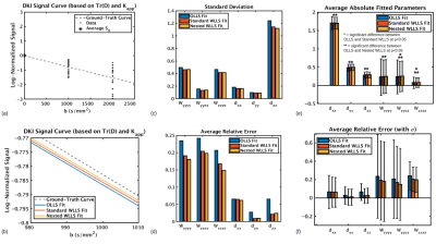 |
Weighted linear least squares in multi-shell diffusion MRI: Should high b-shells always be weighted less?
Jordan A. Chad1,2 and Ofer Pasternak3
1Rotman Research Institute, Baycrest Health Sciences, Toronto, ON, Canada, 2Department of Medical Biophysics, University of Toronto, Toronto, ON, Canada, 3Departments of Psychiatry and Radiology, Brigham and Women's Hospital, Harvard Medical School, Boston, MA, United States
Fitting diffusion MRI signal models with the standard weighted linear least squares (WLLS) approach necessarily places lower weight on data with lower SNR, therefore placing lower weight on shells with higher b-values. This can be non-optimal for fitting signal models that rely on information from high b-shells. In this work, we propose a “nested” WLLS approach where each shell is assigned a relative weight, with standard WLLS applied within each shell. We demonstrate that weighting shells equally may be beneficial for fitting signal models dependent on multiple shells.
|
|
4416.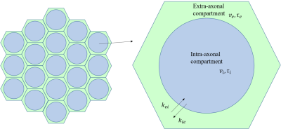 |
Exploring effects of membrane permeability on dMRI metrics with Analytical solutions and Monte Carlo simulations
Zihan Zhou1, Qiuping Ding1, Hongjian He1, and Jianhui Zhong1,2
1Center for Brain Imaging Science and Technology, Key Laboratory for Biomedical Engineering of Ministry of Education, College of Biomedical Engineering and Instrumental Science, Zhejiang University, Hangzhou, China, 2School of Medicine and Dentistry, University of Rochester Medical Center, New York, NY, United States
White Matter Tract Integrity (WMTI) is a biophysical model with specificity to underlying tissue microstructures. However, recent work has suggested that inter-compartmental water exchange may affect outcomes of the model metrics. In this work, we analytically relate the WMTI-derived dMRI metrics to membrane permeability, and validate our predictions using Monte Carlo simulations. Our results show that the water exchange has a non-trivial effect on the metrics and needs to be carefully considered in WMTI.
|
|
4417.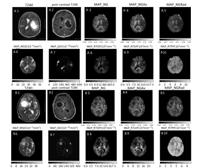 |
Application of MAP diffusion modelin differential diagnosis between high-grade glioma and single brain metastatic tumor
BO LI1, YANG GAO1, SHAOYU WANG2, YAN XU2, GUANG YANG3, and HUAPENG ZHANG2
1Department of Radiology, The Affiliated Hospital of Inner Mongolia Medical University, Hohhot, China, 2MR Sicentific Marketing, Siemens Healthineers, Shanghai, China, 3Shanghai Key Laboratory of Magnetic Resonance, East China Normal University, Shanghai, China This study aimed at using mean-apparent-propagator (MAP) diffusion model in differential diagnosis of high-grade glioma and single brain metastatic tumor.the findings show that multiple parameters quantified by DSI quantitative model MAP MRI, especially MSD and QIV has high applicatical value and provides more information for preoperative diagnosis of patients. |
|
4418.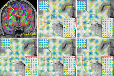 |
A New Method for Reconstructing the Orientation Distribution Function in HARDI
Diwei Shi1, Xuesong Li2, Ziyi Pan2, Hua Guo2, and Quanshui Zheng1
1Center for Nano & Micro Mechanics, Tsinghua University, Beijing, China, 2Center for Biomedical Imaging Research, Department of Biomedical Engineering, School of Medicine, Tsinghua University, Beijing, China We propose a novel definition of orientation distribution function (ODF), which also represents diffusion coefficients for non-Gaussian situations, thus ODF has physical meaning. Then an expression is derived to calculate ODF values through diffusion-weighted signals. Next a new HARDI (high angular resolution diffusion imaging) method called “g-OPDT” (grids orientation probability density transform) is designed for practical use. Numerical simulations are performed to verify its accuracy. ISMRM-2015-Tracto-challenge data are used to quantitatively compare g-OPDT with other methods. Results show that g-OPDT has superior performance on most indicators. In-vivo experiments are conducted to show that its glyph representations have less redundant lobes. |
|
4517. |
Quantification of uncertainty in parameterisations of cardiac myofibre orientations from diffusion-weighted imaging
Bianca Freytag1, Vicky Y Wang2, Alistair A Young3, and Martyn P Nash1
1Auckland Bioengineering Institute, The University of Auckland, Auckland, New Zealand, 2Veterans Affairs Medical Center, San Francisco, CA, United States, 3Biomedical Engineering Department, King's College London, London, United Kingdom
Electro-mechanical models of heart function rely upon accurate representation of microstructural orientations in the myocardium. We assessed the sensitivity of a finite element parameterisation of the myofibre orientation field derived from high-resolution DWI data of a canine heart. Eigenanalysis of the indifference region in the neighbourhood of the optimal fit of the myofibre field enabled quantification of helix angle uncertainty consistent with the DWI data. This method can be used to propagate the uncertainty in myofibre parameterisation to electro-mechanics simulation results.
|