
Digital Poster Session
Diffusion: Diffusion Microstructure, Modeling and Tractography
Diffusion
4419 -4433 Diffusion Microstructure, Modeling and Tractography - Diffusion: Microstructure 1
4434 -4448 Diffusion Microstructure, Modeling and Tractography - Diffusion: Microstructure 2
4449 -4463 Diffusion Microstructure, Modeling and Tractography - Diffusion: Microstructure 3
4464 -4478 Diffusion Microstructure, Modeling and Tractography - Orientation Modelling & Fibre Tractography 1
4479 -4494 Diffusion Microstructure, Modeling and Tractography - Orientation Modelling & Fibre Tractography 2
4495 -4509 Diffusion Microstructure, Modeling and Tractography - Diffusion: Phantoms, Simulation & Histological Evaluation
4419.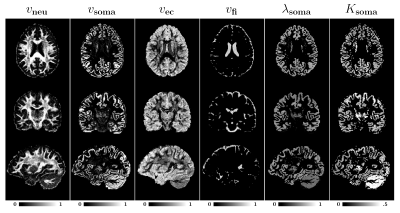 |
Quantifying Intra-Soma Diffusion Properties via Spherical Mean Spectrum Imaging
Khoi Minh Huynh1,2, Ye Wu2, Kim-Han Thung2, Sahar Ahmad2, Hoyt Patrick Taylor IV2, Weili Lin2, and Pew-Thian Yap2
1Department of Biomedical Engineering, UNC-Chapel Hill, Chapel Hill, NC, United States, 2Department of Radiology and BRIC, UNC-Chapel Hill, Chapel Hill, NC, United States
We propose a novel model to quantify intra-soma diffusion properties, including volume fractions and kurtosis. The model is flexible, allowing simultaneous modeling of multiple tissue compartments without the need for assumptions on the number of compartments and their diffusivities. We demonstrate that our method provides biologically meaningful contrasts that agree well with histological data.
|
|
4420.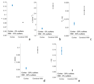 |
Characterisation of microvascular blood velocity, exchange and structure using multi-diffusion time DWI
Lauren Scott1, Ben Dickie1, Damien McHugh2, John McFadden1, Andrew N Priest3, and Laura M Parkes1
1Division of Neuroscience and Experimental Psychology, University of Manchester, Manchester, United Kingdom, 2Quantitative Biomedical Imaging Laboratory, University of Manchester, Manchester, United Kingdom, 3Radiology, Cambridge University Hospitals NHS Foundation Trust, Cambridge, United Kingdom
Diffusion-time (∆) dependence in the bi-exponential intra-voxel incoherent motion model of diffusion-weighted imaging (DWI) signal decay is generally not considered. However, by using multi-∆ signal acquisitions, blood flow dynamics across different flow regimes can be accessed. In this study, we use multi-∆ DWI and an adapted velocity autocorrelation model to estimate blood velocity (v), capillary segment length (l) and transvascular water exchange. Human cerebral cortex and white matter (WM) estimates of v (cortex: 1.85 ± 0.21 mms-1; WM: 2.67 ± 1.01 mms-1) and l (cortex: 43.4 ± 19.5 µm; WM: 93.3 ± 51.1 µm) are consistent with literature values.
|
|
4421.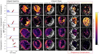 |
Data-Driven Multi-Contrast Spectral Microstructure Imaging with InSpect: INtegrated SPECTral Component Estimation and Mapping
Paddy J. Slator1, Jana Hutter2,3, Razvan V. Marinescu1, Marco Palombo1, Laurence Jackson2,3, Alison Ho4, Lucy C. Chappell4, Mary A. Rutherford2, Joseph V. Hajnal2,3, and Daniel C. Alexander1
1Centre for Medical Image Computing, Department of Computer Science, University College London, London, United Kingdom, 2Centre for the Developing Brain, School of Biomedical Engineering and Imaging Sciences, King's College London, London, United Kingdom, 3Biomedical Engineering Department, School of Biomedical Engineering and Imaging Sciences, King's College London, London, United Kingdom, 4Women's Health Department, King's College London, London, United Kingdom
We introduce a novel spectroscopic imaging technique - termed InSpect - for analysing multi-contrast microstructural MRI experiments. Such data potentially supports estimation of multidimensional correlation spectra via a regularised inverse Laplace transform, but this is an ill-posed calculation. InSpect addresses these limitations in a data-driven way. The algorithm simultaneously estimates a canonical basis of spectral components for the whole data set, and maps their spatial distribution across images. Unlike standard approaches, InSpect shares information across voxels, implementing data-driven regularisation of the inverse Laplace transform. We demonstrate the method on combined diffusion-relaxometry placental MRI scans, revealing anatomically-relevant substructures, and identifying dysfunctional placentas.
|
|
4422.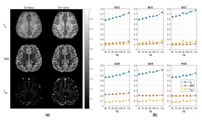 |
TE-dependence of NODDI-derived parameters and its modelling with compartmental T2 relaxation times
Ting Gong1,2, Qiqi Tong1, Hongjian He1, Jianhui Zhong1,3, and Hui Zhang2
1Center for Brain Imaging Science and Technology, College of Biomedical Engineering and Instrumental Science, Zhejiang University, Hangzhou, China, 2Department of Computer Science & Centre for Medical Image Computing, University College London, London, United Kingdom, 3Department of Imaging Sciences, University of Rochester, Rochester, NY, United States
Compartment-based models of diffusion MRI signals have become popular for probing tissue microstructure. However, the standard models do not explicitly model compartment-specific T2 relaxation. This has been shown to cause a TE-dependence of DTI-derived measures in white matter, which can confound the interpretation and quantification of results. Here we explored the TE-dependence of the widely used NODDI model and proposed a technique to determine parameter estimates that are TE-independent. This could be useful in investgateing diseases where changes of T2 and signal fraction interact.
|
|
4423.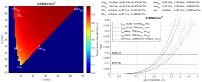 |
Optimisation of Temporal Diffusion Ratio (TDR) to maximise its potential to map large axons: Insight from simulations
William Richard Warner1, Marco Palombo1, Flavio Dell'Acqua2, and Ivana Drobnjak1
1CMIC, Computer Science Department, University College London, London, United Kingdom, 2NatBrainLab, King's College London, London, United Kingdom
Temporal Diffusion Ratio (TDR) is a novel technique introduced by Dell’Acqua et al. at ISMRM 2019, with potential for mapping areas with large diameter axons in the human brain. We aim to maximise TDR signal in practical applications by optimizing the sequence parameters used. Working in simulation, it is found that the highest TDR signal for a given axon diameter is produced by contrasting signal from sequences with as disparate as possible shapes: a tall/narrow gradient shape should be contrasted with a short/wide gradient to produce the best contrast.
|
|
4424.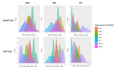 |
Single-shell derived tissue signal fraction maps show increased contrast between hippocampal subfields compared to multi-shell analysis.
Benjamin T Newman1,2, Thijs Dhollander3,4, and T. Jason Druzgal1,2
1Department of Radiology & Medical Imaging, Division of Neuroradiology, University of Virginia Health System, University of Virginia, Charlottesville, VA, United States, 2Brain Institute, University of Virginia, Charlottesville, VA, United States, 3The Florey Department of Neuroscience, University of Melbourne, Melbourne, Australia, 4The Florey Institute of Neuroscience and Mental Health, Melbourne, Australia
Recent advances in the analysis of diffusion MRI have allowed for the estimation of 3 tissue compartments in the brain from data with only a single non b=0 shell. There is currently no published quantitative comparison between signal fractions derived from either single- or multi-shell methods. Applying both single-shell analysis and multi-shell analysis to the same dataset shows high b-value single-shell analysis may increase contrast between different hippocampal subfields. While this effect may occur due to differences in microstructure between ROIs it should be a noted factor when applying either model and deserving of further study.
|
|
4425.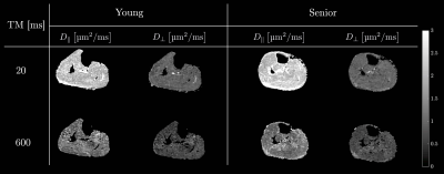 |
Skeletal Muscle Diffusion Modeling to Identify Age Related Remodeling of Muscle Microstructure
Vadim Malis1, Shantanu Sinha2, Edward Smitaman2, and Usha Sinha3
1Physics, UC San Diego, La Jolla, CA, United States, 2Radiology, UC San Diego, La Jolla, CA, United States, 3Physics, San Diego State University, San Diego, CA, United States
Diffusion modelling of the time dependence of the skeletal muscle diffusion eigenvalues derived from DTI allows one to probe tissue microstructure. We applied the Random Permeable Barrier Model to the time dependent diffusion data to identify age related remodeling in skeletal muscle microstructure. Model derived volume fraction (measure of the membrane’s ability to hinder diffusion) decreased with age while diffusion time and residence time in a cell increased with age while other model parameters such as the free diffusion coefficient, the fiber size, and membrane permeability did not.
|
|
4426.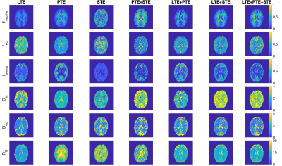 |
Improving neural soma imaging using the power spectrum of the free gradient waveforms
Maryam Afzali1, Marco Palombo2, Lars Mueller 1, Hui Zhang2, Daniel C Alexander2, Markus Nilsson3, and Derek K Jones1,4
1Cardiff University Brain Research Imaging Centre (CUBRIC), School of Psychology, Cardiff University, Cardiff, United Kingdom, 2Centre for Medical Image Computing, Department of Computer Science, University College London, London, United Kingdom, 3Clinical Sciences Lund, Radiology, Lund University, Lund, Sweden, 4Mary MacKillop Institute for Health Research, Faculty of Health Sciences, Australian Catholic University, Melbourne, Victoria, 3065, Australia
Diffusion magnetic resonance imaging is a non-invasive technique to probe the microstructural features of tissue. Conventional diffusion encoding is unable to disentangle different microstructural features; therefore, multidimensional diffusion encoding has been proposed previously to solve this problem. Here we investigate different combinations of b-tensor encoding in a three-compartment model called SANDI. To estimate the size of soma in this model, we use frequency domain analysis because optimized b-tensor encoding waveforms do not provide a well-defined diffusion-time. The results show that different combinations of linear, planar, and spherical tensor encoding can improve the estimation of a specific parameter.
|
|
4427.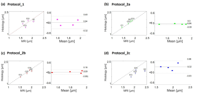 |
Accuracy of axon diameter estimation using diffusion MRI in fiber space
Mohammad Ashtarayeh1, Tobias Streubel1,2, Francisco Javier Fritz1, Joao Periquito3, Andreas Pohlmann3, Thoralf Niendorf3, Henriette Rusch4, Markus Morawski4, Carsten Jäger2, Stefan Geyer2, Bibek Dhital5, Muhamed Barakovic6,7, Simona Schiavi 8,
Alessandro Daducci8, and Siawoosh Mohammadi1,2
1Department of Systems Neurosciences, University Medical Center Hamburg-Eppendorf, Hamburg, Germany, 2Department of Neurophysics, Max Planck Institute for Human Cognitive and Brain Sciences, Leipzig, Germany, 3Berlin Ultrahigh Field Facility (B.U.F.F.), Max-Delbrueck-Center for Molecular Medicine in the Helmholtz Association, Berlin, Germany, 4Paul Flechsig Institute of Brain Research, University of Leipzig, Leipzig, Germany, 5Department of Radiology, University Medical Center Freiburg, Freiburg, Germany, 6Signal Processing Lab (LTS5), Ecole Polytechnique Federale de Lausanne, Lausanne, Switzerland, 7Translational Imaging in Neurology (ThINk) Basel, Department of Medicine and Biomedical Engineering, University Hospital Basel and University of Basel, Basel, Switzerland, 8Department of computer science, University of Verona, Verona, Italy
We investigated the effect of different diffusion MRI acquisition protocols (incl. varying gradient-strength and signal-to-noise ratio) on the accuracy of axon diameter index (ADI) estimation in fiber space for human specimens. We used the Bland-Altman plot to estimate the error in the MRI-based ADI measurement. We found that gradient strengths above 900mT/m and b-values larger than b=50k s/mm2 provided accurate estimation of ADI (error smaller than 0.13 µm). The error for the best protocol was about 0.04 µm, allowing to distinguish white matter tracts with varying effective axon diameters (e.g. corpus callosum, genu, 1.5 µm and posterior body, 1.93 µm).
|
|
4428.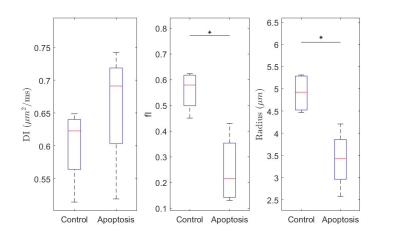 |
Using stimulated echo diffusion MRI to elucidate cellular changes during cell death
Daniel Djayakarsana1,2, Gregory J Czarnota1,2,3, and Colleen Bailey1,2
1Medical Biophysics, University of Toronto, Toronto, ON, Canada, 2Physical Sciences, Sunnybrook Research Institute, Toronto, ON, Canada, 3Radiation Oncology, Sunnybrook Health Sciences Centre, Toronto, ON, Canada
Morphological changes caused by cellular death alter the movement of water, which diffusion MRI has the potential to detect. Cellular death can be easily isolated with an in vitro model. I find that the ADC, kurtosis and the ball-sphere model showed significant changes between the control and the treated groups. Likely sources of parameter changes are the increased membrane permeability, organelle/macromolecule breakdown and differences in cellular size.
|
|
4429.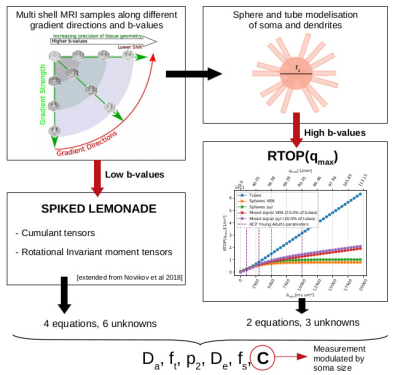 |
Indetermination-free cytoarchitecture measurements in brain gray matter via a forward diffusion MRI signal separation method
Maëliss Jallais1 and Demian Wassermann1
1Université Paris-Saclay, Inria, CEA, Palaiseau, France Non-invasive imaging at the cellular level could lead us to quantify grey matter tissue cytoarchitecture, which has so far been accessible only through histology, or by indeterminate-prone approaches. We propose a new dMRI-based index to study modulated by the size of the somas. This index can be extracted without indeterminations from any acquisition including three b-values superior to 3 ms/μm2. Simulations were experimentally confirmed by tests on the HCP MGH data set. |
|
4430.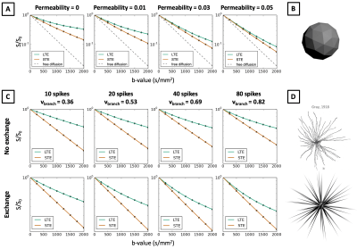 |
Discrepancy between In-vivo Measurements and Monte-Carlo Simulations with Spherical Structures in B-tensor Encoding
Noemi G Gyori1,2, Matt G Hall2, Christopher A Clark2, Daniel C Alexander1, and Enrico Kaden1
1Centre for Medical Image Computing, University College London, London, United Kingdom, 2Great Ormond Street Institute of Child Health, University College London, London, United Kingdom
In in-vivo B-tensor encoding measurements, linear tensor encoding (LTE) and spherical tensor encoding (STE) waveforms estimate the same mean diffusivity in brain white and grey matter. Contrary to this, we show that in Monte-Carlo simulations of quasi-spherical restrictions, LTE and STE signal decay curves are markedly different and lead to different mean diffusivity estimates. We investigate this discrepancy in the presence of variable compartment sizes, geometric distortions in cell shapes, permeable cell membranes, and water exchange between connected cellular geometries. Our results suggest that a strict model of restricted diffusion may not be suitable for brain tissue.
|
|
4431.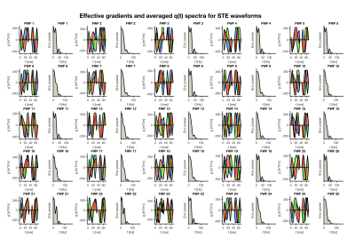 |
Varying the frequency-content of high b-value spherical diffusion encoding improves the characterisation of isotropic restricted compartment
Malwina Molendowska1, Mark Drakesmith1, Derek K. Jones1, and Chantal M. W. Tax1
1Cardiff University Brain Research Imaging Centre (CUBRIC), Cardiff University, Cardiff, United Kingdom
The use of ultra-strong diffusion gradient systems facilitates high b-values to suppress the signal from biological compartments with significant mobility, and as such the complexity of the model fit can be reduced. Here, we adopt isotropic encoding at high b-values to isolate spherical restricted compartments, and explore whether adding waveforms with different frequency-characteristics give an improvement in parameter-estimation of the spherical compartment.
|
|
4432.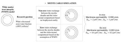 |
Decreased axon water fraction can result from either axonal loss and/or demyelination, reflecting different ex-vivo and in-vivo conditions
Zihan Zhou1, Qiqi Tong1, Qiuping Ding1, Junye Yao1, Hongjian He1, and Jianhui Zhong1,2
1Center for Brain Imaging Science and Technology, Key Laboratory for Biomedical Engineering of Ministry of Education, College of Biomedical Engineering and Instrumental Science, Zhejiang University, Hangzhou, China, 2School of Medicine and Dentistry, University of Rochester Medical Center, New York, NY, United States
White Matter Tract Integrity (WMTI) is specific to tissue microstructure. However, answers to a question of what a decreased axon water fraction (AWF), a metric of WMTI, reflects seem contradictory among previous in-vivo and ex-vivo studies. To our knowledge, there have been no studies that compare the difference in AWF decline under both ex-vivo and in-vivo conditions. Here we use Monte Carlo (MC) simulation to investigate such question and results show that AWF decline indicated demyelination under ex-vivo conditions while it also related to axonal loss in-vivo besides demyelination. Results highlighted the non-negligible effect of membrane permeability on the difference.
|
|
4433.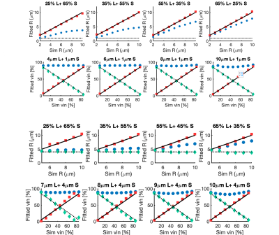 |
A simulation study of the robustness of cell size and volume mapping for tissue with two underlying cell populations using diffusion weighted MRI
Shu Xing1,2 and Ives R. Levesque1,2,3,4
1Department of Physics, McGill University, Montreal, QC, Canada, 2Medical Physics Unit, McGill University, Montreal, QC, Canada, 3McGill University Health Center Research Institute, Montreal, QC, Canada, 4Department of Oncology, McGill University, Montreal, QC, Canada
Advanced diffusion-weighted MRI allows non-invasive cell size and volume mapping in cancerous tumours. Existing methods assume that a given voxel contains one cell population. This work investigates the feasibility of estimating the radii and volume fractions of 2 cell populations co-existing in the same voxel and introduces techniques to improve the robustness and stability of fitting. The influence of noise on the fitted parameters is also examined for a range of SNR values. Reliable estimation of cell size and volume for tissue with 2 cell populations opens exciting avenue of potential applications in both tumor diagnosis and treatment monitoring.
|
 Watch the Video
 View the Poster
4434.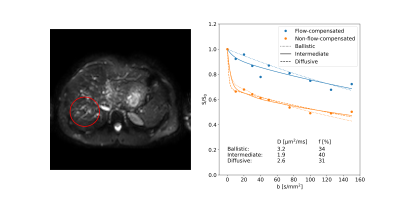 |
On the bias introduced by assuming different intravoxel incoherent motion regimes
Oscar Jalnefjord1,2, Nicolas Geades3, and Maria Ljungberg1,2
1Department of Radiation Physics, University of Gothenburg, Gothenburg, Sweden, 2Department of Medical Physics and Biomedical Engineering, Sahlgrenska University Hospital, Gothenburg, Sweden, 3Philips Clinical Science, Gothenburg, Sweden
Intravoxel incoherent motion (IVIM) analysis typically assumes that the motion of blood caused by microcirculation mimics a random walk with several steps taken during the diffusion encoding (diffusive regime). Some studies have suggested use of the other extreme regime where no direction changes occur during diffusion encoding (ballistic regime). However, data available suggest that an intermediate regime is more likely. In this study, we explore the impact of assuming different IVIM regimes on modeling and parameter estimation. Results on healthy liver indicate that substantial bias may be introduced unless proper modeling is used.
|
|
4435.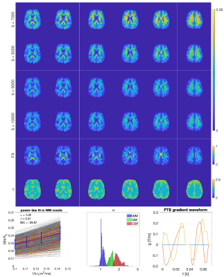 |
Power-Law Behaviour of the Diffusion-Weighted Signal Under Different B-Tensor Encoding Schemes
Maryam Afzali1, Santiago Aja-Fernandez1,2, and Derek K Jones1,3
1Cardiff University Brain Research Imaging Centre (CUBRIC), School of Psychology, Cardiff University, Cardiff, United Kingdom, 2Laboratorio de Procesado de Imagen, ETSI Telecomunicacion Edificio de las Nuevas Tecnologias, Campus Miguel Delibes s/n, Universidad de Valladolid, Valladolid, Spain, 3Mary MacKillop Institute for Health Research, Faculty of Health Sciences, Australian Catholic University, Melbourne, Victoria, Australia
It has been shown previously that for the linear (LTE), as well as planar tensor encoding (PTE) and in tissue with 'stick-like' geometry, the diffusion-weighted signal at high b-values follows a power-law. Specifically, the signal decays as $$$1/\sqrt{b}$$$ in LTE and $$$1/b$$$ in PTE. Here, we investigate whether power-law behaviors occur with other encodings and geometries. The results show that using an axisymmetric b-tensor a power-law only exists for stick-like geometries, using LTE and PTE. Finally, using ultra-strong gradients, we confirm –for the first time in vivo– that a power-law exists for PTE in white matter of the human brain.
|
|
4436.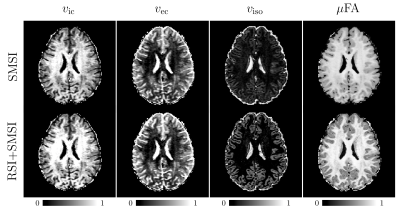 |
Tackling Degeneracy in Linear Tensor Encoding Diffusion MRI
Khoi Minh Huynh1, Ye Wu2, Hoyt Patrick Taylor IV2, Weili Lin2, and Pew-Thian Yap2
1Biomedical Engineering, UNC-Chapel Hill, Chapel Hill, NC, United States, 2Department of Radiology and BRIC, UNC-Chapel Hill, Chapel Hill, NC, United States
Model degeneracy in diffusion MRI could lead to misestimation of microstructural properties. This work presents a metric to characterize the degree of degeneracy and proposes a strategy to break degeneracy by utilizing both diffusion-weighted signal and its spherical mean. We will demonstrate that breaking the degeneracy results in more accurate quantification of microstructure.
|
|
4437.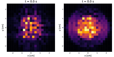 |
Toolbox for spin-level MRI simulations: an application to cardiac diffusion
Carlos Castillo-Passi1,2,3, Gabriel della Maggiora1,2,4, and Pablo Irarrazaval1,2,3,4
1Millennium Nucleus for Cardiovascular Magnetic Resonance, Santiago, Chile, 2Biomedical Imaging Center, Pontificia Universidad Católica de Chile, Santiago, Chile, 3Institute for Biological and Medical Engineering, Pontificia Universidad Católica de Chile, Santiago, Chile, 4Electrical Engineering Department, Pontificia Universidad Católica de Chile, Santiago, Chile
Cardiac diffusion imaging is a field with multiple challenges to overcome. The main challenge, compared to other techniques, is that the diffusion encoding sequence is prone to artifacts1. In this work, we show the first version of an MRI simulator that includes the most relevant effects for this type of contrast. And could be a useful tool for the development and testing of new pulse sequences. It is programmed in Julia2, which provides an intuitive object-oriented framework for the pulse sequences and creation of digital phantoms. We show two examples with results that correspond well with the theory.
|
|
4438.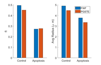 |
Using free waveform diffusion MRI to investigate time-dependence of water movement in an isotropic in vitro system
Daniel Djayakarsana1,2, Gregory J Czarnota1,2,3, and Colleen Bailey1,2
1Medical Biophysics, University of Toronto, Toronto, ON, Canada, 2Physical Sciences, Sunnybrook Research Institute, Toronto, ON, Canada, 3Radiation Oncology, Sunnybrook Health Sciences Centre, Toronto, ON, Canada
Signal attenuation due to diffusion is affected by several different acquisition parameters, such as gradient duration, spacing, strength and shape. In this study, I used free waveforms to investigate the effect of non-rectangular pulses. I chose an isotropic in vitro system such that the signal represents time and spatial scales relevant to human imaging and is independent of orientation. By varying only the gradient shape to target certain q-space frequencies, the signal measured could represent different combinations of diffusion time.
|
|
4439.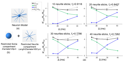 |
Quantification of soma compartment in cerebral cortical mean kurtosis with diffusion MRI
Tianjia Zhu1,2 and Hao Huang1,3
1Radiology, Children's Hospital of Philadelphia, Philadelphia, PA, United States, 2Bioengineering, University of Pennsylvania, Philadelphia, PA, United States, 3Radiology, University of Pennsylvania, PHILADELPHIA, PA, United States
Unlike white matter composed mostly of neurites, cerebral cortex includes a significant amount of somas from neurons or glial cells besides neurites. Mean kurtosis (MK) of diffusion kurtosis imaging characterizes cortical microstructural complexity contributed by both neurites and somas, but the exact contribution of somas to cortical MK is unknown. Neuronal density plays a vital role in neurodegenerative disorders. Quantitative delineation of the soma compartment is critical for assessment and therapeutic monitoring of soma compartment with noninvasive diffusion MRI. We for the first time proved and quantified soma and neurite compartmental contributions to cerebral cortical MK.
|
|
4440. |
Behaviors of high b-value direction-averaged DWI signal decay in human gray matter
Chu-Yu Lee1, In-Young Choi1,2,3, and Phil Lee1,4
1Hoglund Brain Imaging Center, University of Kansas Medical Center, Kansas City, KS, United States, 2Department of Neurology, University of Kansas Medical Center, Kansas City, KS, United States, 3Department of Molecular & Integrative Physiology, University of Kansas Medical Center, Kansas City, KS, United States, 4Department of Radiology, University of Kansas Medical Center, Kansas City, KS, United States
Neurite microenvironment has been approximated as impermeable, thin cylindrical tubes. This assumption has been validated in human white matter by a power law with the exponent of around 0.5 at b-values above 4000 s/mm2. However, the decay exponent in gray matter deviates from 0.5, suggesting that the cylindrical tube approximation does not apply in gray matter. This study aimed to study the whole-brain gray matter distribution of the decay exponent, and demonstrated an apparent contrast in the decay exponent between cortical gray matter and deep gray matter. This suggests that inherent microstructural differences may exist between these gray matter regions.
|
|
4441.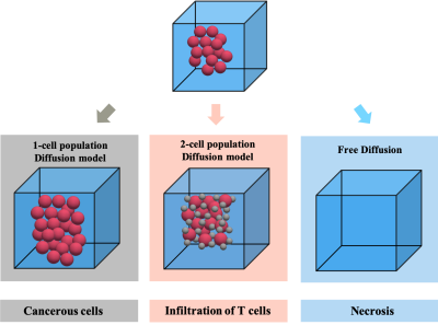 |
Detecting the evolving tumor microenvironment with diffusion microstructure modeling: a simulation study
Shu Xing1,2 and Ives R. Levesque1,2,3,4
1Department of Physics, McGill University, Montreal, QC, Canada, 2Medical Physics Unit, McGill University, Montreal, QC, Canada, 3McGill University Health Center Research Institute, Montreal, QC, Canada, 4Department of Oncology, McGill University, Montreal, QC, Canada
Diffusion MRI with microstructure modeling can nominally map voxelwised cell radii and relative cell volume fractions for the entire tumour. This work proposes a model selection method that chooses the most suitable diffusion model among the two microstructural models and the monoexponential ADC model. This technique can potentially distinguish tissue microstructure featuring one or two underlying cell populations, and acellular necrosis. Microstructural parameters were also reliably estimated. These observations suggest the possible differentiation of three tumour microenvironments resulting from cancer therapy.
|
|
4442.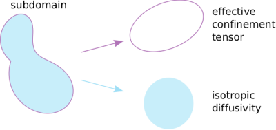 |
Compartment-specific diffusivity: A new dimension in multidimensional diffusion MRI?
Deneb Boito1,2, Cem Yolçu1, and Evren Özarslan1,2
1Department of Biomedical Engineering, Linköping University, Linköping, Sweden, 2Center for Medical Image Science and Visualization CMIV, Linköping, Sweden
We extend the diffusion tensor distribution imaging framework so that each subdomain making up the tissue is represented by an effective confinement (rather than diffusion) tensor. The confinement tensor is determined by the subdomain’s shape as well as the acquisition parameters and is the physical quantity that leads to microscopic diffusion anisotropy. In this approach, the scalar-valued diffusivity remains as an independent parameter, which could be different in different subdomains. We demonstrate the difficulties in estimating compartment-specific diffusivity alongside the confinement tensor distribution when typical free gradient waveforms are employed.
|
|
4443.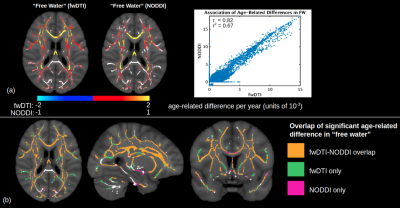 |
Mapping diffusion in aging white matter: A comparison of two free-water modeling approaches
Jordan A. Chad1,2, Ofer Pasternak3, and J. Jean Chen1,2
1Rotman Research Institute, Baycrest Health Sciences, Toronto, ON, Canada, 2Department of Medical Biophysics, University of Toronto, Toronto, ON, Canada, 3Departments of Psychiatry and Radiology, Brigham and Women's Hospital, Harvard Medical School, Boston, MA, United States
To gain more insight into age-effects on white matter diffusivity, advanced diffusion MRI models attempt to separate the effect of cellular diffusion from extracellular isotropic water movement by modeling the latter as free water (FW). Using these models, increased diffusivity with age is typically attributed to an enlargement of the FW compartment with age. The current study compares the sensitivity of FW to age using two common modeling approaches: single-shell fwDTI and multi-shell NODDI.
|
|
4444.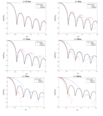 |
What’s the FWAF? A Finite Width Adjustment Factor for the effect of finite pulse duration on the diffusion signal in impermeable cylinders
Nadia A S Smith1, Jessica E Talbott1, Chris A Clark2, and Matt G Hall1,2
1National Physical Laboratory, Teddington, United Kingdom, 2UCL Great Ormond Street Institute of Child Health, University College London, London, United Kingdom
A common approach to microstructure imaging in diffusion MRI is to fit the signal with a weighted sum of geometric compartments. This approach is widespread but the need for analytical closed-form solutions necessitates highly restrictive assumptions about the underlying physics which are rarely met in practice. In particular, violation of the narrow-pulse approximation is a significant potential source of bias. This abstract investigates the effect of violating the narrow pulse approximation numerically and proposes a simple effect correction factor to reduce apparent bias as a scale factor on q as a function of δ.
|
|
 |
4445.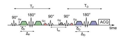 |
Apparent exchange rate (AXR) mapping: Influence of extracellular volume fraction and membrane permeability
Julian Rauch1,2, Tristan Anselm Kuder1, Frederik Bernd Laun3, Karel D. Klika4, Peter Bachert1,2, and Dominik Ludwig1,2
1Department of Medical Physics in Radiology, German Cancer Research Center (DKFZ), Heidelberg, Germany, 2Faculty of Physics and Astronomy, Heidelberg University, Heidelberg, Germany, 3Institute of Radiology, University Hospital Erlangen, Friedrich-Alexander-Universität Erlangen-Nürnberg (FAU), Erlangen, Germany, 4Molecular Structure Analysis, German Cancer Research Center (DKFZ), Heidelberg, Germany
Apparent exchange rate (AXR) mapping can be used to non-invasively investigate water exchange between the intra- and extracellular compartment, which might yield insight into cell membrane permeability. However, the measured AXR is also significantly influenced by intra- and extracellular water fractions. In this study, using simulations and experiments with yeast cells, we show that – for low cell concentrations – the AXR exhibits only a moderate dependence on the cell concentration, while for high concentrations, a stronger influence of the packing density on AXR occurs so that it becomes more difficult to disentangle these two influencing parameters.
|
4446.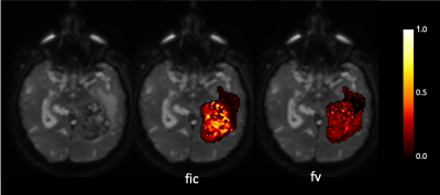 |
Estimation of the vascular fraction in brain tumors by VERDICT correlated with Perfusion MRI
Matteo Figini1, Antonella Castellano2, Valentina Pieri2, Samira Bouyagoub3, Andrea Falini2, Daniel C Alexander1, Mara Cercignani3, and Eleftheria Panagiotaki1
1Centre for Medical Image Computing, Department of Computer Science, University College London, London, United Kingdom, 2Neuroradiology Unit and CERMAC, Vita-Salute San Raffaele University and IRCCS San Raffaele Scientific Institute, Milano, Italy, 3Clinical Imaging Sciences Centre, Brighton and Sussex Medical School, Brighton, United Kingdom
We used the VERDICT framework to find clinically useful microstructural models to characterize vasculature in brain tumors. We correlated the vascular fractions, estimated by all possible three-compartment models, with perfusion MRI metrics (plasma volume and cerebral blood volume) derived from independent measurements on the same patients. The models with the strongest correlation with the perfusion MRI and clinical data incorporate spherical restriction for the intracellular compartment, isotropic diffusion for the extracellular compartment, and isotropically hindered or restricted pseudo-diffusion for the vascular compartment.
|
|
4447.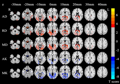 |
Microstructural brain abnormalities and reorganization of early-blind adolescents: a voxel-based diffusion kurtosis imaging study
Zhifeng Zhou1, Xia Liu2, Long Qian3, Gangqiang Hou2, Wentao Jiang2, and Hengguo Li4
1Radiology, Shenzhen Mental Health Center/Shenzhen Kangning Hospital, Shenzhen, China, 2Shenzhen Mental Health Center/Shenzhen Kangning Hospital, Shenzhen, China, 3GE Healthcare, Beijing, China, 4The First Affiliated Hospital of Jinan University, Guangzhou, China
An important focus of blind brain research, especially the early-blind brain, is how to identify the specific neural plasticity patterns. Neuroimaging studies, particularly the diffusion MRI, are powerful probes for characterizing the microstructural changes in human brain. Additionally, previous study indicated that the Diffusion Kurtosis Imaging (DKI) is an advanced diffusion model without the assumption of Gaussian distribution. Taken together, it is feasible to utilize the DKI to investigate the structural neuroplasticity in early-blind brain. Our results demonstrated that the neural reorganization and compensatory development process induced by visual deprivation are coexisted in early-blind adolescents. Furthermore, the diffusion kurtosis metrics are more sensitive to detect the pathology and development related brain regions than diffusion tensor metrics.
|
|
4448.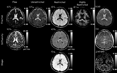 |
Comprehensive analysis of full-spectrum diffusion weighted MR images with Multimodal Anomalous Diffusion Method (MAD)
Frederick C. Damen1, Mirko Vukelich1, Nitu Saran1, and Kejia Cai1
1Radiology, University of Illinois at Chicago, CHICAGO, IL, United States
In physiological and pathological conditions in which multiple underlying tissue properties are expected to vary the diffusion weighted signal, the diagnostic value of conventional apparent diffusion coefficient (ADC) notably decreases. By fully dissecting the whole spectrum of diffusion weighted imaging (DWI) data from low to high b‑values, we developed a method to extract the anomalous diffusion parameters for four water milieus: flow, and, unrestricted, restricted, and highly restricted diffusion, thus, expounding upon the diverse diffusivity within a voxel.
|
4449.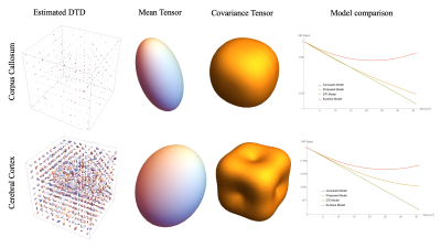 |
Diffusion Tensor Distribution (DTD) MRI Reimagined
Magdoom Kulam Najmudeen1, Michal E Komlosh1,2, Dario Gasbarra3, and Peter J Basser1
Video Permission Withheld
1SQITS/NICHD, National Institute of Health, Bethesda, MD, United States, 2The Henry M. Jackson Foundation for the Advancement of Military Medicine, Inc., Bethesda, MD, United States, 3University of Helsinki, Helsinki, Finland
A new DTD imaging method is presented with novel data acquisition schemes for higher rank b-matrices, and analysis pipeline to estimate the mean and covariance of diffusion tensor. The developed pulse sequence is simple, easy to implement with well defined diffusion, mixing and pulsed gradient dwell times and immune to concomitant fields. The method is validated using PDMS phantom and excised rat brain tissue. Estimated DTD was used to compute the microscopic FA and AD. The phantom results agreed as expected with zero FA and uFA and accurate AD. The approach was able to capture the heterogeneity in the brain.
|
|
4450.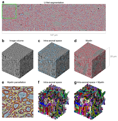 |
Automated segmentation of human axon and myelin from electron microscopy data using deep learning for microstructural validation and simulation
Qiyuan Tian1,2, Chanon Ngamsombat1, Hong-Hsi Lee3,4, Daniel R. Berger5, Yuelong Wu5, Qiuyun Fan1,2, Berkin Bilgic1,2, Dmitry S. Novikov3,4, Els Fieremans3,4, Bruce R. Rosen1,2, Jeff W. Lichtman5, and Susie Y. Huang1,2
1Martinos Center for Biomedical Imaging, Massachusetts General Hospital, Charlestown, MA, United States, 2Department of Radiology, Harvard Medical School, Boston, MA, United States, 3Department of Radiology, Center for Biomedical Imaging, New York University School of Medicine, New York, NY, United States, 4Center for Advanced Imaging Innovation and Research (CAI2R), New York University School of Medicine, New York, NY, United States, 5Department of Molecular and Cellular Biology, Harvard University, Cambridge, MA, United States
Diffusion microstructural metrics represent inferences of axonal size and morphology rather than directly imaged quantities, validation of these metrics is essential. With novelty of multibeam-serial electron microscopy, high-resolution images of human white matter can be acquired at nanometer resolution over volumes of tissue large enough to capture the diffusion-MRI dynamics extending over length scales comparable to MRI voxel size. This work presents automated segmentation of serial EM of a sub-volume of human white matter using a 3D convolutional neural network studying variations in axonal diameter over the longest axons within the volume of tissue.
|
|
4451.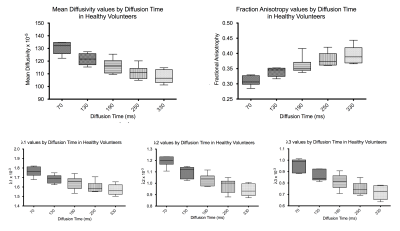 |
Time-dependent diffusion in healthy human calf muscle in a clinically feasible acquisition time
Amy R McDowell1, Matthew G Hall1,2, Thorsten Feiweier3, and Chris A Clark1
1GOS UCL Institute of Child Health, London, United Kingdom, 2Medical Radiation Physics, National Physical Laboratory, London, United Kingdom, 3Siemens Healthcare GmbH, Erlangen, Germany
Muscle fibres have a complex hierarchical internal structure which leads to a marked decrease in the observed MR diffusion coefficient with diffusion time. We expect the form of this diffusion time dependence to be highly sensitive to changes in this internal structure. However, characterising the diffusion time dependence in tissue can be highly demanding of total acquisition time. We examine the time-dependence of diffusion indices in human calf muscle in healthy volunteers in a clinically feasible scan time. We find significant time dependence and diffusion restriction in all directions, including longitudinally, due to the internal structure of muscle fibres.
|
|
4452.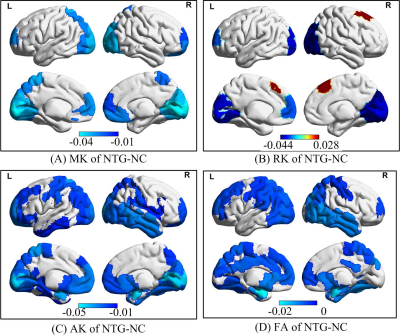 |
Brodmann atlas based non-Gaussion water diffusion analysis of normal tension glaucoma: A diffusion kurtosis imaging study
Xiaoxia Qu1, Lizhi Xie2, Qian Wang1, Ting Li1, Jian Guo1, and JunFang Xian1
1Radiology Department, Beijing Tongren Hospital, Capital Medical University, Beijing, China, 2GE Healthcare, MR Research China, Beijing, Beijing, China
To access the non-Gaussion water diffusion characteristic of normal tension glaucoma (NTG), we analyzed the diffusion parameters generated from diffusion kurtosis imaging (DKI). The parameters derived from DKI within changed brain regions of NTG group were reduced and involved the occipital lobe. The visual cortex in NTG group had lower degree of diffusional heterogeneity and microstructural complexity compared to normal control (NC) group.
|
|
4453.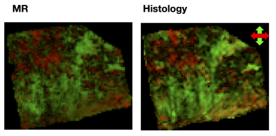 |
Imaging meningioma tumor microstructure: a comparison between quantitative histopathology and high-resolution diffusion tensor imaging
Jan Brabec1, Filip Szczepankiewicz2,3,4, Elisabet Englund5, Johan Bengzon6, Linda Knutsson1,7, Carl-Fredrik Westin2,3, Pia Sundgren4,8, and Markus Nilsson4
1Clinical Sciences Lund, Medical Radiation Physics, Lund University, Lund, Sweden, 2Department of Radiology, Brigham and Women's Hospital, Boston, MA, United States, 3Harvard Medical School, Boston, MA, United States, 4Clinical Sciences Lund, Diagnostic Radiology, Lund University, Lund, Sweden, 5Clinical Sciences, Oncology and Pathology, Lund University, Lund, Sweden, 6Clinical Sciences, Neurosurgery, Lund University, Lund, Sweden, 7Russell H. Morgan Department of Radiology and Radiological Science, Johns Hopkins University School of Medicine, Baltimore, MD, United States, 8Lund University Bioimaging Center, Lund University, Lund, Sweden
We explore anisotropy of meningioma tumors by using structure tensor analysis of histological images and high-resolution diffusion MRI scans. We observed a good visual agreement between the structure- and diffusion-based anisotropy measures, but a more modest quantitative agreement. The results highlight the need to image microscopic rather than voxel-level fractional anisotropy in meningioma tumors.
|
|
4454. |
In search of the right solution for Ex-Vivo Diffusion Weighted Imaging
Shijia Teo1, Jochen Leupold1, Juergen Hennig1, Marco Reisert1, and Valerij G. Kiselev1
1Dept. of Radiology, Medical Physics, Medical Center - University of Freiburg, Faculty of Medicine, University of Freiburg, Freiburg, Germany
Utilising the right solution in immersing an ex-vivo sample is the first and yet important step in optimising dWI, while preserving the biological and structural integrity of the sample for effective and meaningful repeated scans. In so doing, using 1%PFA, while toxic in preparation, has many other benefits in optimizing diffusion-weighted imaging (dWI). For a safer handling option, PBS + 0.005%NaN3 is also a viable alternative.
|
|
4455.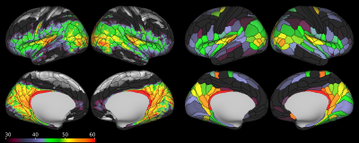 |
Parcellation of the human brain cortex using a model free, sparse acquisition diffusion MRI approach («S-index»)
Tetsuya Yamamoto1, Masaki Fukunaga1, Norihiro Sadato1, and Denis Le Bihan1,2,3
1Department of System Neuroscience, National Institute for Physiological Sciences, Okazaki, Japan, 2Human Brain Research Center, Graduate School of Medicine, Kyoto University, Kyoto, Japan, 3NeuroSpin/Joliot, CEA-Saclay Center, Paris-Saclay University, Gif-sur-Yvette, France
We have used a model free diffusion MRI approach (S-index) to classify brain tissue types from the “proximity” or resemblance of their diffusion MRI signal profile at a sparse set of key b values (maximizing sensitivity to tissue microstructure) to a library of “signature” signal profiles (e.g. typical brain grey and white matter). 3D S-index maps have been generated and overlaid on a brain parcellation atlas from the Human Connectome Project showing differences among cortical brain areas.
|
|
4456.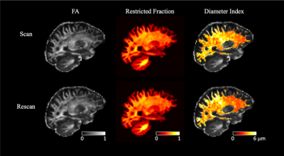 |
Scan-Rescan Reliability of Microstructural Imaging Metrics Derived from High-Gradient Diffusion MRI
Maya Polackal1, Qiyuan Tian1,2, Chanon Ngamsombat1,2,3, Aapo Nummenmaa1,2, Thomas Witzel1,2, Eric C. Klawiter2,4, Susie Y. Huang1,2,5, and Qiuyun Fan1,2
1Department of Radiology, Athinoula A. Martinos Center for Biomedical Imaging, Massachusetts General Hospital, Charlestown, MA, United States, 2Harvard Medical School, Boston, MA, United States, 3Department of Radiology, Faculty of Medicine, Siriraj Hospital, Mahidol University, Thailand, 4Department of Neurology, Massachusetts General Hospital, Boston, MA, United States, 5Harvard-MIT Division of Health Sciences and Technology, Massachusetts Institute of Technology, Cambridge, MA, United States
Scan-rescan reliability of axon diameter index, restricted volume fraction, and DTI metrics derived from high-gradient diffusion MRI was assessed in seven healthy volunteers. Maps of all three metrics were visually comparable from scan/rescan sessions, and moderate to high voxel-wise correlation coefficients were obtained for all metrics. Intraclass correlation coefficients were high for well-defined white matter tracts across all subjects. Our preliminary findings support the robustness and reliability of high-gradient diffusion MR metrics for use in clinical research studies.
|
|
4457.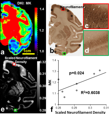 |
Neuroanatomical underpinning of cerebral cortical mean kurtosis from diffusion MRI
Tianjia Zhu1,2 and Hao Huang1,3
1Radiology, Children's Hospital of Philadelphia, Philadelphia, PA, United States, 2Bioengineering, University of Pennsylvania, Philadelphia, PA, United States, 3Radiology, University of Pennsylvania, Philadelphia, PA, United States
Cortical mean kurtosis (MK) of diffusion MRI characterizes microstructural complexity in the cerebral cortex. In this study, we tested a linear model that soma and neurite densities jointly contributed to MK with different weights. The actual soma and neurite densities were quantified from digitization of macaque brain histological images of Nissl and neurofilament staining. DKI was acquired from 6 postmortem macaque brains. We found actual MK measurement was accurately predicted by a weighted linear combination of soma and neurite densities from histological images with higher neurite density weight.
|
|
4458.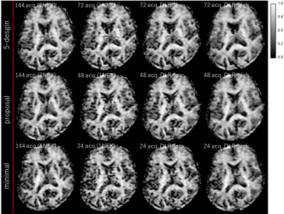 |
Evaluation of Denoising Deep Convolutional Neural Network for Double Diffusion Encoding Technique
Hiroshi Kusahara1, Masanori Ozaki2, Masahiro Abe1, Koji Kamagata3, Masaaki Hori4, and Shigeki Aoki3
1Advanced MRI development PJ Team, Canon Medical Systems Corporation, Kanagawa, Japan, 2Research&Development Center, Canon Medical Systems Corporation, Kanagawa, Japan, 3Department of Radiology, Juntendo University School of Medicine, Tokyo, Japan, 4Department of Radiology, Toho University Omori Medical Center, Tokyo, Japan Poster Permission Withheld
Double Diffusion Encoding (DDE) is a diffusion measurement technique that applies two directions of diffusion encoding in parallel and orthogonal directions and can calculate μFA that can evaluate detailed information of anisotropy in voxels. However, since the diffusion encoding method generally acquired many directions, the acquisition time becomes long. In this study, we evaluated DDE technique applying denoising DLR recently developing. It was demonstrated that the dDLR techniques are capable of generating DDE with higher SNR compared to normal DDE, with the additional benefit of being able to optimize the acquisition time and number of acquisitions without affecting μFA.
|
|
4459.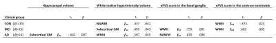 |
Exploring the vascular and neurodegenerative origin of increased interstitial fluid diffusion in intravoxel incoherent motion
Merel M. van der Thiel1,2,3, Whitney M. Freeze2,3,4, Alida A. Postma1, Inge I.C.M. Verheggen1,2,3, Sau M. Wong1, Joost J.A. de Jong1, Frans R.J. Verhey2,3, Inez H.G.B. Ramakers2,3, Walter H. Backes1,3, and Jacobus F.A. Jansen1,3
1Department of Radiology and Nuclear Medicine, Maastricht University Medical Center, Maastricht, Netherlands, 2Department of Psychiatry and Neuropsychology, Maastricht University, Maastricht, Netherlands, 3School for Mental Health and Neuroscience, Alzheimer Center Limburg, Maastricht, Netherlands, 4Department of Radiology, Leiden University Medical Center, Leiden, Netherlands
Spectral analysis using non-negative least squares in intravoxel incoherent motion enables estimation of an intermediate component in the diffusion spectrum between parenchymal diffusion and microvascular pseudodiffusion. However, the precise source of the intermediate component remains unknown. By studying the relation of the intermediate component with vascular (i.e. WMH volume) and neurodegenerative (i.e. hippocampal atrophy) biomarkers, the current study aimed to gain more knowledge about the interpretation of the intermediate peak in relation to cognitive status and its underlying pathology. The intermediate peak seems to be a promising imaging biomarker for microvascular and neurodegenerative pathology in Alzheimer’s disease and its prestages.
|
|
4460.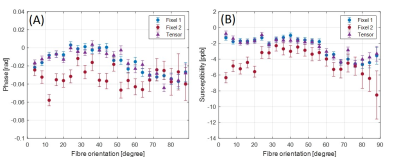 |
Influence of WM fibre orientation on gradient-echo derived tissue parameters
Tonima S Ali1, Elisabeth C van der Voort1,2, and Markus Barth3,4
1University of Queensland, Brisbane, Australia, 2Eindhoven University of Technology, Eindhoven, Netherlands, 3The University of Queensland, University of Queensland, Brisbane, Australia, 4The ARC Training Centre for Innovation in Biomedical Imaging Technology, University of Queensland, Brisbane, Australia
This study investigates the effect of white matter fibre orientation with respect to main magnetic field on gradient-echo (GRE) derived tissue phase, susceptibility, magnetization transfer ratio, R2* and water fraction of myelin water compartments. Spherical deconvolution is used to identify multiple fibre bundles within each imaging voxel. We observed that these GRE derived parameters show moderate sensitivity towards the changes in fibre orientations. Using bi- instead of mono-exponential fitting, it additionally showed that myelin R2* seems more sensitive to the fibre orientation than previously anticipated.
|
|
4461.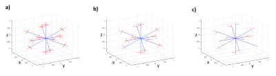 |
Preliminary study of diffusion encoding scheme for double diffusion encoding in clinical application on 3T.
Masanori Ozaki1, Hiroshi Kusahara2, Masahiro Abe2, Masaaki Hori3, Koji Kamagata4, and Shigeki Aoki4
1Research and Development Center, Canon Medical Systems Corporation, Kanagawa, Japan, 2Advanced MRI development PJ Team, Canon Medical Systems Corporation, Kanagawa, Japan, 3Department of Radiology, Toho University Omori Medical Center, Tokyo, Japan, 4Department of Radiology, Juntendo University School of Medicine, Tokyo, Japan Poster Permission Withheld
Double diffusion encoding (DDE) can measure the microscopic anisotropy (μFA), however one of problems for clinical application is that the acquisition time is prolonged due to the many diffusion encoding patterns necessary. Recently, DDE with a reduced number of acquisitions has been proposed as a solution and been validated as a solution at 9.4T. The aim of this study is to validate the proposed solution for clinical application on 3T clinical scanners. Our results show that our proposed solution can obtain more accurate μFA than previous reported solutions on 3T clinical scanners.
|
|
4462. |
Precision and goodness of fit of diffusion-based microstructure models in brain tumours: an integrated information theory approach
Umberto Villani1,2, Erica Silvestri1,2, Marco Castellaro1,2, Simona Schiavi3, Mariagiulia Anglani4, Silvia Facchini1,5, Elena Monai1,5, Domenico Davella1,5, Alessandro Della Puppa6, Diego Cecchin7, Maurizio Corbetta1,5,8, and Alessandra Bertoldo1,2
1Padova Neuroscience Center, University of Padova, Padova, Italy, 2Department of Information Engineering, University of Padova, Padova, Italy, 3Department of Computer Science, University of Verona, Verona, Italy, 4Neuroradiology Unit, University of Padova, Padova, Italy, 5Department of Neuroscience, University of Padova, Padova, Italy, 6Departments of Neurosurgery, Neuroscience, Psychology, Pharmacology, and Child Health, University of Firenze, Firenze, Italy, 7Department of Medicine, Unit of Nuclear Medicine, University of Padova, Padova, Italy, 8Departments of Neurology, Radiology, Neuroscience, Washington University School of Medicine, St.Louis, MO, United States
Diffusion-based microstructure modeling techniques potentially provide significant biomarkers to characterize the tumoral architecture in the human brain. While clinical studies focus on the application of these technique, not enough care is being devoted to understand whether the employed models provide precise and reliable parameter estimates when fitted on the cancerous tissues. The present works tackles these issues on a cohorts of 11 patients diagnosed with different types of brain tumours by quantifying the variance of parameter estimates and the goodness-of-fit in an integrated view borrowing concepts from information theory.
|
|
4463.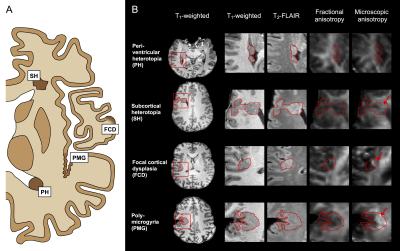 |
Diffusion MRI with tensor-valued diffusion encoding as a marker of axonal content within malformations of cortical development
Björn Lampinen1, Ariadne Zampeli2, Filip Szczepankiewicz3,4, Kristina Källén5, Maria Compagno Strandberg2, Isabella Björkman-Burtscher6, and Markus Nilsson3
1Clinical Sciences Lund, Medical Radiation Physics, Lund University, Lund, Sweden, 2Clinical Sciences Lund, Neurology, Lund University, Lund, Sweden, 3Clinical Sciences Lund, Diagnostic Radiology, Lund University, Lund, Sweden, 4Brigham and Women's Hospital, Harvard Medical School, Boston, MA, United States, 5Clinical Sciences Lund, AKVH-Neurology Helsingborg, Lund University, Lund, Sweden, 6Radiology, Sahlgrenska university hospital, Gothenburg, Sweden
MRI is central to presurgical workup for malformations of cortical development (MCD) but findings are ambiguous on morphological imaging. For example, assessments on cortical thickness may disagree with histology. We investigated whether this gap may be bridged by diffusion MRI (dMRI) with the novel ‘tensor-valued diffusion encoding’ technique. Results from thirteen patients showed WM-like content with high microscopic anisotropy within lesions that were uniform and GM-like in T1- and T2-weighted images and on conventional dMRI. As a marker of axonal content less confounded by myelination and orientation dispersion, tensor-valued diffusion encoding may improve MCD characterization and evaluation of cortical thickness.
|
4464.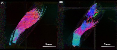 |
Diffusion Tensor Tractography of Tendons as a Tool for Assessing MRgFUS Ablation
William Chu Kwan1, Warren Foltz2, Ben Keunen1, Matt Walker3, Karolina Piorkowska1, Adam Waspe1, and James Drake4
1Center for Image Guided Innovation and Therapeutic Intervention, Hospital for Sick Children, Toronto, ON, Canada, 2Radiation Oncology, University Health Network, Toronto, ON, Canada, 3Krembil Research Institute, University Health Network, Toronto, ON, Canada, 4Department of Surgery, Center for Image Guided Innovation and Therapeutic Intervention, Hospital for Sick Children, Toronto, ON, Canada
MRgFUS ablation of tendons is a promising treatment to disrupt tendons in conditions such as musculotendinous contractures. In this study, DTI and tractography were proposed as tools to assess tendons before and after MRgFUS ablation, providing a quantitative, directional, and visual assessment of tendon tract architecture and integrity. Diffusivity parameters (FA and ADC) changed in accordance to the tendon fascicle integrity. Fiber tractography visually delineated healthy tendon fascicles and confirmed tract disruptions at the same location as registered with b0 and T1-weighted images. This study demonstrates the potential for DTI and tractography as assessment tools for MRgFUS ablation treatments.
|
|
4465.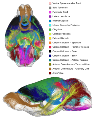 |
A novel ex vivo structural connectome atlas of the C57Bl6 mouse brain using ultra-high field diffusion MRI at 17.2T
Ivy Uszynski1, Roxane Golgolab1, David A. Barrière1, Tomokazu Tsurugizawa1, Michel Simonneau2,3,4,5, Luisa Ciobanu1, and Cyril Poupon1
1NeuroSpin, CEA, Gif-sur-Yvette, France, 2Centre Psychiatrie & Neurosciences, INSERM U894, Paris, France, 3Ecole Normale Supérieure Paris-Saclay & LAC-CNRS, Institut d’Alembert, Cachan, France, 4LBPA, Institut d’Alembert, Ecole Normale Supérieure Paris-Saclay, Université Paris-Saclay, Cachan, France, 5Département de Biologie, Ecole Normale Supérieure Paris-Saclay, Université Paris-Saclay, Cachan, France
Diffusion MRI is a powerful tool to investigate the structural connectivity of the brain. Ultra-high field preclinical MRI systems are equipped with strong gradients that allow to reach higher spatial and angular resolutions in animal models, enabling to segment white matter bundles in a similar way to what was achieved in humans. In this study, we propose to adapt the clustering approach of Guevara to rodents to establish a novel atlas of the connectivity of C57BI6 mice brains. 3D Hybrid diffusion imaging was performed ex-vivo allowing the reconstruction of a novel white matter atlas including 25 well-described fiber bundles.
|
|
4466.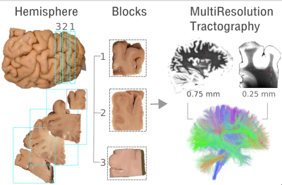 |
Investigating SLFI anatomy using multi-resolution dMRI
Chiara Maffei1, Robert Jones1, Connor Johnson2, Hui Wang1, and Anastasia Yendiki1
1Athinoula A. Martinos Center for Biomedical Imaging, Massachusetts General Hospital and Harvard Medical School, Charlestown, MA, United States, 2Brown University, Providence, RI, United States
The ability of tractography methods to discern between different fiber populations in the presence of challenging anatomical architectures remains limited, and it is hampered by the low spatial resolution achievable by diffusion MRI (dMRI). As a result, tractography often fails to reliably reconstruct some major white matter connections of the human brain. Here we incorporate high-resolution ex-vivo dMRI into the tractography process to understand whether this additional information can improve tractography results.
|
|
4467.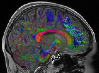 |
Cortical projections of the superoanterior fasciculus (SAF)
Szabolcs David1, Michel Thiebaut de Schotten2, Fenghua Guo1, Flavio Dell’Acqua3, Alexander Leemans1, and Alberto de Luca1
1Image Sciences Institute, UMC Utrecht, Utrecht, Netherlands, 2Brain Connectivity and Behaviour Laboratory, Sorbonne Universities, Paris, France, 3NatBrainLab, Department of Forensics and Neurodevelopmental Sciences, Sackler Institute for Translational Neurodevelopment, King's College London, London, United Kingdom
The superoanterior fasciculus (SAF) is defined as a bilateral tract in the frontal lobe that resembles the structure of the anterior cingulum, but is located superior, anterior and lateral. In this work, we investigated the cortical projections of the structure utilizing the latest multi-shell multi tissue (MSMT) constrained spherical deconvolution (CSD) method to analyze diffusion MRI data from the Human Connectome Project (HCP). Our results show that the paracentral lobular, mid cingulate cortex and the orbital- and polar frontal cortex show high SAF termination prevalence, suggesting a novel connection in the frontal lobe.
|
|
4468.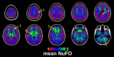 |
Diffusion MRI analysis of complex fiber configurations in human brain white matter and cortex
Szabolcs David1, Hamed Y Mesri1, Fenghua Guo1, Alexander Leemans1, and Alberto de Luca1
1Image Sciences Institute, UMC Utrecht, Utrecht, Netherlands
Fiber configurations in the human brain white matter and cortex are dominant with multiple orientations. In this work, we investigated the prevalence of crossing fibers in the brain, utilizing the latest multi-shell multi tissue (MSMT) constrained spherical deconvolution (CSD) method to analyze diffusion MRI data from the Human Connectome Project (HCP). Voxel-wise number of fiber orientation (NuFO) was calculated from 56 subjects, using the Generalized Richardson Lucy (GRL) framework, which offers robust fiber orientation distribution (FOD) estimation. Our results suggest that 83% of the voxels have at least one and 37% of the voxels have at least two fiber orientations.
|
|
4469.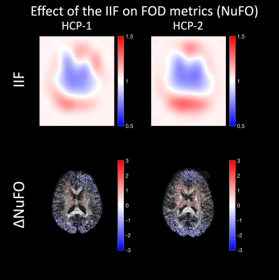 |
Does the intensity inhomogeneity field in diffusion MRI data affect constrained spherical deconvolution results?
Alberto De Luca1, Hamed Yousefi Mesri1, Szabolcs David1, Martijn Froeling2, and Alexander Leemans1
1Image Sciences Institute, University Medical Center Utrecht, Utrecht, Netherlands, 2Department of Radiology, University Medical Center Utrecht, Utrecht, Netherlands
The correction for the intensity inhomogeneity field (IIF) is becoming de-facto a pre-processing step in diffusion MRI, but its effect on CSD remains unclear. In this study, we investigated the fundamental effects of the IIF on CSD and the effectiveness of corrections based on a tool commonly applied to structural MRI (N4ITK). We show that the IIF modulates the amplitude of the fiber orientation distribution (FOD), influencing not only the estimation of the response function, but also subsequent uses of the FOD amplitude, as tractography termination criteria or the estimation of the number of fibers (NuFO).
|
|
4470.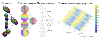 |
RIPPLE: A new framework for estimating rotationally-invariant with paired-ODF spatial correlations in fiber tracking using MRI
Ying-Chia Lin 1,2, Steven Baete1,2, Xiuyuan Wang1,2, and Fernando Boada1,2
1Center for Biomedical Imaging, Department of Radiology, NYU School of Medicine, New York, NY, United States, 2Center for Advanced Imaging Innovation and Research (CAI2R), NYU School of Medicine, New York, NY, United States
Quantification of internal structure (i.e., microstructure) is central to for the modeling of diffusion signals. In recent years, rotationally-invariant measures have generated significant attention because of their tremendous potential for characterizing the underlying structural information contained in the orientation-distribution-function (ODF). We demonstrate the use of a new approach for the analysis of long-range structural connectivity based on the use of pairwise correlations between ODF’s. This approach is shown to better capture differences between the underlying morphological features present within a fiber bundle.
|
|
4471.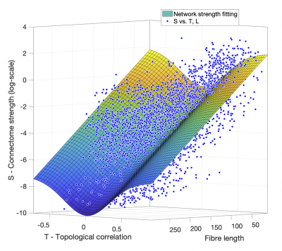 |
The investigation of topological contributions: A novel approach for modelling brain networks
Xiaoyun Liang1,2, Chun-Hung Yeh2, Govinda Poudel1, Juan F Domínguez D1, and Karen Caeyenberghs1
1Mary Mackillop Institute for Health Research, Australian Catholic University, Melbourne, Australia, 2Florey Institute of Neuroscience and Mental Health, University of Melbourne, Melbourne, Australia
Diffusion MRI streamline tractography offers a unique approach to probe into such mechanisms underlying structural brain network. In this study, we propose modelling network strength based on both fibre length and topological profile of brain network. The proposed model leads to more robust fitting outcomes. Meanwhile, our results show that network thresholding could dramatically alter the relationship between the connection strength and physical length; this would inevitably compromise further network analyses. Further results demonstrate that within- and between-hemisphere connections bear distinct patterns in terms of the relationship between network strength and topological correlation matrix.
|
|
4472.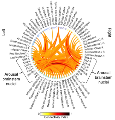 |
Redundancy of arousal brainstem structural connectivity pathways in humans by 7 Tesla HARDI
María Guadalupe García-Gomar1, Kavita Singh1, and Marta Bianciardi1
1Department of Radiology, Athinoula A. Martinos Center for Biomedical Imaging, Massachusetts General Hospital and Harvard Medical School, Boston, MA, United States
Arousal plays a crucial role in wakefulness/sleep, autonomic function, affect and attention. Recent meta-analysis of rat neuroanatomical macroconnectome based on pathway-tracing studies showed redundancy of arousal mechanism through the presence of multiple connectivity pathways and of high interconnectivity among arousal brainstem nuclei. Using 7 Tesla HARDI MRI and a recently developed brainstem nuclei atlas, we build the structural connectome of brainstem arousal nuclei in living humans. In line with rat studies, this connectome showed high redundancy through the presence of multiple interconnected pathways. This connectome might be used to better understand arousal and its impairment.
|
|
4473.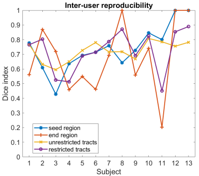 |
What factor influences most the reproducibility of MRI-based white matter tract estimation using probabilistic tractography?
Irène Brumer1,2,3, Enrico De Vita1, Jonathan Ashmore2,4, Jozef Jarosz2, and Marco Borri2
1Department of Biomedical Engineering, School of Biomedical Engineering and Imaging Sciences, King’s College London, London, United Kingdom, 2Department of Neuroradiology, King’s College Hospital, London, United Kingdom, 3Computer Assisted Clinical Medicine, Medical Faculty Mannheim, Heidelberg University, Mannheim, Germany, 4Department of Medical Physics and Bioengineering, NHS Highland, Inverness, United Kingdom
MRI-based white matter tract estimation using probabilistic tractography is valuable for presurgical planning. In this work, we investigate three factors influencing the reproducibility of probabilistic tractography: the tract threshold used to visualise the analysis results in the final tractography image, the manually drawn seed region, and the end region of the tracts. A compromise between low risk and high reproducibility has to be found for the tract threshold. The end region definition was found to be the most important parameter for the inter-user reproducibility of restricted tracts. Good inter-user reproducibility for the unrestricted tracts is achievable for similar seed regions.
|
|
4474. |
Mapping white matter specificity captured by diffusion tractography through deep learning on structural MRI
Qi Yang1, Colin Hansen1, Francois Rheault2, Bramsh Qamar3, Owen Williams4, Susan Resnick4, Eleftherios Garyfallidis3, Adam W Anderson5,6, Maxime Descoteaux2, Bennett A Landman5,6,7,8, and Kurt G Schilling5
1Computer Science, Vanderbilt University, Nashville, TN, United States, 2Sherbrooke Connectivity Imaging Laboratory (SCIL), Universite de Sherbrooke, Sherbrooke, QC, Canada, 3Intelligent Systems Engineering, Indiana University Bloomington, Bloomington, IN, United States, 4Laboratory of Behavioral Neuroscience, National Institute on Aging, Baltimore, MD, United States, 5Vanderbilt University Institute of Imaging Science, Nashville, TN, United States, 6Biomedical Engineering, Vanderbilt University, Nashville, TN, United States, 7Radiology and Radiological Sciences, Vanderbilt University Medical Center, Nashville, TN, United States, 8Electrical Engineering, Vanderbilt University, Nashville, TN, United States
We investigate the value added by diffusion fiber tractography dissection of white matter pathways over standard T1w images in which WM is largely homogenous. We analyze structural differences in pathway segmentations between a deep learning network trained to label WM directly on T1w images and the same pathways dissected using tractography, and find that although the core of the stem of many fiber bundles is accurately delineated, the value of tractography is in delineating terminal connections and determining fine-scale stem geometrical properties. Thus, while localizing regions-of-interest is possible in structural images, tractography is needed for increased pathway and connection information.
|
|
4475. |
Clinical Recovery of intracellular volume fraction and fiberODF for a patient with asymptomatic temporal-occipital lesion using Deep Learning
Sudhir Kumar Pathak1, Vishwesh Nath2, Sandip Panesar3, Kurt G. Schilling4, Juan Carlos Fernandez-Miranda3, Bennett A. Landman2,5, and Walter Schneider1
1Learning Research and Development Center, University of Pittsburgh, Pittsburgh, PA, United States, 2Electrical Engineering & Computer Science, Vanderbilt University, Nashville, TN, United States, 3Department of Neurosurgery, Stanford University, Palo Alto, CA, United States, 4Radiology, Vanderbilt University Medical Center, Nashville, TN, United States, 5Biomedical Engineering, Vanderbilt University, Nashville, TN, United States
Diffusion-weighted magnetic resonance imaging (DW-MRI) offers a unique insight on microarchitecture of the in-vivo human brain. Multiple well-known reconstruction methods that model geometrical and micro-structural properties of the tissue such as multi-tissue constrained spherical deconvolution (MT-CSD) and spherical mean technique (SMT) rely on high quality acquisitions (more than 2 shells and 45 gradient directions) which is a constraint. We propose recovery of fiber-ODFs, compartment diffusivities and volume-fractions using a two-stage deep learning framework by training on human-connectome-project dataset. The proposed approach can predict fiber-ODFs using single shell DW-MRI on a tumor patient and assess the diseased region of interest.
|
|
4476.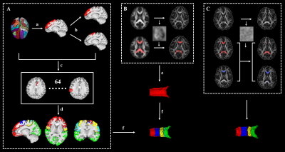 |
Longitudinal white matter changes in the callosal subsections in Parkinson’s Disease using connectivity-based parcellation.
Jingjing Wu1, Xiaoujun Guan1, Tao Guo1, Cheng Zhou1, Ting Gao2, Peiyu Huang1, Xiaojun Xu1, and Minming Zhang1
1Department of Radiology, The Second Affiliated Hospital, Zhejiang University School of Medicine, Hangzhou, China, 2Department of Neurology, The Second Affiliated Hospital, Zhejiang University School of Medicine, Hangzhou, China
Corpus callosum (CC) is the most important association fiber intrinsically connecting with different cortical regions. Studies reported the CC and its subsections could be used to differentiate different phenotypes in PD, PD and PDS. In this study, 39 PD patients with a mean time interval of 21m and 82 NC were recruited. We segmented the whole CC into five subsections according to their functional connectivity with predefined cortices. As a result, we observed that the microstructure and structure were fairly preserved in PD at baseline, but widespread changes occur in the corpus callosum during PD evolvement.
|
|
4477.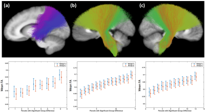 |
Gender differences in brain white matter discovered by whole brain suprathreshold fiber cluster statistics
Fan Zhang1, Suheyla Cetin-Karayumak1, Yang Song2, Weidong Cai3, James J Levitt1, Nikos Makris1, Carl-Fredrik Westin1, and Lauren J O'Donnell1
1Harvard Medical School, Boston, MA, United States, 2The University of New South Wales, Sydney, Australia, 3The University of Sydney, Sydney, Australia
We propose to study whole-brain white matter connectivity differences between females and males using diffusion MRI (dMRI) tractography. We leverage a well-established data-driven fiber clustering pipeline and a novel suprathreshold fiber cluster statistical method. We study a large cohort of 707 healthy adult subjects from the Human Connectome Project. We find multiple fiber tracts that are significantly different between the female and male groups in terms of the fractional anisotropy and/or the trace measures of the fiber tracts. These tracts include the corticospinal tract, the arcuate fasciculus, and the corpus callosum tract.
|
|
4478.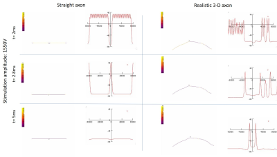 |
MODELLING NEURAL EXCITABILITY OF REALISTIC DTI-DERIVED AXON TRAJECTORIES
Sulagna Sahu1, Munish Chauhan1, Saurav Zaman Khan Sajib1, Stephen Helms Tillery1, Vikram D. Kodibagkar1, and Rosalind J. Sadleir1
1Arizona State University, Tempe, AZ, United States
Studying neural activation patterns is critical in the understanding of neuromodulation in transcranial electrical stimulation methods (TES). This research focuses on simulations of neural excitability of a realistic axon model obtained from DTI tractography of the trigeminal nerve, in comparison to a linear axon model obtained from literature, in presence of induced electric fields found from realistic FEM (finite element method) modelling of TES. The differences observed highlight the need for use of realistic trajectories in neural simulations and provide means for patient specific personalized studies.
|
4479.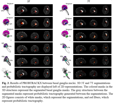 |
Comparison of interconnected basal ganglia probabilistic tractography between 3T and 7T MRI Images
Jae-Hyuk Shim1 and Hyeon-Man Baek1
1Gachon University, Incheon, Republic of Korea
Basal ganglia structures, globus pallidus internal, globus pallidus external, subthalamic nucleus, substantia nigra, red nucleus and striatum were automatically segmented on 3T and 7T HCP preprocessed diffusion weighted images. Connectivity between each basal ganglia structure was observed using probabilistic tractography generated with FSL's diffusion tools such as BEDPOSTX and PROBTRACKX. Tractography between basal ganglia was compared between 3T and 7T to observe differences that arise from tradeoffs of each acquisition.
|
|
4480.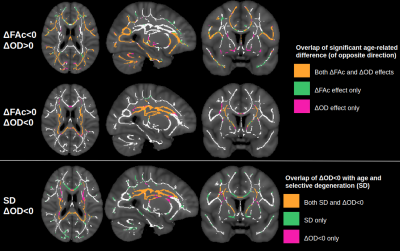 |
Investigating age-related differences in fractional anisotropy in relation to complex fibre architecture
Jordan A. Chad1,2, Ofer Pasternak3, and J. Jean Chen1,2
1Rotman Research Institute, Baycrest Health Sciences, Toronto, ON, Canada, 2Department of Medical Biophysics, University of Toronto, Toronto, ON, Canada, 3Departments of Psychiatry and Radiology, Brigham and Women's Hospital, Harvard Medical School, Boston, MA, United States
It is well established that white matter diffusion fractional anisotropy (FA) is sensitive to age, but interpreting this effect in terms of cellular microstructure has posed a challenge. Here we investigate how age-related differences in FA relate to measures of crossing and dispersing fibres derived from advanced diffusion MRI models. We find that, in aging, decreased FA is associated with increased fibre dispersion, and increased FA is associated with selective degeneration of crossing fibres. Importantly, we show that age-related differences in FA more closely reflect age-related differences in fibre architecture when applying free water elimination.
|
|
4481.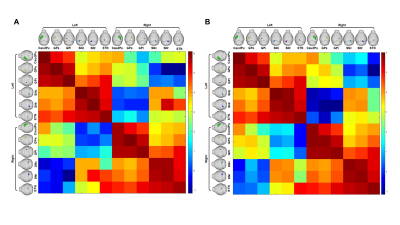 |
Comparison of Basal Ganglia-based Structural Connectome between 6-OHDA-induced Parkinson Mouse Model and Normal Mouse Model
Ayoon Kim1 and Hyeon-Man Baek1
1Department of Health Science and Technology, GAIHST, Gachon University, gachon university, Incheon, Republic of Korea, Republic of Korea
The study on probabilistic connectivity of basal ganglia have not reported in mouse model. However, we have quantified fiber connectivity and visualized specific tractography pathway. The basal ganglia is a complex system of a subcortical nuclei network which plays a fundamental role in a wide range of processes related to motor and limbic functions. Altered neural connectivity of basal ganglia may contribute to a number of neurologic and psychiatric disease such as Parkinson’s disease. This study shows that probabilistic diffusion tractography allows for detailed 3D reconstruction of the projections of basal ganglia in ex vivo control and PD mouse brain.
|
|
4482.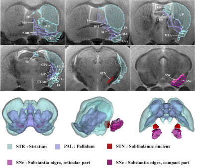 |
Detecting neuronal connectivity using diffusion tensor imaging in a Parkinson`s disease mouse basal ganglia using 9.4T MRI
Sang-Jin Im1 and Hyeon-Man Baek1
1Gachon university, Incheon, Korea, Republic of
Parkinson's disease (PD) is a neurodegenerative disorder that affects motor and cognition, resulting from dopaminergic cell death in substantia nigra (SN). Basal ganglia, a neural circuit involved in executive functions such as motor control and have been studied extensively in PD mouse models. Mouse brain tractography using MRI have seen increasing use to study neural networks but more comprehensive analysis is needed to establish a stronger consensus. The purpose of this study is to provide a comprehensive analysis of basal ganglia neuronal connectivity in control and PD mouse models.
|
|
4483.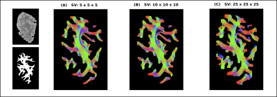 |
Quantitative assessment of multi-scale tractography: bridging the resolution gap with 3D-Polarized Light Imaging
Abib Alimi1, Matteo Frigo1, Samuel Deslauriers-Gauthier1, and Rachid Deriche1
1Athena Project-Team, Inria Sophia Antipolis Méditerranée, Université Côte d'Azur, Sophia Antipolis, France
Three-dimensional Polarized Light Imaging (3D-PLI) is an optical technique presented as a good candidate for validation of diffusion Magnetic Resonance Imaging (dMRI) results such as orientation estimates and tractography. We previously demonstrated a multi-scale tractography approach using 3D-PLI. Here, we show how tractograms obtained at different spatial scales from 3D-PLI human brain datasets can be inspected using homology theory in order to perform a quantitative comparison between them. This paves the way for the validation of tractography obtained from dMRI using multi-scale fiber tracking based on 3D-PLI.
|
|
4484.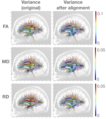 |
Aligning Shapes of Along-tract Diffusion Profiles across Populations
David Soobin Lee1, Antoni Kubicki1, Ashish Kaul Sahib1, Katherine L Narr1, and Shantanu H Joshi1
1UCLA, Los Angeles, CA, United States
Various tools exist to extract diffusion measures from tract profiles. However, they lack the precision in terms of statistical analysis since the tract shapes are registered to a common template. Although this analysis leads to meaningful results, we postulate that there is a significant geometric information in the along-tract diffusion profiles that is unaccounted for. Here we propose a novel method for aligning shapes of diffusion profiles across populations. This method offers improved matching and statistical analysis of along-tract diffusion profiles. Furthermore, this method could be used to perform multivariate statistical analysis of along-tract diffusion profiles in an invariant manner.
|
|
4485.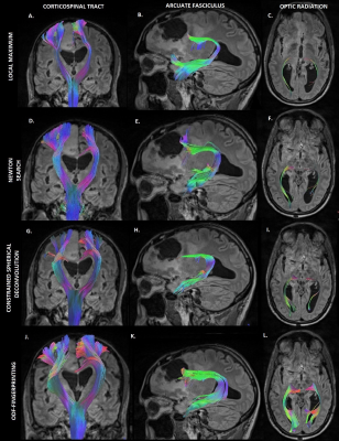 |
Application of Diffusion MRI ODF-Fingerprinting for Neurosurgical Tractography
Genevieve Barroll1,2, Dimitris Placantonakis, M.D., Ph.D.3, Fernando E. Boada, Ph.D. 1, Timothy Sheperd, M.D., Ph.D.1,2, and Steven Baete, Ph.D.1,2
1Center for Biomedical Imaging, Dept. of Radiology, NYU School of Medicinie, New York, NY, United States, 2Center for Advanced Imaging Innovation and Research (CAI2R), NYU School of Medicine, New York, NY, United States, 3Department of Neurosurgery, Perlmutter Cancer Center, Neuroscience Institute, Kimmel Center for Stem Cell Biology, NYU School of Medicinie, New York, NY, United States
The ODF-Fingerprinting method has proven to be successful in improving detection of fiber pairs with small crossing fibers [1]. We evaluated the performance of ODF-Fingerprinting and other algorithms used to detect fiber directions, to determine whether the clinical application contains the same success that was experienced in a healthy control. The performance of these competing algorithms was examined in a healthy control and post-operative scan of a tumor patient. With the success of ODF-Fingerprinting in comparison to other algorithms, we hope that the application of this algorithm will lead to improved neurosurgical precision.
|
|
4486.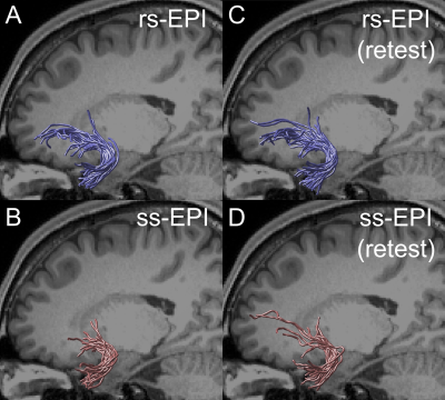 |
Advantage of readout-segmented EPI in simultaneous multi-slice diffusion MRI measurements for identifying uncinate fasciculus
Hiromasa Takemura1,2, Wei Liu3, Hideto Kuribayashi4, and Ikuhiro Kida1,2
1Center for Information and Neural Networks, National Institute of Information and Communications Technology, Suita, Japan, 2Graduate School of Frontier Biosciences, Osaka University, Suita, Japan, 3Siemens Shenzhen Magnetic Resonance Ltd., Shenzhen, China, 4Siemens Healthcare K.K., Tokyo, Japan
We evaluated the performance of diffusion MRI (dMRI) acquisition with simultaneous multi-slice (SMS) readout-segmented echo-planar imaging (rs-EPI) in measuring the uncinate fasciculus, which is typically affected by susceptibility-induced distortion or signal dropout. We found that SMS rs-EPI data provides a larger number of streamlines supported by dMRI data and larger fractional anisotropy in uncinate, as compared to conventional SMS single-shot EPI data. However, such an advantage was not consistent across subjects in the forceps major. Taken together, our results demonstrate that dMRI acquisition with SMS rs-EPI has advantages in measuring specific white matter tracts affected by susceptibility-induced artifacts.
|
|
4487.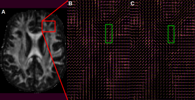 |
Does introduction of priors improve reliability to segment hyperdirect connections using tractography for Deep Brain Stimulation ?
Tristan MOREAU1, Maxime PERALTA1, John BAXTER1, and Pierre JANNIN1
1UMR 1099 LTSI, RENNES, France
A better knowledge of subthalamic nucleus connections could help predict the clinical outcome of deep brain stimulation in the context of Parkinson’s disease. However, diffusion weighted images used in clinical context are generally of poor resolution. In this study, we compared a whole brain versus an a-priori-based tractography method in order to segment hyperdirect connections linking subthalamic nucleus to cortex on normal controls acquired in clinical context. In systematically comparing our hyperdirect connections segmentation results with high resolution data set, we showed better reliability when using the a-priori-based tractography compared to the whole-brain method.
|
|
4488.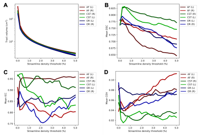 |
Assessing scan-rescan reproducibility of surgically relevant white matter tractography reconstructions
Kan Yan Chloe Li1,2, Leevi Kerkelä1, Jonathan D Clayden1, Kiran K Seunarine1, Matt G Hall1,2, and Chris A Clark1
1UCL Great Ormond Street Institute of Child Health, London, United Kingdom, 2National Physical Laboratory, Teddington, United Kingdom
Tractography is regularly used in neuroscientific research and neurosurgical planning for its ability to delineate fibre bundles non-invasively. The construction and analysis of tractograms often requires user-defined parameter choices such as streamline density thresholding for estimating tract volume. In this study, we assessed the reproducibility of a manual tractography pipeline that is applied in neurosurgical planning for oncology and epilepsy operations, and quantified the effect of streamline density thresholding on reproducibility. The pipeline was found to be robust in terms of reproducibility over the range of investigated thresholding levels with some variability between tracts.
|
|
4489.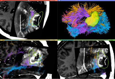 |
SlicerDMRI: a suite of clinician-accessible tools for neurosurgical planning research using diffusion MRI and tractography
Fan Zhang1, Thomas Noh1, Parikshit Juvekar1, Sarah F Frisken1, Laura Rigolo1, Isaiah Norton1, Tina Kapur1, Sonia Pujol1, William Wells III1,2, Alex Yarmarkovich3, Gordon Kindlmann4, Demian Wassermann5, Raul San Jose Estepar1,
Yogesh Rathi1, Ron Kikinis1,6, Hans J Johnson7, Carl-Fredrik Westin1, Steve Pieper3, Alexandra J Golby1, and Lauren J O'Donnell1
1Harvard Medical School, Boston, MA, United States, 2Massachusetts Institute of Technology, Boston, MA, United States, 3Isomics, Inc., Cambridge, MA, United States, 4University of Chicago, Chicago, IL, United States, 5Université Paris-Saclay, Palaiseau, France, 6University of Bremen and Fraunhofer MEVIS, Bremen, Germany, 7University of Iowa, Iowa City, IA, United States
We present an open-source software suite, SlicerDMRI (dmri.slicer.org), that enables neurosurgical planning research using diffusion magnetic resonance imaging (dMRI). SlicerDMRI is built upon and deeply integrated with 3D Slicer, an NIH-supported open-source platform for medical image informatics, image processing, and three-dimensional visualization. In this work, we give a demonstration of SlicerDMRI to enable end-to-end dMRI analyses in two retrospective imaging datasets from patients with high-grade glioma. Analyses demonstrated here include conventional diffusion tensor imaging (DTI) analysis, advanced multi-fiber tractography, automated identification of critical fiber tracts, and integration of multimodal imagery with dMRI.
|
|
4490.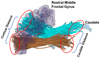 |
Assessing brain wiring patterns using tractography fiber clustering to study sex differences of the frontostriatal white matter connections
Fan Zhang1, Mark Vangel1, Marek Kubicki 1, Martha E Shenton1, Lauren J O'Donnell1, and James J Levitt1
1Harvard Medical School, Boston, MA, United States
We studied structural connectivity differences between females and males in frontostriatal white matter connections. We proposed a novel method to measure the topographical organization of the frontostriatal circuits, leveraging a data-driven fiber clustering of whole-brain dMRI tractography. The proposed topographical measure provides important information to assess the segregated and integrative brain wiring patterns in the human brain. We applied this technique to test for sex differences in a cohort of 100 healthy subjects. We identified significant group differences in the frontostriatal white matter connections, potentially related to the sex-specific brain wiring patterns.
|
|
4491.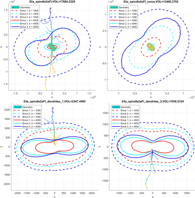 |
Diffusion MRI simulation of realistic neurons with SpinDoctor and the Neuron Module
Van-Dang Nguyen1, Chengran Fang2,3, Demian Wassermann3, and Jing-Rebecca Li2
1Department of Computational Science and Technology, KTH Royal Institute of Technology, Stockholm, Sweden, 2INRIA-Saclay, Equipe DEFI, Palaiseau, France, 3INRIA Saclay, Equipe Parietal, Palaiseau, France
The dMRI simulation arising from realistic neurons can help investigate the cellular microstructure. Ensuring correct connectivity between distinct cellular compartments while minimizing the computational burden is one of the main challenges. The design of simulation algorithm and the construction of high-quality neuron meshes are crucial. The NeuronModule can perform HARDI simulation of realistic neurons at various b-values and diffusion sequences with high accuracy. The computational time per gradient-direction is around 30 seconds which is faster than some Monte-Carlo simulators. The linearity between the diffusion-direction-averaged signal and one over square root of b for tubular structures is validated by our simulations.
|
|
4492.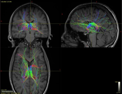 |
Structural connectivity analysis of anterior nuclei of the thalamus in Papez circuit based on DW-MRI fiber tractography for ANT-DBS in epilepsy
Ruhunur Özdemir1,2, Kai Lehtimäki3, Jukka Peltola1,3, and Hannu Eskola1,2
1Department of Medicine and Health Technology, Tampere University, Tampere, Finland, 2Department of Radiology, Tampere University Hospital, Tampere, Finland, 3Department of Neuroscience and Rehabilitation, Tampere University Hospital, Tampere, Finland
The white matter organization of anterior nuclei of thalamus (ANT) has not been comprehensively examined before. We visualized white matter tracks in Papez circuit determining different ROIs in ANT, cingulate gyrus, mammillothalamic body, hippocampus and parahippocampal region for ANT-DBS in epilepsy. We employed constrained spherical deconvolution (CSD), multi-shell multi-tissue (MSMT) CSD, and DTI model to characterize white matter tissue organization in healthy controls and one patient diagnosed with refractory epilepsy in DW-MRI. We demonstrate a connectivity pattern of ANT. CSD model tractography may provide consistent insights on the alteration of the connections in patients with refractory epilepsy for performing ANT-DBS.
|
|
4493.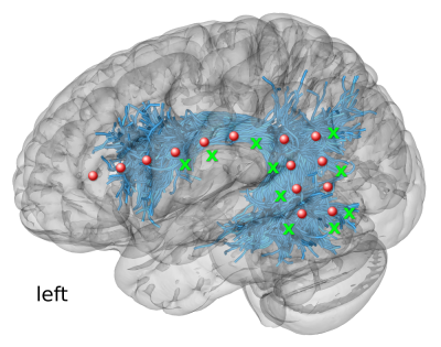 |
Diffusion MRI-based Connectivity Enriched with Microstructure Information Predicts the Propagation of Cortico-Cortical Evoked Potentials
Patryk Filipiak1, Fabien Almairac2, Théodore Papadopoulo1, Denys Fontaine2, Lydiane Mondot3, Stéphane Chanalet3, Rachid Deriche1, Maureen Clerc1, and Demian Wassermann4
1INRIA Sophia Antipolis - Méditerranée, Valbonne, France, 2Service de Neurochirurgie, Centre Hospitalier Universitaire de Nice, Université Côte d’Azur, Nice, France, Nice, France, 3Service de Radiologie, Centre Hospitalier Universitaire de Nice, Université Côte d’Azur, Nice, France, Nice, France, 4INRIA, CEA, Université Paris-Saclay, Paris, France, Paris, France
The propagation of Cortico-Cortical Evoked Potentials (CCEPs) varies depending on numerous structural features of brain tissue. In this work, we show that combined dMRI-based connectivity enriched with microstructure data has the potential to measure cortico-cortical communication as it predicts CCEP-based effective connectivity. Our multiple linear regression model incorporates q-space indices like Q-space Inverse Variance, Non-Gaussianity and Return to Plane Probability with minimum streamline lengths obtained from tractography to predict delays and amplitudes of the P1 peaks in CCEPs. In our experiment, we use presurgical dMRI and intrasurgical ECoG recordings of 9 patients operated on brain tumor in the awake condition.
|
|
4494.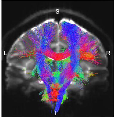 |
Structural connectome of autonomic and sensory brainstem nuclei in humans based on 7 Tesla high spatial and angular resolution diffusion imaging
KAVITA SINGH1, María Guadalupe García-Gomar1, Jeffery P Staab2,3, Simone Cauzzo4, Iole Indovina5,6, and Marta Bianciardi1
1Brainstem Imaging Laboratory, Department of Radiology, Athinoula A. Martinos Center for Biomedical Imaging, Massachusetts General Hospital and Harvard Medical School, Boston, MA, United States, 2Department of Psychiatry and Psychology, Mayo Clinic, Rochester, MN, United States, 3Department of Otolaryngology, Head and neck Surgery, Mayo clinic, Rochester, MN, United States, 4Institute of Life Sciences, Sant'Anna School of Advanced Studies, Pisa, Italy, 5Laboratory of neuromotor physiology, IRCCS, Santa Lucia Foundation, Rome, Italy, 6Centre of SpaceBiomedicine, University of Rome for Vergata, Rome, Italy
With the advancement of imaging technologies and signal processing tools, ample progress has been made in cortical and sub-cortical brain structural connectivity mapping; however, this is still missing in living humans for brainstem nuclei. Through high spatial-resolution 7 Tesla HARDI MRI and a recently developed probabilistic brainstem nuclei atlas, we built a structural connectome of autonomic and sensory brainstem nuclei in living humans. Interestingly, our connectome corresponded well with established non-human connectivity data. We foresee this connectome as basis for structural and functional studies of autonomic and sensory circuits in health and disease.
|
4495.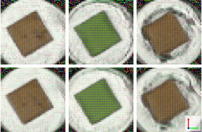 |
3D-printed phantom for validating diffusion MRI models
Michael Woletz1, Franziska Gantner2,3, Benedikt Hager4, Peter Gruber2,3, Siawoosh Mohammadi5,6, Zoltan Nagy7, Aleksandr Ovsianikov2,3, and Christian Windischberger1
1Center for Medical Physics and Biomedical Engineering, Medical University of Vienna, Vienna, Austria, 2Institute of Materials Science and Technology, Technical University Vienna, Vienna, Austria, 3Austrian Cluster for Tissue Regeneration, Vienna, Austria, 4Department of Biomedical Imaging and Image-guided Therapy, Medical University of Vienna, Vienna, Austria, 5Department of Systems Neuroscience, University Medical Center Hamburg-Eppendorf, Hamburg, Germany, 6Department of Neurophysics, Max Planck Institute for Human Cognitive and Brain Sciences, Leipzig, Germany, 7Laboratory for Social and Neural Systems Research (SNS Lab), University of Zurich, Zurich, Switzerland
Here we show the first DTI phantom manufactured by advanced high-resolution 3D-printing methods. The phantom consists of hollow, 12μm thin, liquid filled channels, that can be constructed in arbitrary configurations, ideally suited for validating diffusion sequences and analysis models. A simple configuration with orthogonal channel directions is presented. Diffusion weighted images were acquired and a diffusion tensor model employed. The main direction of the resulting tensors is accurately able to capture the directions of the channels with an average fractional anisotropy of 0.46. This method for creating diffusion phantoms will help to test and validate different models in the future.
|
|
4496.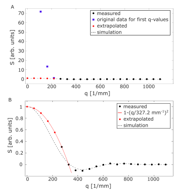 |
Water diffusion pore imaging on a 14.1 T spectrometer using glass capillary phantoms in the presence of extraporal fluid
Dominik Ludwig1,2, Frederik Bernd Laun3, Karel D. Klika4, Mark Edward Ladd1,2,5, Peter Bachert1,2, and Tristan Anselm Kuder1
1Medical Physics in Radiology, German Cancer Research Center (DKFZ), Heidelberg, Germany, 2Faculty of Physics and Astronomy, Heidelberg University, Heidelberg, Germany, 3Institute of Radiology, University Hospital Erlangen, Friedrich-Alexander-Universität Erlangen-Nürnberg (FAU), Erlangen, Germany, 4Molecular Structure Analysis, German Cancer Research Center (DKFZ), Heidelberg, Germany, 5Faculty of Medicine, Heidelberg University, Heidelberg, Germany
Diffusion pore imaging (DPI) can be used to retrieve the pore space function of arbitrary closed pores. In this study we show that DPI of glass capillaries is possible even under difficult experimental conditions. By separating the long gradient into a CPMG-like gradient echo train and matching the magnetic susceptibility, it was possible to acquire diffraction patterns of glass capillaries that were placed orthogonal to the main magnetic field. Furthermore, the feasibility of doing DPI in the presence of water outside the pores was demonstrated for the first time.
|
|
4497.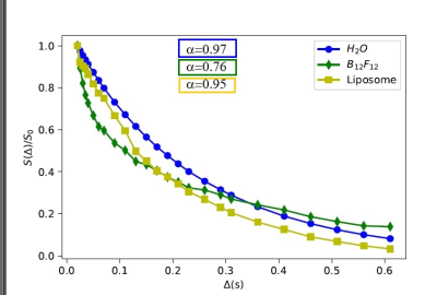 |
Validation of NMR transient subdiffusion in controlled samples
Silvia Capuani1, Fabio Micalizzi2, Simona Sennato3, Giulio Costantini3, Roberto Matassa4, Dante Rotili5, Lorenzo Correale2, Francesca Giuffrida2, Annalisa Caligiuri2, Giovanni Familiari4, and Andrea Gabrielli3
1Physics Dpt. Sapienza, CNR ISC, Rome, Italy, 2Physics Dpt., Sapienza University of Rome, Rome, Italy, 3Physics Dpt. Sapienza Roma, CNR ISC, Rome, Italy, 4Anatomical, Histological, Forensic Medicine and Orthopedics Sciences, Sapienza University of Rome, Rome, Italy, 5Drug chemistry and technology department, Sapienza University of Rome, Rome, Italy The quantification of transient anomalous diffusion (tAD) parameters by NMR could provide higher sensitivity, resolution and complementary detailed information for detecting early changes due to pathological conditions than the conventional metric does. However, there are some questions and controversial issues which prevent the take-off of tAD NMR investigations in soft condensed matter and medical diagnostic field. To overcome these obstacles, we planned to investigate the potential and limits of NMR subdiffusion in calibration standards characterized by porous structured materials and diffusing probes matching or not characteristics length-scales of the porous matrix.
|
|
4498.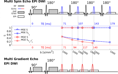 |
Phantom validation of diffusion weighted T2- and T2*-relaxometry EPI sequences
Yu Zidan1,2, Jelle Veraart1,2, Gregory Lemberskiy1,2, Ying-Chia Lin1,2, Tiejun E. Zhao3, Martijn Cloos1,2, Dan Iosifescu4,5, and Steven H. Baete1,2
1Center for Biomedical Imaging, Dept. of Radiology, NYU School of Medicine, New York, NY, United States, 2Center for Advanced Imaging Innovation and Research (CAI2R), NYU School of Medicine, New York, NY, United States, 3Siemens Medical Solutions, New York, NY, United States, 4Dept. of Psychiatry, NYU School of Medicine, New York, NY, United States, 5Clinical Research Division, Nathan Kline Institute for Psychiatric Research, Orangeburg, NY, United States
Combination of diffusion weighted MRI with orthogonal measures such as T2- or T2*-weighting has been proposed to overcome the fit degeneracy found in microstructure modeling of diffusion signals. However, the repetition of diffusion measurements at different TE leads to unacceptably long acquisition times, hindering clinical applicability of this approach. Here, we propose accelerated acquisitions using diffusion weighted multi-spin and multi-gradient echo trains which sample the signal at several TEs after a standard diffusion encoding spin echo. In the current configurations these sequences speed up the acquisitions by 2.6x or 3.6x respectively. We validate these approaches on a phantom.
|
|
4499.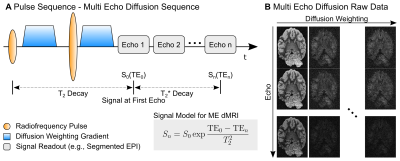 |
Sensitivity Gain from Multi-Echo Acquisitions in Ex-Vivo Diffusion Imaging: Numerical Simulations and Experimental Verification
Cornelius Eichner1, Michael Paquette1, Toralf Mildner2, Torsten Schlumm2, Catherine Crockford3, Roman Wittig3, Carsten Jäger4, Markus Morawski4, Harald E. Möller2, Angela D. Friederici1, and Alfred Anwander1
1Department of Neuropsychology, Max Planck Institute for Human Cognitive and Brain Sciences, Leipzig, Germany, 2NMR Unit, Max Planck Institute for Human Cognitive and Brain Sciences, Leipzig, Germany, 3Max Planck Institute for Evolutionary Anthropology, Leipzig, Germany, 4Paul Flechsig Institute of Brain Research, University Leipzig, Leipzig, Germany
Ex-vivo dMRI acquisitions achieve very high resolutions from increased SNR at the cost of prolonged scan times. Due to highly-segmented acquisition strategies, the ex-vivo dMRI signal is often not fully decayed when signal encoding progresses. Using numerical simulations and real-dMRI data from a wild chimpanzee, we show that this circumstance can be effectively employed using an optimized noise informed combination of multi-echo acquisitions from segmented EPI trains. The additional sampling comes at a small price and leads to increased SNR and a general reduction of signal bias.
|
|
4500.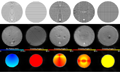 |
Characterization of orientation dispersion’s impact on diffusion kurtosis and NODDI using an axon-mimetic 3D printed phantom
Tristan K. Kuehn1,2, Farah N. Mushtaha2, Omar El-Deeb3, Amanda Moehring3, Corey A. Baron1,2,4, and Ali R. Khan1,2,4,5
1School of Biomedical Engineering, Western University, London, ON, Canada, 2Centre for Functional and Metabolic Mapping, Robarts Research Institute, Western University, London, ON, Canada, 3Department of Biology, Western University, London, ON, Canada, 4Department of Medical Biophysics, Western University, London, ON, Canada, 5The Brain and Mind Institute, Western University, London, ON, Canada
Existing phantoms used to validate diffusion MRI models of white matter microstructure struggle to represent the complex fibre configurations found in vivo. Here we demonstrate a 3D printed phantom that realizes several complex fibre configurations inexpensively. We prepare a set of phantoms and use them to characterize the change in diffusion MRI model parameters with fibre curvature and crossing fibres. Most parameters computed by DTI, kurtosis, and NODDI had relationships with fibre crossing angle. These phantoms are a promising tool for evaluating the effect of orientation dispersion on diffusion MRI models of white matter.
|
|
4501.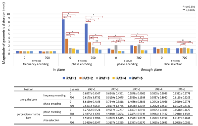 |
Geometric Distortion Evaluation of Integrated Slice Dependent Shimming (iShim) Using ACR MRI Phantom
Cindy Xue1, Gladys Lo1, Raymond Lee1, Chi Wai Michael Liu1, Oi Lei Wong1, and Jing Yuan1
1Hong Kong Sanatorium and Hospital, Hong Kong, Hong Kong
Integrated slice dependent shimming (iShim) has been found to be able to reduce the magnetic field inhomogeneity and geometric distortion. In this study, we aim to evaluate, optimize and quantify the performance of the in-plane and through-plane geometric distortion in EPI-DWI using iShim. ACR large MRI phantom was scanned in two positions (a: along the bore, b: perpendicular to the bore) to evaluate the in-plane and through-plane geometric distortion. The phantom was separately scanned at 5 GRAPPA acceleration factors (iPAT 1-5) twice. In-plane geometric distortion along phase encoding direction with iShim may be minimized by increasing the acceleration factor.
|
|
4502.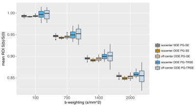 |
Double-Diffusion-Encoding MRI: phantom evaluation of concomitant gradient effects with single- and twice-refocused sequences
Lisa Novello1, Stefano Tambalo1, Thorsten Feiweier2, and Jorge Jovicich1
1CIMeC, Center for Mind/Brain Sciences, University of Trento, Rovereto, Italy, 2Siemens Healthcare GmbH, Erlangen, Germany
In Double-Diffusion-Encoding sequences, concomitant gradients may introduce spatial bias in the measured MRI signal. It is important to characterize such biases since they can affect the accuracy of quantitative microstructural metrics in brain studies. In this work, we assess the signal deviations at different positions along the scanner z-axis in an isotropic phantom for both single- and twice-refocused spin-echo sequences.
|
|
4503.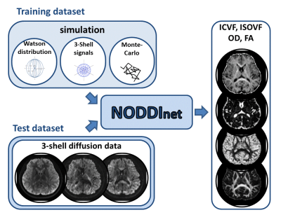 |
NODDInet: Diffusion parameter mapping using deep neural network trained by computer simulation data
Juhyung Park1, Woojin Jung1, Eun-jung Choi1, and Jongho Lee1
1Seoul National University, Korea, Republic of Korea
A deep neural network, NODDInet, was developed to generate NODDI parameters (ICVF, ISOVF, OD, and FA) in 1 min. This network was trained using a computer simulation-generated training dataset only, and, therefore, is unbiased to experimental data and covers a wide range of the parameters. For the network input, the diffusion measurements of each shell were projected onto three 2D plains to reduce the input data size while preserving the geometric information of the diffusion measurements. The results demonstrate higher accuracy and faster processing time (x14) than a previous method (AMICO).
|
|
4504.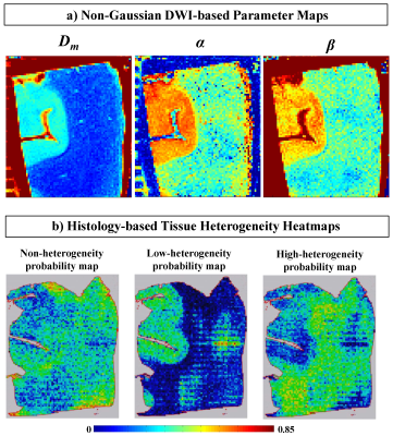 |
Intravoxel Tissue Heterogeneity: One-to-one Correspondence between Non-Gaussian Diffusion MRI Parameters and Histologic Features
Muge Karaman1,2, Lingdao Sha3, Tingqi Shi1, Dan Schonfeld2,3,4, Tibor Valyi-Nagy5, and X Joe Zhou1,2,6
1Center for Magnetic Resonance Research, University of Illinois at Chicago, Chicago, IL, United States, 2Department of Bioengineering, University of Illinois at Chicago, Chicago, IL, United States, 3Department of Electrical and Computer Engineering, University of Illinois at Chicago, Chicago, IL, United States, 4Department of Computer Science, University of Illinois at Chicago, Chicago, IL, United States, 5Department of Pathology, University of Illinois at Chicago, Chicago, IL, United States, 6Departments of Radiology and Neurosurgery, University of Illinois at Chicago, Chicago, IL, United States
The parameters of the continuous-time random-walk (CTRW) model have been shown to be related to the underlying tissue heterogeneity. However, it has been challenging to establish a rigorous correlation between the CTRW parameters and the gold-standard histology. This is primarily because the histopathological evaluation of tissue heterogeneity is a labor-intensive task. We develop a machine-learning algorithm that quantifies the microscopic tissue heterogeneity revealed by histology as probability maps; and demonstrate a one-to-one correspondence between the CTRW parameters and tissue structural heterogeneity. Our results have provided evidence towards establishing a correlation between the CTRW parameter values and tissue heterogeneity.
|
|
4505.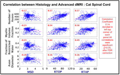 |
Validation of Diffusion Propagator Imaging measures in White Matter using Histology
Madhura Baxi1,2, Lipeng Ning2, Suheyla Cetin-Karayumak2, Marek Kubicki2,3, and Yogesh Rathi2,3
1Graduate Program for Neuroscience, Boston University, Boston, MA, United States, 2Psychiatry Neuroimaging Lab, HMS, BWH, Boston, MA, United States, 3Department of Psychiatry, MGH, Boston, MA, United States
This study is the first attempt towards histological validation of three advanced diffusion MRI measures derived from the 3D diffusion propagator in white matter tissue: i) return-to-origin-probability (RTOP), ii) return-to-axis-probability (RTAP) and iii) mean squared displacement (MSD), using ex-vivo cat spinal cord tissue. We compared these dMRI measures voxel-wise with the underlying histological properties of the tissue. RTOP and RTAP were found to be significantly correlated with the following biological characteristics: i) Number of axons, ii) Myelin volume fraction and iii) Restricted water fraction, showing that the diffusion propagator imaging measures are sensitive to the underlying white matter microstructural properties.
|
|
4506.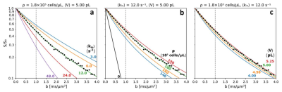 |
Fundamental Cell Biology Properties Underlying In Vivo DWI
Brendan Moloney1, Eric M. Baker1, Xin Li1, Erin W. Gilbert2, and Charles S. Springer, Jr.1
1Advanced Imaging Research Center, Oregon Health & Science University, Portland, OR, United States, 2Surgery, Oregon Health & Science University, Portland, OR, United States
With cellular ensembles featuring stochastic [“Voronoi”] geometries, Monte Carlo random walk simulated DWI b-space decays exhibit sensitivity to cell biology parameters measuring membrane Na+,K+‑ATPase [NKA] activity, cell density, ρ, and voxel average cell volume, <V>. Furthermore, the simulations matching disparate in vivo tissue [murine xenograft colorectal cancer, human cerebral cortex, and human bladder] experimental b‑space decays have parameters [cellular water efflux rate constant <kio>, ρ, and <V>] in near absolute agreement with the most pertinent literature. Inspecting the common, empirical early decay measure, ADC, of these simulations provides insights into acute and chronic tissue property changes in vivo.
|
|
4507.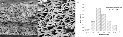 |
Co-electrospun spinal cord phantom for diffusion MRI
Fenglei Zhou1,2, Francesco Grussu1,3, Zhanxiong Li4, and Geoff Parker1,5
1Centre for Medical Image Computing, Department of Computer Science, University College London, London, United Kingdom, 2School of Pharmacy, University College London, London, United Kingdom, 3Queen Square MS Centre, Queen Square Institute of Neurology, Faculty of Brain Sciences, University College London, London, United Kingdom, 4College of Textile and Clothing Engineering, Soochow University, Suzhou, China, 5Bioxydyn Limited, Manchester, United Kingdom
A spinal cord-mimicking fibre phantom was developed to evaluate its potential for validating dMRI. Microfibres were co-electrospun with two polymer solutions and characterized by SEM. The phantom comprised two material samples designed with 0o and 90o crossings. SEM revealed that fibres were uniaxially aligned and hollow, having similar sizes to spinal cord axons. Diffusion tensor analysis of a ZOOM dMRI acquisition demonstrated the difference in alignment of the two samples. Diffusion kurtosis analysis demonstrated differences in axial and radial diffusion restriction, with parameter values consistent with published spinal cord data. Relaxation time constants were similar in two samples.
|
|
4508.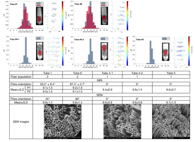 |
Validating pore size estimates in a biomimetic microfibre architecture with a stochastic distribution of pore-sizes and cross-sectional shapes
Chih-Chin Heather Hsu1, Chun-Chung Huang2,3, Slawomir Kusmia4, Mark Drakesmith4, Feng-Lei Zhou5, Geoff Parker5, Ching-Po Lin2,3,6, and Derek Jones4
1Department of Biomedical Imaging and Radiological Science, National Yang Ming University, Taipei, Taiwan, 2Institute of Science and Technology for Brain Inspired Intelligence, Fudan University, Shanghai, China, 3Aging and Health Research Center, National Yang-Ming University, Taipei, Taiwan, 4Cardiff University Brain Research Imaging Center (CUBRIC), Cardiff University, Cardiff, United Kingdom, 5Centre for Medical Image Computing, University College London, London, United Kingdom, 6Institute of Neuroscience, National Yang-Ming University, Taipei, Taiwan
Previous studies showed that AxCaliber-like frameworks produce reliable orientation and inner diameter estimates in idealised phantoms (i.e., highly parallel hollow cylinders with uniform circular cross-section). We extend this work to ‘biomimetic’ phantoms, having stochastic pore-size distributions, non-circular cross sections and complex (i.e., crossing) fibre configurations. Using a Connectom scanner, and assuming a Poisson pore-size distribution, inner diameter and crossing angle estimates were in excellent agreement with electron-microscopy measurements in the same sample. To our knowledge, this is the first validation of pore-size estimates in complex geometries on a human scanner, lending support to the promise of mapping these parameters in-vivo.
|
|
4509.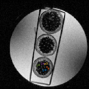 |
Comparison of Cylindrical and Spherical Geometric Models to Infer Cell Sizes in a Garlic Stem
Melissa Sarah Lillian Anderson1, Henri Sanness Salmon1, Guneet Uppal1,2, Jarrad Perron3, Lisa Bako4, Gong Zhang5, Sheryl Lyn Herrera1,6, and Melanie Martin1
1Physics, University of Winnipeg, Winnipeg, MB, Canada, 2University of Manitoba, Winnipeg, MB, Canada, 3Physics, University of Manitoba, Winnipeg, MB, Canada, 4Cubresa, Inc., Winnipeg, MB, Canada, 5Brain Engineering Centre, Anhui University, China Physics, University of Winnipeg, Winnipeg, MB, Canada, 6Cubresa, Winnipeg, MB, Canada
Temporal diffusion spectroscopy (TDS) can be used to infer sizes of cells in samples. It relies on a geometric model to relate the MRI signal to the cell sizes. Garlic stem collenchyma tissue has long cells which might be modelled as cylinders. We compared a cylindrical and spherical geometric model in temporal diffusion spectroscopy to determine how important the geometrical model was for garlic stems. The inferred diameters of cells in the garlic stem (4μm-6μm) were not statistically different when using the two different geometric models. This is the first step toward understanding the importance of geometric models for TDS.
|