
Digital Poster Session
Diffusion: Diffusion Applications
Diffusion
4510 -4523 Diffusion Applications - Diffusion: Brain Applications 1
4524 -4539 Diffusion Applications - Diffusion: Brain Applications 2
4540 -4555 Diffusion Applications - Diffusion: Brain Applications 3
4556 -4569 Diffusion Applications - Diffusion: Applications Beyond the Brain
Session Topic: Diffusion Applications
Session Sub-Topic: Diffusion: Brain Applications 1
Digital Poster
Diffusion
4510.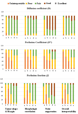 |
Radiological Qualitative Assessment of IVIM Parameters for Total Variation Penalty Function Approach: A Pilot Study in Osteosarcoma
Amit Mehndiratta1, Esha Badiya Kayal1, Kedar Khare 2, Sameer Bakhshi3, Raju Sharma4, and Devasenathipathy Kandasamy4
1Centre for Biomedical Engineering, Indian Institute of Technology Delhi, New Delhi, India, 2Department of Physics, Indian Institute of Technology Delhi, New Delhi, India, 3Dr. BRA Institute-Rotary Cancer Hospital, All India Institute of Medical Sciences, New Delhi, India, 4Department of RadioDiagnosis, All India Institute of Medical Sciences, New Delhi, India
Radiological based qualitative assessment of evaluated IVIM parametric maps using state-of-the-art IVIM analysis methods Bi-exponential (BE) model with adaptive Total Variation Penalty function (BE+TV) and BE with adaptive Huber Penalty function (BE+HPF) has been performed in comparison to the commonly used IVIM analysis methodologies like BE model, and segmented BE methods. BE+TV/BE+HPF preserve spatial homogeneity in the parametric images by updating them iteratively during data fitting. Experimental results showed all IVIM analysis methods showed BE+TV and BE+HPF produced comparative better noise suppression and diagnostic quality and interpretability for D* and f than other methods.
|
|
4511.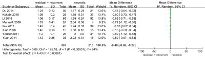 |
Utility of DWI with quantitative ADC in diagnosing residual or recurrent HCCs after TACE: A systematic review and meta-analysis
Hai-Feng Liu1 and Wei Xing1
1Department of Radiology, Third Affiliated Hospital of Soochow University, changzhou, China
This meta-analysis was to investigate the accuracy of DWI and ADC value in diagnosing residual or recurrent HCCs after TACE. This study suggested DW have high diagnostic efficacy, and ADC value can be used to differentiate residual or recurrent HCCs after TACE.
|
|
4512. |
Impact of processing pipelines on biological findings of large scale multicenter DTI studies
Chung-Man Moon1, Amritha Nayak1,2, M. Okan Irfanoglu1, and Carlo Pierpaoli 1
1National Institute of Biomedical Imaging and Bioengineering, National Institutes of Health, Bethesda, MD, United States, 2Henry M. Jackson Foundation for the Advancement of Military Medicine, Rockville, MD, United States
Different post-processing pipelines might influence the research or even clinical conclusions obtained from diffusion MRI. Our study investigated the impact of alternative pre-processing techniques and spatial normalization approaches on the outcome of a large multicenter study in schizophrenia that was originally analyzed using the ENIGMA pipeline. Our main finding is the discovery of profound effects of the spatial normalization approach used, on both biological conclusions and inter-site harmonization of the results.
|
|
4513. |
Rigorous Prospective Reduction of Inter-scanner Variance of Diffusion Imaging: Initial Experience
Vincent Kyu Lee1, Benjamin Meyers1, William T Reynolds2, Rafael Ceschin1, Vincent Schmithorst1, Jeffrey Berman3, Thomas Chenevert4, Borjan Gagoski5, Peter LaViolette6, Deqiang Qiu7, Sudhir Pathak1, Ashok Panigrahy1, and Walter Schneider8
1Radiology, University of Pittsburgh, Pittsburgh, PA, United States, 2Biomedical Informatics, University of Pittsburgh, Pittsburgh, PA, United States, 3Radiology, Children's Hospital of Philadelphia, Philadelphia, PA, United States, 4Radiology, University of Michigan, Ann Arbor, MI, United States, 5Radiology, Harvard Medical School, Boston, MA, United States, 6Radiology, Medical College of Wisconsin, Milwaukee, WI, United States, 7Radiology, Emory University, Atlanta, GA, United States, 8Psychology, University of Pittsburgh, Pittsburgh, PA, United States
Our study shows for the first time that synthetic phantoms that simulate fiber anatomical characteristics can provide in vitro corrections factors for reducing in-vivo inter-scanner variance specific to discrete segments of cortical association fiber tracts. The overall goal of our evolving rigorous harmonization approach is to reduce inter-scanner variance that can confound biological variance derived from multi-center discovery and neuroprotection clinical trials in the developing human brain.
|
|
4514.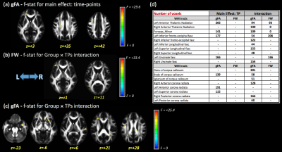 |
Evaluation of longitudinal changes in white matter structural integrity and grey matter volume in Parkinson’s disease
Maurizio Bergamino1, Elizabeth Keeling1,2, Ryan R Walsh3, and Ashley M Stokes1
1Division of Neuroimaging Research, Barrow Neurological Institute, Phoenix, AZ, United States, 2Neuroscience Department, Arizona State University, Phoenix, AZ, United States, 3The Muhammad Ali Parkinson Center, Barrow Neurological Institute, Phoenix, AZ, United States
In this study, we investigated longitudinal changes in white matter (WM) structural integrity and gray matter (GM) volume in Parkinson’s disease (PD). More specifically, free-water diffusion tensor imaging (FW-DTI), generalized q-sampling imaging (GQI), and voxel-based morphometry (VBM) were assessed in data obtained from the Parkinson Progression Markers Initiative (PPMI) database. We hypothesize that the use of advanced DTI metrics and VBM analysis will improve the sensitivity and specificity for the detection of WM alterations and GM volumetric changes in PD over time.
|
|
4515.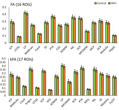 |
Are DKI Measures Relatively More Sensitive In Identifying HIV Infection Related Pathologies Than DTI Measures?
Sameer Vyas1, Teddy Salan2, Paramjeet Singh1, Sulaiman Sheriff2, Mahendra Kumar2, and Varan Govind2
1Postgraduate Institute of Medical Education and Research,, Chandigarh, India, 2University of Miami, Miami, FL, United States
DTI has shown evidence of alterations to the micro-structural integrity of the brain due to infection from HIV. However this finding is not consistent across all studies. In this work, we compare metrics obtained from DTI and diffusion kurtosis imaging (DKI) to determine if DKI can provide a more reliable and sensitive measurement of structural integrity.
|
|
4516.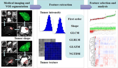 |
Application Study of DWI Radiomics Features with Transurethral Resection on Assessing the Muscular Infiltrating of Bladder Carcinoma
guiqin Liu1, shuaishuai XU1, yongming Dai2, Guangyu WU1, and jianrong XU1
1Radiology, Renji Hospital,Shanghai Jiaotong University School of Medicine, Shanghai, China, 2United Imaing Healthcare, Shanghai, China
To investigate the value of radiomics features from diffusion-weighted imaging (DWI) in differentiating muscle-invasive bladder cancer (MIBC) from non-muscle-invasive bladder cancer (NMIBC). Combining DWI radiomics features with TUR could improve the sensitivity and accuracy in discriminating the presence of muscle invasion in bladder cancer for clinical practice. Multi-center, prospective studies are needed to confirm our results.
|
|
4518.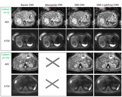 |
Evaluation of simultaneous multislice acquisition with advanced processing vs. conventional sequence in free-breathing DWI for liver patients
Mihaela Rata1, Katja De Paepe1, Matthew R Orton1, Erica Scurr1, Julie Hughes1, Alto Stemmer2, Marcel Dominik Nickel2, and Dow-Mu Koh1
1Royal Marsden Hospital and Institute of Cancer Research, London, United Kingdom, 2Siemens Healthcare, Erlangen, Germany
Diffusion Weighted Imaging (DWI) in combination with simultaneous multislice (SMS) acquisition has the potential to decrease acquisition time and improve image quality in abdominal MRI. In this study, we evaluated the image quality of free-breathing DWI acquired from 25 patients with liver metastases and compared SMS (with/without an advanced processing option) DWI with conventional bipolar echo planar DWI. We found that free-breathing liver DWI based on a SMS-accelerated protocol with advanced processing methods was faster and demonstrated better image quality when compared with a conventional bipolar DWI protocol.
|
|
4519.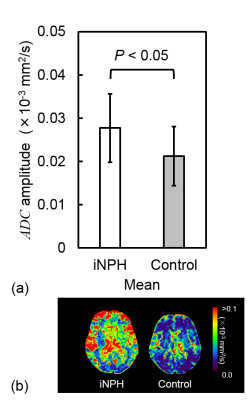 |
Fourier analysis of dynamic diffusion changes during cardiac cycle in idiopathic normal pressure hydrocephalus
Yuya Yasuda1, Tosiaki Miyati1, Naoki Ohno1, Mitsuhito Mase2, Ryo Yagawa1, Rika Saito1, Masatomo Uehara1, Harumasa Kasai2, Yuta Shibamoto2, and Satoshi Kobayashi1
1Graduate School of Medical Sciences, Kanazawa University, Kanazawa, Japan, 2Nagoya City University Hospital, Nagoya, Japan Poster Permission Withheld
In this study, we evaluated the frequency characteristics of the apparent diffusion coefficient (ADC) wave form in the cardiac cycle of the brain in idiopathic normal pressure hydrocephalus (iNPH). iNPH is associated with higher ADC amplitude with a wide frequency range. The Fourier analysis of ADC change in the cardiac cycle in iNPH makes it possible to noninvasively obtain a more detailed information regarding the intracranial state in iNPH.
|
|
4520.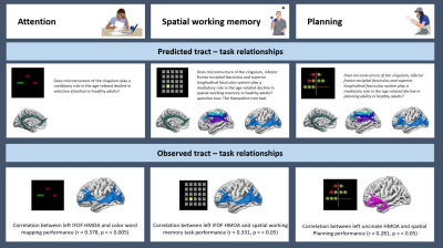 |
Which frontal white matter pathways mediate executive decline in healthy ageing?
Anoushka Leslie1, Ahmad Beyh1,2, Marco Catani3, Flavio Dell'Acqua3, Ceriesse Gunasinghe4, Henrietta Howells5, Richard Parker6, Andy Simmons1, Michel Thiebaut de Schotten7,8, Steve Williams1, and Mitul Mehta1
1Department of Neuroimaging, King's College London, London, United Kingdom, 2NatBrainLab, King's College London, London, United Kingdom, 3Natbrainlab, Department of Neuroimaging, King's College London, London, United Kingdom, 4Department of Psychological Medicine, King's College London, London, United Kingdom, 5Dipartimento di Biotecnologie Mediche e Medicina Traslazionale, Universita degli studi di Milano, Milano, Italy, 6IXICO plc, London, United Kingdom, 7Groupe d’Imagerie Neurofonctionnelle, Institut des Maladies Neurodégénératives-UMR 5293, Universite de Bordeaux, Bordeaux, France, 8Brain Connectivity and Behaviour Laboratory, BCBlab, Sorbonne Universities, Paris, France
This study aimed to expand our understanding of how changes to frontal white matter pathways might influence executive decline in healthy ageing. We selected three cognitive components of executive function, attention, spatial working memory and planning and predicted that changes to microstructure of the cingulum, IFOF and SLFI -III would play a mediatory role in age related cognitive decline of 86 healthy adults. Contrary to our predictions, no mediation effects were found within the proposed tract - task groupings. Instead, during exploratory analysis, HMOA of the left uncinate demonstrated a small to medium indirect effect on age-related decline in planning performance.
|
|
4521.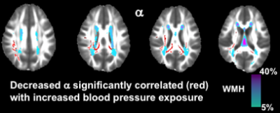 |
Increased non-Gaussian subdiffusion in white matter is associated with increased longitudinal blood pressure exposure in adults at midlife
Carson Ingo1,2, Shawn Kurian3, James Higgins4, Lisanne Jenkins5, Donald Lloyd-Jones6, and Farzaneh Sorond1
1Department of Neurology, Northwestern University, Chicago, IL, United States, 2Department of Physical Therapy and Human Movement Sciences, Northwestern University, Chicago, IL, United States, 3Department of Neurology, Chicago, IL, United States, 4Department of Radiology, Northwestern University, Chicago, IL, United States, 5Department of Psychiatry and Behavioral Sciences, Northwestern University, Chicago, IL, United States, 6Department of Preventative Medicine, Northwestern University, Chicago, IL, United States
The increased presence of non-Gaussian subdiffusive dynamics, possibly reflecting the presence of increased neuronal and glial microstructural heterogeneity, is sensitive increased vascular risk exposure, which was not observed with traditional DTI metrics such as FA.
|
|
4522. |
Along-tract correlation analysis of diffusion metrics and white matter lesions in a 70-year old birth cohort
Carole Hélène Sudre1,2, Chiara Maffei3, Josephine Barnes2, David Thomas2, David Cash2, Tom Parker2, Chris Lane2, Marcus Richards2, Hui Zhang2, Sebastien Ourselin1, Jonathan Schott2, Anastasia Yendiki3, and M. Jorge Cardoso1
1King's College London, London, United Kingdom, 2University College London, London, United Kingdom, 3Massachusetts General Hospital, Boston, MA, United States
In a population of 260 elderly individuals presenting white matter hyperintensities of presumed vascular origin, lesion profile along reconstructed tracts correlated strongly with diffusion metrics obtained from multishell acquisition (notably intracellular volume fraction, orientation dispersion index, axial radial and mean kurtosis). Results appeared stronger when the tractography was performed using data from the highest b-value. Furthermore, changes to the diffusion signal was observed consistently on the reconstructed tracts at the vicinity of the lesions potentially indicative of tissue vulnerability beyond the lesion border identified from FLAIR images.
|
|
4523.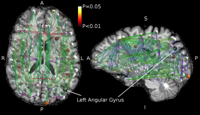 |
Microstructural and structural connectivity alterations in dexmedetomidine-induced loss of consciousness
Timo Roine1,2, Oskari Kantonen3, Jaakko Langsjö3,4, Kimmo Kaskinoro5, Roosa Kallionpää2,5,6, Annalotta Scheinin3,5, Katja Valli2,5,6,7, Timo Laitio5, Antti Revonsuo2,6,7, and Harry Scheinin3,5,8
1Department of Neuroscience and Biomedical Engineering, Aalto University School of Science, Espoo, Finland, 2Turku Brain and Mind Center, University of Turku, Turku, Finland, 3Turku PET Centre, University of Turku and the Hospital District of Southwest Finland, Turku, Finland, 4Department of Intensive Care, Tampere University Hospital, Tampere, Finland, 5Division of Perioperative Services, Intensive Care and Pain Medicine, Turku University Hospital, University of Turku, Turku, Finland, 6Department of Psychology and Speech-Language Pathology, University of Turku, Turku, Finland, 7Department of Cognitive Neuroscience and Philosophy, School of Bioscience, University of Skövde, Skövde, Sweden, 8Department of Pharmacology, Drug Development and Therapeutics, University of Turku, Turku, Finland
We used diffusion MRI to investigate brain microstructure and structural connectivity in 10 healthy subjects before and during dexmedetomidine-induced loss of consciousness. We found rapid local changes both in the microstructural properties and in the structural brain connectivity networks, most prominently in the left angular gyrus and its connections indicating possible involvement of the area in consciousness. Moreover, our results indicate that conventional high b-value diffusion MRI acquisitions, in addition to sequences specifically designed to capture functional changes, are sensitive to at least major brain state changes.
|
 View the Poster
 Watch the Video
Session Topic: Diffusion Applications
Session Sub-Topic: Diffusion: Brain Applications 2
Digital Poster
Diffusion
4524.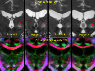 |
Presurgical planning of MRgFUS for Essential Tremor (ET) with protocol for high quality DTI
Amritha Nayak1,2, Angela Bissoli3, Okan M Irfanoglu2, Guiseppe Ricciardi3, Elisa Ciceri3, and Carlo Pierpaoli Pierpaoli2
1Henry Jackson Foundation for advancement in Military Medicine Inc, Rockville, MD, United States, 2National Insitutes of Health, Bethesda, MD, United States, 3Azienda Ospedaliera Universitaria Integrata, Verona, Italy
This study evaluates the role of a high quality DTI in presurgical planning of MRgFUS for initial target localization.
|
|
4525. |
Abnormal insula white matter tracts in smokers
Chao Wang1, Shuyue Wang1, Peiyu Huang1, and Minming Zhang1
1Department of Radiology, the Second Affiliated Hospital, Zhejiang University School of Medicine, Hangzhou, China
The insula, a cortical region that is thought to play a central role in this reward circuitry, has been implicated as an important role in the maintenance of nicotine addiction. However, it remains largely unclear about the alterations in white-matter tracts of insula circuits in nicotine addiction. Here, we further investigated the differences of insula white-matter tracts between smokers and nonsmokers. We found abnormal white matter tracts of insula subregions in smokers. These altered insula microstructural connectivity could interfere with the normal neural circuitry of reward processing, which might be the underlying neurobiology of nicotine addiction.
|
|
4526.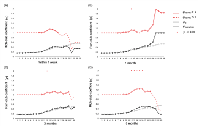 |
Rich-Club Organizational Changes Over the Course of Motor Recovery after First-Time Acute Stroke
Lu Wang1, Hing-Chiu Chang1, Peng Cao1, and Edward.S Hui1
1Department of Diagnostic Radiology, The University of Hong Kong, Hong Kong, China
In this study we acquired diffusion data and motor functions from stroke patients at within 1 week, 1, 3 and 6 months and found rich-club organization remerged one month after stroke and high order regions were found at 6 months when patients functionally recovered. Feeder connections showed middle cost and high capacity while rich-club connections showed high cost and middle capacity at 6 months after stroke. Together these results suggest that there could be a potential relation between reemergence of rich-club organization and brain motor recovery.
|
|
4527.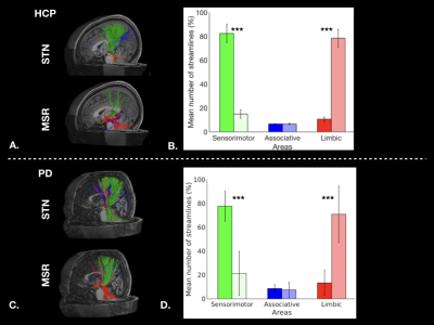 |
Tractography based organization of the hyperdirect pathway to the subthalamic area in HCP subjects and parkinsonian patients
Gizem Temiz1, Sophie Sébille1, Chantal François1, Eric Bardinet1, and Carine Karachi1,2
1CENIR, Institut du Cerveau et de la Moelle Epinière, Paris, France, 2AP-HP, Hôpital de la Pitié-Salpêtrière, Department of Neurosurgery, Paris, France, Paris, France
The aim of this study is to analyze differences in the hyperdirect cortical connectivity between the subthalamic nucleus and its medial region, and the anatomo-functional organization of these regions using tractography. These analyzes were performed in healthy subjects and parkinsonian patients. Our results show that a dominant motor cluster was located in the posterolateral STN, a limbic cluster located medially in the MSR, and an intermediate motor-limbic cluster located in between for both cohorts. In conclusion, tractography based sub-parcellisation of subthalamic regions could be helpful to refine individual targeting for functional neurosurgery.
|
|
4528.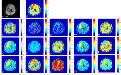 |
Differentiating Low- and High-Grade Adult glioma Using Multi-diffusion Models
Junqi Xu1, He Wang1,2, Xueying Zhao1, Hui Zhang1, Xiaoyuan Feng3, and Ren Yan3
1Institute of Science and Technology for Brain-Inspired Intelligence, Fudan University, Shanghai, China, 2Human Phenome Institute, Fudan University, Shanghai, China, 3Radiology, Huashan Hospital, Fudan University, Shanghai, China
To offer a potential multiparameter-mapping platform for clinical pathological diagnosis using multi-diffusion models including mono-exponentional model, IVIM, SEM, FROC, CTRW, SM and DKI. The U-test and ROC analysis show the superiority of FROC, CTRW and DKI models in diagnostic accuracy for grading brain tumors (at 0.770, 0.780 and 0.817 respectively).
|
|
4529. |
Evaluating advanced multi-shell diffusion MRI microstructural biomarkers of Alzheimer’s disease
Julio Ernesto Villalon Reina1, Talia Miriam Nir2, Sophia Thomopoulos2, Lauren E Salminen3, Neda M Jahanshad2, Rutger Fick4, Matteo Frigo5, Rachid Deriche5, and Paul M Thompson2
1USC Imaging Genetics Center, University of Southern California, Los Angeles, CA, United States, 2USC Stevens Neuroimaging and Informatics Institute, University of Southern California, Marina del Rey, CA, United States, 3USC Imaging Genetics Center, University of Southern California, Marina del Rey, CA, United States, 4Therapanacea, Paris, France, 5Athena Project Team, Inria Sophia-Antipolis Méditerranée, Université Côte d'Azur, Nice, France
To identify microstructure-based biomarkers sensitive to cognitive impairment, we used ADNI-3 multi-shell dMRI data to estimate 18 measures from seven dMRI models and assessed their ability to predict mild cognitive impairment (MCI). For each measure, we used TV-L1 regularized logistic regression to find cohesive clusters of brain tissue that contribute to correct classification. We found that tensor-based (DTI) diffusivity and multi-compartment spherical mean technique (MC-SMT) measures showed the highest prediction accuracy, but differential anatomical distributions of classifying voxels. MC-SMT may offer greater sensitivity and specificity to MCI than DTI as MC-SMT resulted in the highest recall and fewest classifying voxels.
|
|
4530. |
Topological alterations in structural brain connectivity networks are associated with survival after out-of-hospital cardiac arrest
Timo Roine1,2, Oskari Kantonen3, Ulrika Roine1, Sami Virtanen4, Jani Saunavaara4,5, Riitta Parkkola4, Ruut Laitio6, Olli Arola6, Marja Hynninen7, Juha Martola8, Heli M Silvennoinen8, Marjaana Tiainen9, Risto O. Roine10, Harry
Scheinin6, and Timo Laitio6
1Department of Neuroscience and Biomedical Engineering, Aalto University School of Science, Espoo, Finland, 2Turku Brain and Mind Center, University of Turku, Turku, Finland, 3Turku PET Centre, University of Turku and the Hospital District of Southwest Finland, Turku, Finland, 4Department of Radiology, Turku University Hospital, University of Turku, Turku, Finland, 5Department of Medical Physics, Turku University Hospital, University of Turku, Turku, Finland, 6Division of Perioperative Services, Intensive Care and Pain Medicine, Turku University Hospital, University of Turku, Turku, Finland, 7Division of Intensive Care Medicine, University of Helsinki and Helsinki University Hospital, Helsinki, Finland, 8Department of Radiology, University of Helsinki and Helsinki University Hospital, Helsinki, Finland, 9Department of Neurology, University of Helsinki and Helsinki University Hospital, Helsinki, Finland, 10Division of Clinical Neurosciences, Turku University Hospital, University of Turku, Turku, Finland
Mortality after out-of-hospital cardiac arrest is high, and there is a substantial need for new biomarkers to improve the identification of patients with poor outcome. Therefore, we investigated structural brain connectivity networks in patients after out-of-hospital cardiac arrest in order to detect differences related to survival. We found decreased global efficiency and strength from MRI scans acquired in a median of 53 hours (IQR 47-64) after OHCA to be related to mortality at 6 months after OHCA. In addition, several regions with decreased strength and local efficiency were found, most significantly in the pallidum, and superior frontal and supramarginal cortices.
|
|
4531.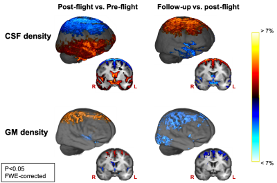 |
Diffusion MRI reveals macro- and microstructural changes in cosmonauts' brains after long-duration spaceflight
Steven Jilings1, Angelique Van Ombergen2, Elena Tomilovskaya3, Alena Rumshiskaya4, Liudmila Litvinova4, Inna Nosikova3, Ekaterina Pechenkova5, Ilya Rukavishnikov3, Inessa Kozlovskaya3, Stefan Sunaert6, Paul M Parizel7, Valentin Sinitsyn8, Victor Petrovichev4,
Steven Laureys9, Peter zu Eulenburg10, Jan Sijbers11, Floris Wuyts1, and Ben Jeurissen11
1Lab for Equilibrium Investigations and Aerospace, Dept. of Physics, University of Antwerp, Antwerp, Belgium, 2Translations Neuroscience, Dept. of Medicine, University of Antwerp, Antwerp, Belgium, 3Institute of Biomedical Problems, Russian Academy of Sciences, Moscow, Russian Federation, 4Dept. of Radiology, Federal Center of Treatment and Rehabilitation, Moscow, Russian Federation, 5Laboratory for Cognitive Research, National Research University Higher School of Economics, Moscow, Russian Federation, 6Dept. of Imaging and Pathology, KU Leuven, Leuven, Belgium, 7Dept. of Radiology, Royal Perth Hospital and University of Western Australia, Perth, Australia, 8Faculty of Fundamental Medicine, Lomonosov Moscow State University, Moscow, Russian Federation, 9Coma Science Group, Dept. of Neurology, University (Hospital) of Liège, Liège, Belgium, 10German Center for Vertigo and Balance Disorders, Dept. of Neurology, Ludwig-Maximilians-University Munich, Munich, Germany, 11imec - Vision Lab, Dept. of Physics, University of Antwerp, Antwerp, Belgium
There is currently limited information on the effects of spaceflight on the human brain. Therefore, we investigated longitudinal changes in gray matter (GM), white matter (WM), and cerebrospinal fluid (CSF) density and mass using diffusion MRI data acquired pre-flight, post-flight and at follow-up. Our results show redistributed CSF and concomitant GM morphological changes post-flight, which reverse at follow-up. At the same time, our results show no evidence of neurodegeneration. Moreover, GM and WM mass increased in sensorimotor brain regions post-flight, which largely persisted at follow-up. These results indicate, for the first time, sensorimotor neuroplasticity after spaceflight.
|
|
4532.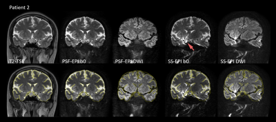 |
High-resolution Distortion-free DWI of Pituitary Adenomas and Rathke Cleft Cysts Using Point-spread-function Encoded EPI
Jieying Zhang1, Chunjie Guo2, Xinrui Liu3, Yishi Wang1,4, Huimao Zhang2, and Hua Guo1
1Center for Biomedical Imaging Research, Department of Biomedical Engineering, School of Medicine, Tsinghua University, Beijing, China, 2Department of Radiology, the First Hospital of Jilin University, Changchun, China, 3Department of Neurosurgery, the First Hospital of Jilin University, Changchun, China, 4Philips Healthcare, Beijing, China
Differentiation between pituitary adenomas (PAs) and Rathke cleft cysts (RCCs) is important for treatment planning. Diffusion weighted imaging (DWI) has been reported for the differentiation. However, traditional single-shot echo planar imaging (EPI) DWI has limited resolution and suffers from image distortion near the skull base. High-resolution distortion-free DWI should be more suitable. We apply a recently developed fast point-spread-function encoded EPI on pituitary imaging. It generates high-resolution distortion-free DWI, in which the signals of the microadenoma and the RCCs are hyperintense and hypointense, respectively. This study shows the potential of this technique to distinguish between PAs and RCCs.
|
|
4533.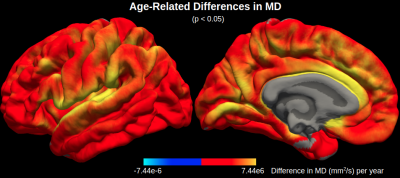 |
Can mapping cortical diffusivity provide unique microstructural insight into aging?
Graham A. D. Archibald1, Jordan A. Chad1,2, David H. Salat3,4, and J. Jean Chen1,2
1Rotman Research Institute, Baycrest Health Sciences, Toronto, ON, Canada, 2Department of Medical Biophysics, University of Toronto, Toronto, ON, Canada, 3MGH/HST Athinoula A. Martinos Center for Biomedical Imaging, Massachusetts General Hospital, Harvard Medical School, Charlestown, MA, United States, 4Neuroimaging Research for Veterans Center, VA Boston Healthcare System, Boston, MA, United States
The aging process in the cerebral cortex is typically measured in-vivo via MRI-based cortical thickness. Here we show that mean diffusivity (MD), a parameter derived from diffusion MRI, serves as a more sensitive measure of aging than thickness across the cortex. MD shows distinct age-related differences than thickness, suggesting that MD can provide insight into microscopic degeneration that cannot be detected with macroscopic structural MRI measures. While diffusion MRI microstructural analyses are typically limited to the white matter, this work suggests that additionally assessing age-related differences in cortical gray matter diffusivity can offer potentially valuable information on early cortical degeneration.
|
|
4534. |
Hemodialysis can contribute to acute changes in cerebral volume and white matter structure
Madeleine T Dacey1,2,3, Stefan E Poirier1,3, Janice Gomes2,4, Udunna C Anazodo1,3, and Christopher W McIntyre1,2
1Medical Biophysics, Western University, London, ON, Canada, 2Kidney Clinical Research Unit, Lawson Health Sciences Center, London, ON, Canada, 3Imaging, Lawson Health Research Insitute, London, ON, Canada, 4Pathology and Laboratory Medicine, Western University, London, ON, Canada
Cognitive impairment and white matter degeneration are common in hemodialysis patients. Hemodialysis can severely impede blood flow and create osmotic imbalances in the brain. This may cause brain injury by a mechanism similar to that of stroke. To investigate the acute effects of hemodialysis on the brain, we used a novel system to perform diffusion and T1 weighted MRI scans during hemodialysis. Several tracts exhibit diffusion tensor imaging markers for cytotoxic and ionic edema. Increased white and grey matter volume during hemodialysis further support the presence of ionic edema. Ionic and cytotoxic edema are evidence of acute brain injury.
|
|
4535. |
Clinical monitoring of axonal loss in multiple sclerosis using advanced diffusion MRI
Scott Kolbe1, Meaghan Clough1, Frederique Boonstra1, Myrte Strik2, Anneke van der Walt1, Helmut Butzkueven1, Owen White1, Joanne Fielding1, and Meng Law1
1Monash University, Prahran, Australia, 2University of Melbourne, Parkville, Australia
Brain atrophy is currently the accepted method for measuring neurodegeneration in multiple sclerosis (MS) but is insensitive over short times and ignores changes in cellular density. Recent advances in diffusion weighted imaging provide markers of axonal fibre density and atrophy. This study aimed to study the longitudinal sensitivity of these markers in comparison to brain atrophy based on data acquired in a standard clinical MS study. Annualised within-patient change in fibre density was around seven times more sensitive than brain volume change. This supports the development of fibre density based methods for clinical monitoring of axonal loss in MS.
|
|
4536.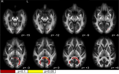 |
White matter fiber density and cross-section alterations in neuropathic pain after spinal cord injury
Shana Black1,2, Andrew Janson1,2, and Christopher R Butson1,2,3
1Biomedical Engineering, University of Utah, Salt Lake City, UT, United States, 2Scientific Computing and Imaging Institute, University of Utah, Salt Lake City, UT, United States, 3Neurology, Neurosurgery, and Psychiatry, University of Utah, Salt Lake City, UT, United States
Fiber-specific white matter changes were compared using fixel-based analysis between spinal cord injured subjects with chronic neuropathic pain (n=17) and spinal cord injured subjects without any pain symptoms (n=15). Fiber density, fiber cross-section (FC), and a combined measurement of fiber density and cross section (FDC) were calculated and compared between groups using multi-shell, 3-tissue constrained spherical deconvolution and connectivity-based fixel enhancement. Statistically significant increases (FWE-corrected p<0.1) in FC and FDC were identified in a posterior-inferior commissural white matter pathway, corresponding to the splenium and major forceps of the corpus callosum and regions of the retrosplenial complex.
|
|
4537. |
Brain white matter abnormalities and correlation with severity in amyotrophic lateral sclerosis: An atlas-based diffusion tensor imaging study
XiaoQiang Du1, YunJing Xue1, HuaJun Chen1, and ZhongShuai Zhang2
1Fujian Medical University Union Hospital, Fuzhou, China, 2SIEMENS Healthcare, Shanghai, China
This study used atlas-based region of interest analysis to assess WM microstructure in ALS by combining intra-voxel metrics, which included FA and MD, and an inter-voxel metric, i.e., LDH. We found WM abnormalities extending from the motor to the extra-motor regions in ALS and observed a correlation between distinct diffusion metrics and various clinical variables, supporting DTI metrics may be utilized as the diagnostic biomarker of ALS. Meanwhile, the extent of WM abnormalities in several tracts (e.g. ATR and LIFOF) were better revealed by LDH measurement, suggesting the supplementary role in reflecting ALS-related pathological process.
|
|
4538.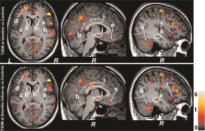 |
Diffusion Tensor Imaging Detects Cross-Sectional and Longitudinal Brain Changes in Type 2 Diabetes Mellitus
Bhaswati Roy1, Sarah E Choi2, Milena Lai3, Luke Ehlert3, Rashmi Mullur4, Matthew J. Freeby4, and Rajesh Kumar3,5,6,7
1University of California at Los Angeles, LOS ANGELES, CA, United States, 2UCLA School of Nursing, University of California at Los Angeles, Los Angeles, CA, United States, 3Anesthesiology, University of California at Los Angeles, Los Angeles, CA, United States, 4Medicine, University of California at Los Angeles, Los Angeles, CA, United States, 5Radiology, University of California at Los Angeles, Los Angeles, CA, United States, 6Bioengineering, University of California at Los Angeles, Los Angeles, CA, United States, 7Brain Research Institute, University of California at Los Angeles, Los Angeles, CA, United States
Patients with Type 2 diabetes mellitus (T2DM) show brain tissue changes in mood and cognitive control sites, functions that are deficient in the condition. However, the nature and extent of brain injury in T2DM, and their progression with time along with functional deficits are unclear. Using diffusion tensor imaging based MD procedures, we showed wide-spread chronic tissue changes in T2DM subjects and their continued progression after 6 months follow-up in areas involve in mood and cognitive regulatory functions. These findings may have resulted from underlying metabolic dysfunction associated with the condition.
|
|
4539.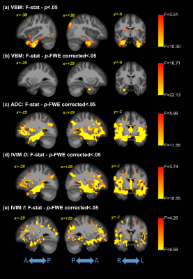 |
Preliminary Assessment of Intravoxel Incoherent Motion Diffusion-Weighted MRI (IVIM-DWI) Metrics in Alzheimer’s Disease
Maurizio Bergamino1, Ashley Nespodzany1, Leslie C Baxter1,2, Anna Burke3, Richard Caselli2, Marwan N Sabbagh4, Ryan R Walsh5, and Ashley M Stokes1
1Division of Neuroimaging Research, Barrow Neurological Institute, Phoenix, AZ, United States, 2Mayo Clinic Arizona, Phoenix, AZ, United States, 3Department of Neuropsychiatry, Barrow Neurological Institute, Phoenix, AZ, United States, 4Lou Ruvo Center for Brain Health, Cleveland Clinic, Las Vegas, NV, United States, 5The Muhammad Ali Parkinson Center, Barrow Neurological Institute, Phoenix, AZ, United States
The objective of this study was to assess complementary metrics from voxel-based morphometry (VBM) and intravoxel incoherent motion diffusion-weighted images (IVIM-DWI) MRI methods in aging populations. Using voxel-based analysis, grey matter (GM) and white matter (WM) differences were analyzed across Alzheimer’s disease (AD), mild cognitive impairment (MCI), and cognitively normal (HC) individuals. IVIM-DWI demonstrated early significant differences between MCI and HC groups, while VBM did not. In addition, voxel-based correlations between neuroimaging metrics and cognitive assessments were assessed in the cognitively impaired groups (AD and MCI).
|
Session Topic: Diffusion Applications
Session Sub-Topic: Diffusion: Brain Applications 3
Digital Poster
Diffusion
4540.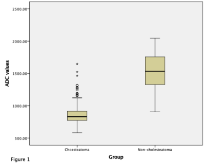 |
A comparison of RESOLVE DWI with delayed gadolinium-enhanced T1-weighted MRI in detecting cholesteatoma based on 531 patients’ data
Yaru Sheng1, Yan Sha1, Rujian Hong1, Zhongshuai Zhang2, and Wenhu Huang1
1Radiology, EENT Hospital of Fudan University, Shanghai, China, 2Siemens Healthcare, Shanghai, China
This study aimed to compare the performance of readout-segmented echo-planar imaging (RESOLVE) DWI and delayed gadolinium-enhanced T1-weighted MRI to diagnose cholesteatoma on 531 patients. The results showed that RESOLVE DWI can be an excellent tool for the detection of primary cholesteatoma.
|
|
4541.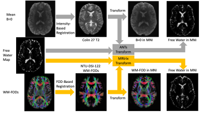 |
A novel diffusion registration method with the NTU-DSI-122 template to transform free water signal fraction maps to stereotaxic space.
Benjamin T Newman1,2, Ana Untaroiu1, and T. Jason Druzgal1,2
1Department of Radiology & Medical Imaging, Division of Neuroradiology, University of Virginia Health System, University of Virginia, Charlottesville, VA, United States, 2Brain Institute, University of Virginia, Charlottesville, VA, United States
We propose the use of the NTU-DSI-122 template as a flexible, diffusion specific, means of registering subject images to stereotaxic MNI-space for further analysis. This allows for registration to be performed based on matching white matter fiber orientation distributions, which create within tissue contrasts, unlike voxel intensity metrics. This advantage is demonstrated by observing the increased consistency of registration versus a leading intensity-based algorithm. The range of b-values present in the NTU-DSI-122 allows for tailoring to selectively register at b-values matching those acquired in an experimental cohort, providing flexibility for both single- and multi-shell acquisitions.
|
|
4542.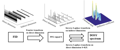 |
Diffusion-Ordered NMR Spectroscopy Reconstruction Based on Low-rank and Sparse Inverse Laplace Transform
Enping Lin1, Yu Yang1, Yuqing Huang1, and Zhong Chen1
1Department of Electronic Science, Xiamen University, Xiamen, China
DOSY (Diffusion-ordered NMR spectroscopy) presents an essential tool for the analysis of compound mixtures. However, existing DOSY reconstruction methods is limited by its relatively low resolution. Here, based on constraints on low rank and sparsity of DOSY data, we propose a reconstruction method to achieve high-resolution DOSY spectrum for measurements on complex mixtures, even when component signals are congested and mixed together along the spectral dimension. Experiment results indicate that our method is robust and possesses high-resolution reconstruction performance.
|
|
4543. |
Diffusion MRI revealed mild optic nerve fiber degeneration during Chimpanzee aging
Chun-Xia Li1, Yumei Yan1, Longchuan Li2, Todd Preuss3, James G Herndon3, Xiaoping Hu4, and Xiaodong Zhang1,3
1Yerkes Imaging Center, Yerkes National Primate Research Center, Emory University, Atlanta, GA, United States, 2Department of Pediatric, Emory University School of Medicine, Atlanta, GA, United States, 3Division of Neuroscience and Neurological diseases, Emory University, Atlanta, GA, United States, 4Department of bioengineering, University of California, Riverside, Riverside, CA, United States
Aging effects on the optic nerve (ON) bundles of chimpanzees were investigated systematically with diffusion tensor imaging (DTI). Mean diffusivity (MD), axial diffusivity (AD or λll), radial diffusivity (RD or λτ) and fractional anisotropy (FA) showed mild and proportional changes from youth to elder adulthood, and significant increase of MD and RD were seen in elder chimpanzees. However, the changes are much milder than previous studies in the ON of monkey and human brain white matter. The unique evolution pattern in chimpanzee white mater aging may grant further investigations to unveil the neural substrate mechanism of the human brain aging.
|
|
4544.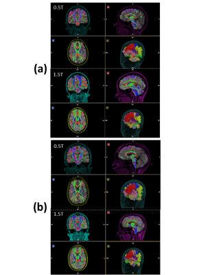 |
Diffusion Tractography at 0.5T: Comparison to 1.5T
Jeff A Stainsby1, Chad T Harris1, Andrew T Curtis1, Philip J Beatty1, and Curtis N Wiens1
1Synaptive Medical, Toronto, ON, Canada
The feasibility of generating diffusion tractography from data obtained on a head-only 0.5T system is demonstrated and results are compared qualitatively to tractography generated from clinical 1.5T data. DTI data from 0.5T compares favorably in quantitative (FA, ADC) measures to literature values, and qualitative (segmented white matter tracts) measures to 1.5T.
|
|
4545. |
Diffusion-weighted imaging at ultra-low field MR for acute stroke detection: what can we see?
Tiago Timoteo Fernandes1, Marc Golub1, Andreia Freitas1, Sairam Geethanath2,3, and Rita Gouveia Nunes1
1ISR, IST - University of Lisbon, Lisbon, Portugal, 2Medical Imaging Research Centre, Dayananda Sagar Institutions, Bangalore, India, 3Magnetic Resonance Research Center, Columbia University, NY, DC, United States
The primary goal of MR in acute stroke imaging is to determine the ischemic tissue at risk. Since a narrow time window is available for treatment, an accessible point-of-care system is desirable. Ultra-low field MR (ULF) shows promise but suffers from low signal-to-noise ratios (SNR). We performed a theoretical study of the SNR and contrast-to-noise (CNR) ratios for a range of B0 fields, with corresponding tissue relaxation times, considering different gradient performances. Illustrative simulated DWI brain images are provided. Results suggest that despite the challenges, ULF is promising for imaging acute stroke lesions.
|
|
4546. |
Ultra-high b-value Diffusion MRI for Evaluation of Single Amyloid Precursor Protein Knock-in Mouse Model of Alzheimer’s Disease
Jin Gao1,2, Zachery Morrissey3, Alex Leow3, Orly Lazarov4, Danilo Erricolo1, Richard Magin5, and Weiguo Li2,5
1Electrical and Computer Engineering, University of Illinois at Chicago, Chicago, IL, United States, 2Research Resources Center, University of Illinois at Chicago, Chicago, IL, United States, 3Department of Psychiatry, University of Illinois at Chicago, Chicago, IL, United States, 4Department of Anatomy and Cell Biology, University of Illinois at Chicago, Chicago, IL, United States, 5Department of Bioengineering, University of Illinois at Chicago, Chicago, IL, United States
Alzheimer’s disease (AD) has a tremendous impact in terms of social and economic cost. White matter damage in the progression of AD and the associated cognitive symptoms and pathophysiology are of crucial interest. Diffusion MRI offers unique insights into the pathophysiology of AD in vivo. This feasibility study aims to assess and visualize the white matter changes using an ultrahigh b-value diffusion MRI.
|
|
4547. |
Cerebrospinal fluid pulsation does not affect on DWI-based thermometry: healthy volunteer study
Koji Sakai1, Jun Tazoe1, Kentaro Akazawa1, Hiroyasu Ikeno2, Toshiaki Nakagawa2, and Kei Yamada1
1Kyoto Prefectural University of Medicine, Kyoto, Japan, 2Kyoto Prefectural University of Medicine Hospital, Kyoto, Japan
Diffusion-weighted imaging (DWI) based thermometry has the potential to be a non-invasive method of temperature measurement for the deep inside of human brain. Nevertheless, the DWI at lateral ventricle in brain might be influenced by the pulsation flow of cerebrospinal fluid (CSF), which is motivated by heartbeat. The purpose of this study was to investigate the influence of pulsation flow on brain DWI thermometry for healthy subjects. Comparisons were performed between magnetic resonance spectroscopy and DWI based thermometry (ΔT) at three CSF speed selections. There was no significant difference on ΔT among the CSF speed and volume on healthy subjects.
|
|
4548. |
T2w-FLAIR generation through deep-learning using distortion-free PSF-EPI DWI
Zhangxuan Hu1, Zhe Zhang2, Yishi Wang3, Yajing Zhang4, and Hua Guo1
1Center for Biomedical Imaging Research, Department of Biomedical Engineering, School of Medicine, Tsinghua University, Beijing, China, 2China National Research Center for Neurological Diseases, Beijing Tiantan Hospital, Capital Medical University, Beijing, China, 3Philips Healthcare, Beijing, China, 4MR Clinical Science, Philips Healthcare (Suzhou), Suzhou, China
MRI examinations usually contain multi-contrast images, which may share redundant information. For example, T2w-FLAIR contrast relies on the property of T2 relaxation and water component of the tissue, which also present in T2- and diffusion-weighted images. T2w-FLAIR acquisition is usually lengthy due to the long inversion time. In this study, point-spread-function (PSF) encoded EPI (PSF-EPI) DWI and T2-weighted images were used to generate T2w-FLAIR images by taking the advantages of high-resolution and distortion-free of PSF-EPI. This method has the potential to improve the acquisition efficiency of MRI.
|
|
4549.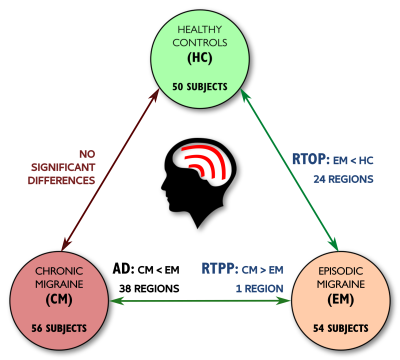 |
AMURA with standard single-shell acquisition can detect changes beyond the Diffusion Tensor: a migraine clinical study
Álvaro Planchuelo-Gómez1, Rodrigo de Luis-García1, Antonio Tristán-Vega1, David García-Azorín2, Ángel Luis Guerrero2, and Santiago Aja-Fernández1
1Imaging Processing Laboratory, Universidad de Valladolid, Valladolid, Spain, 2Headache Unit, Hospital Clínico Universitario de Valladolid, Valladolid, Spain
AMURA (Apparent Measures Using Reduced Acquisitions) is an alternative formulation to drastically reduce the number of samples needed for the estimation of diffusion properties related to the Ensemble Average diffusion Propagator (EAP). Although these measures were initially intended for medium-to-high b-values, in this work we evaluate their performance in DTI-like acquisitions. Fifty healthy controls, 54 episodic migraine (EM) and 56 chronic migraine (CM) patients were compared, using a single-shell diffusion scheme at b=1000 s/mm2. We compare AMURA measures (return-to-origin, return-to-axis and return-to-plane probabilities) to traditional DTI measures. Differences between EM and controls were only detectable using the return-to-origin probability.
|
|
4550.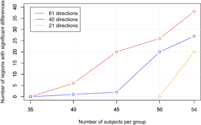 |
Fewer number of gradient directions in diffusion MRI can be counterbalanced with higher sample size: a migraine clinical study
Álvaro Planchuelo-Gómez1, Santiago Aja-Fernández1, David García-Azorín2, Ángel Luis Guerrero2, and Rodrigo de Luis-García1
1Imaging Processing Laboratory, Universidad de Valladolid, Valladolid, Spain, 2Headache Unit, Hospital Clínico Universitario de Valladolid, Valladolid, Spain
The effect of changes in the acquisition parameters on Diffusion Tensor Imaging (DTI) has been studied, but for very specific situations. A whole-brain comparison of 54 episodic migraine (EM) and 56 chronic migraine (CM) patients, using diffusion schemes of 61, 40 and 21 gradient orientations, was performed. Statistical comparisons were repeated reducing the sample size until no significant differences were found. Higher number of regions with significant lower axial diffusivity in CM compared to EM were found using 61 gradient directions. With a larger sample size, results with 40 and 21 directions were equivalent to results acquired with 61 directions.
|
|
4551.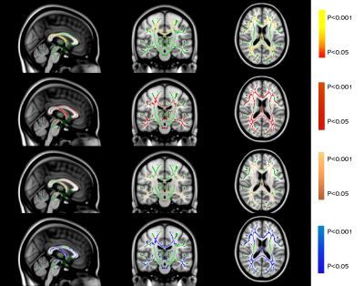 |
A DTI Comparative study – Is demyelination in AD resembling primary demyelinating disease (MS) or secondary demyelinating disease (NPSLE)?
Huiqin Zhang1, Hui Zhang1, Franki Kai-Hei Tse 2, Edward Sai-Kam Hui1, Peng Cao1, Kannie Wai Yan Chan3, Queenie Chan4, Karl HERRUP5, and Henry Ka Fung Mak1
1Department of Diagnostic Radiology, The University of Hong Kong, Hong Kong, Hong Kong, 2Department of Health Technology and Informatics, The Hong Kong Polytechnic University, Hong Kong, Hong Kong, 3Department of Biomedical Engineering, City University of Hong Kong, Hong Kong, Hong Kong, 4Philips Healthcare, Hong Kong, Hong Kong, 5Department of Neurobiology, University of Pittsburgh, Pittsburgh, PA, United States
Demyelination is a known pathology in AD, but it is unclear whether such myelin loss resembles primary or secondary demyelinating disease. Here, we retrospectively analysed the DTI data using voxel-wise TBSS analysis and made pair-wise comparisons between cohorts of AD, Relapsing-remitting MS(RRMS), NPSLE and normal controls (NC). In diffusion metrics analysis, widespread microstructural patterns were similarly found between AD and MS in terms of diffusion metrics, but not between AD and NPSLE. Our results indicate that the microstructural WM changes in AD may share similar pathological mechanisms in demyelinating disease of RRMS.
|
|
4552.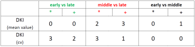 |
A longitudinal study on heterogeneity of diffusional parameters of spontaneously-hypertensive rats.
Jung-Sen Hsiao1, Hung-Yu Fu1, Pei-Lun Yu1, Sheng-Min Huang1, Kung-Chu Ho2, and Fu-Nien Wang1
1Biomedical Engineering and Envionmental Sciences, National Tsing-Hua University, Hsinchu City, Taiwan, 2Chang-Gung Memorial Hospital, Taoyuan City, Taiwan
Diffusion MRI has been regarded as an assessment in characterizing brain tissue integrity. However, the information of tissue heterogeneity is often overlooked. To investigate the feasibility of diffusional heterogeneity in gray matter through DKI derived diffusivities, six spontaneously-hypertensive rats (SHR) in different ages were scanned on a 7T small animal MR scanner. The differences of heterogeneity of diffusivities and kurtosis between different ages were revealed, which may imply its possibility for tissue characterization. Therefore, the diffusional heterogeneity could be suggested to be a potential image-based biomarker for evaluating tissue integrity.
|
|
4553. |
In vivo Diffusion Tensor Magnetic Resonance Imaging of Chronic Cocaine Administered Mouse Brain
Ethan A Cook1, Shannon E Callen2, Shilpa Buch2, and Balasrinivasa R Sajja3
1College of Medicine, University of Nebraska Medical Center, Omaha, NE, United States, 2Pharmacology and Experimental Neuroscience, University of Nebraska Medical Center, Omaha, NE, United States, 3Radiology, University of Nebraska Medical Center, Omaha, NE, United States
In vivo imaging-based biomarkers that can accurately detect the brain structural and functional changes due to chronic drug abuse play a significant role to understand and assess brain damage due to cocaine abuse and to determine the efficacy of the treatment. To this end, we have demonstrated that diffusion tensor MRI could detect brain structural changes, particularly demyelination in white matter structures, in chronic cocaine administered mice.
|
|
4554. |
Observation of the intracellular oxygenation changes in MRI signal
Kei Kikuchi1, Minghui Tang2, Ikuhiro Kida3, and Toru Yamamoto2
1Graduate School of Health Sciences, Hokkaido University, Sapporo, Japan, 2Faculty of Health Sciences, Hokkaido University, Sapporo, Japan, 3National Institute of Information and Communications Technology, Osaka, Japan
Oxygen molecule is paramagnetic and its relaxation-time-shortening effect is strongly enhanced in viscous intracellular environment following the BPP theory. Therefore, DWI signal, which enhances intercellular signal, could reflect oxygen concentration changes in the cell. To verify this property, we formulated an equation of MR signal that includes signal increase/decrease by T1/T2 shortening by oxygen; there exists a TE (TE0) at which the relaxation-time-shortening effect counterbalances showing no signal change. We obtained successive DWI signal of brain under resting-state, and observed the existence of TE0 supporting that intracellular oxygen concentration changes are visible in DWI signal.
|
|
4555.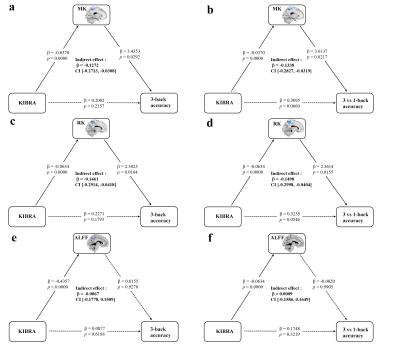 |
Brain microstructural alterations of left precuneus mediate the association between KIBRA rs17070145 and working memory in healthy adults.
Junxia Wang1, Sichu Wu2, Jilei Zhang3, and Bing Zhang2
1Nanjing Drum Tower Hospital Clinical College of Nanjing Medical University, Nanjing, China, 2Drum Tower Hospital, Medical School of Nanjing University, Nanjing, China, 3Clinical Science, Philips Healthcare, Shanghai, Shanghai, China
We used voxel-based independent sample t-test to investigate differences of DKI and resting-state fMRI parametric maps between KIBRA rs17070145 polymorphism in 163 young adults. Mediation analysis was used to explore the association between the KIBRA polymorphism, brain and working memory. We observed that KIBRA C-allele carriers had increased AD, RD, MD and decreased FA, MK, RK, ALFF compared with KIBRA TT homozygotes. The MK, RK of the left precuneus mediated the association between the KIBRA polymorphism and the working memory performance. These findings may be useful in understanding the neural mechanisms of KIBRA and provide a biological pathway of gene-brain-behavior.
|
Session Topic: Diffusion Applications
Session Sub-Topic: Diffusion: Applications Beyond the Brain
Digital Poster
Diffusion
4556. |
A Longitudinal Study on Assessing the Recovery of Spinal Cord on Incomplete Traumatic Spinal Cord Injury using Diffusion Tensor Imaging
Bing Yao1,2, Hannah Ovadia1, Gail Forrest3, and Steven Kirshblum2,4
1Rocco Ortenzio Neuroimaging Center, Kessler Foundation, West Orange, NJ, United States, 2Department of Physical Medicine and Rehabilitation, Rutgers University, Newark, NJ, United States, 3Center for Mobility and Rehabilitation Engineering Research, Kessler Foundation, West Orange, NJ, United States, 4Kessler Institute for Rehabilitation, West Orange, NJ, United States
Physicians rely on self-reports to monitor and evaluate the functional outcome in patients with spinal cord injury during their rehabilitation. These clinical and outcome measurements can be subjective and sometimes impractical if patients have cognitive difficulty. Traditional clinical MRI scans can provide doctors more objective information but they are not sensitive to detect the progression or repair during patient’s recovery. In this study, we investigated the sensitivity of DTI technique in detecting SCI injury and its progression or recovery over the course of rehabilitation in the individuals with SCI.
|
|
4557. |
Quantitative assessment of thyroid and parathyroid lesions by using ZOOMit-based IVIM, DKI and DWI
Bing Liu1,2, Xiangtao Lin1,2, Peng Zhao1, Xianshun Yuan1,2, Mengxiao Liu3, Xiang Feng4, Lei Xue5, Mimi Tian2, Shuai Zhang1,2, Dejuan Shan1,2, and Xiaoli Li1,2
1Department of Radiology, Shandong Provincial Hospital Affiliated to Shandong University, Jinan, China, 2Shandong University, Jinan, China, 3MR Scientific Marketing, Diagnostic Imaging, Siemens Healthcare Ltd, Shanghai, China, 4MR Scientific Marketing, Diagnostic Imaging, Siemens Healthcare Ltd, Beijing, China, 5MR Application, Siemens Healthcare Ltd, Jinan, China
The aim of this study was to evaluate the diagnostic performance of three advanced diffusion techniques (IVIM, DKI and DWI) which based on ZOOMit in demonstrating lesions in thyroid and parathyroid area. Results showed that D* in IVIM, MK in DKI and ADC in DWI were superior than other diffusion parameters (D, F in IVIM, MD in DKI) in accurately locating lesions as well as differentiating between lesions and normal thyroid parenchyma.
|
|
4558.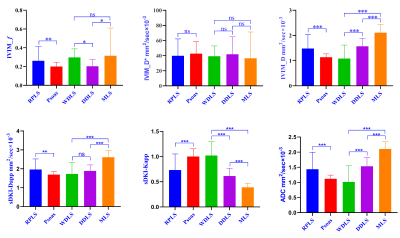 |
Quantitative evaluation of IVIM-DWI combined with simplified DKI for subtype diagnosis and grading prediction in retroperitoneal liposarcoma
Zhang Jiulong1, Shi Nannan1, Zhang Yijun1, Ye Wen1, Zhang yong2, Shan fei1, and Shi Yuxin1
1radiology department, Shanghai public health clinical center, shanghai, China, 2General surgery, Shanghai public health clinical center, shanghai, China
This study used ivim-dwi combined with sDKI models to quantitatively evaluate the differential diagnosis and histological grading prediction of retroperitoneal liposarcoma subtypes. Our results indicated that IVIM-DWI combined with sDKI models could accurately differentiate partial subtypes of RPLS, which was helpful for clinical selection of the optimal treatment strategy.
|
|
4559. |
Is ADC Measurement of Parotid Gland Tumor Sufficient using the Largest Slice rather than Whole Tumor
Shao-Chieh Lin1, Jui-Heng Lin1, Chun-Jung Juan2,3,4, Kai-Min Chien5, Teng-Yi Huang6, Yi-Jui Liu7, Chang Hsien Liu 5, Ya-Hui Li5, Szu Hsien Chou 5, and Chi-Feng Hsieh5
1Master 's Program of Biomedical Informatics and Biomedical Engineering, Feng Chia University, Taichung, Taiwan, 2Department of Medical Imaging, China Medical University Hsinchu Hospital, Hsinchu, Taiwan, 3Department of Radiology, School of Medicine, College of Medicine, China Medical University, Taichung, Taiwan, 4Department of Computer Science and Information Engineering, National Taiwan University, Taipei, Taiwan, 5Department of Medical Imaging, Chinese Medical University Hsinchu Hospital, Hsinchu, Taiwan, 6Department of Electrical Engineering, National Taiwan University of Science and Technology, Taipei, Taiwan, 7Department of Automatic Control Engineering, Feng Chia University, Taichung, Taiwan
The study is to quantitatively compare the diagnostic ability of ADC in distinguishing three types of parotid tumor between the ADC measurement on slice with the largest tumor and whole tumor. This retrospective study enrolled 13 patient for each PMAs, WTs and MTs. All participants underwent 1.5-T fat-saturated EP-DWI. Our results show that ADC and AUC on largest slice and whole tumor were similar in three tumors. ADC of the slice with the largest tumor, which tumor size over 1/3 whole tumor volume, could instead the ADC of whole tumor to diagnosis the PMA, WT and MT in parotid gland.
|
|
4560. |
Intravoxel incoherent motion and diffusion kurtosis imaging in the assessment of pathological grades of clear cell renal cell carcinoma
Qing Xu1, Weiqiang Dou2, and Jing Ye1
1Department of Radiology, Clinical Medical School of Yangzhou University, Northern Jiangsu People’s Hospital, Yangzhou, 457, China, 2GE Healthcare, MR Research China, Beijing, China
In this study, we aimed to investigate whether intravoxel incoherent motion (IVIM) and diffusion kurtosis imaging (DKI) techniques can be used to evaluate the pathological grade of clear cell renal cell carcinoma (ccRCC) patients preoperatively. As a result, the IVIM-related parameters (ADC, apparent diffusion coefficient; D, true diffusivity) and DKI-related parameters (MD, mean diffusivity; MK, mean kurtosis) have shown significant differences between low- and high-grade ccRCC(0.58±0.11 vs 0.48±0.08, 1.39±1.18 vs 0.98±0.21, 2.12±0.35 vs 1.59±0.32, 0.53±0.13 vs 1.59±0.32; p<0.05). Therefore, IVIM and DKI techniques can be used effective tools to differentiate low- and high-grade ccRCC.
|
|
4561.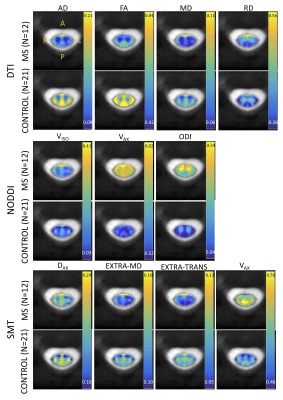 |
Investigating multi-compartment diffusion MRI models in the cervical spinal cord of multiple sclerosis patients
Kurt G Schilling1, Kristin P O'Grady1,2, Samantha By3, Haley Feiler1, Francesca Bagnato4, Bennett A Landman1,5,6,7, and Seth A Smith1,5,8
1Vanderbilt University Institute of Imaging Science, Nashville, TN, United States, 2Radiology and Radiological Sciences, Vanderbilt University Medial Center, Nashvillet, TN, United States, 3Hyperfine Research Inc, Guilford, CT, United States, 4Neurology, Vanderbilt University Medical Center, Nashville, TN, United States, 5Biomedical Engineering, Vanderbilt University, Nashville, TN, United States, 6Radiology and Radiological Sciences, Vanderbilt University Medical Center, Nashville, TN, United States, 7Electrical Engineering, Vanderbilt University, Nashville, TN, United States, 8Radiology and Radiological Sciences, Vanderbilt University, Nashville, TN, United States
We investigate multi-compartment diffusion MRI models in the in vivo spinal cord of multiple sclerosis (MS) patients. We find significant differences in diffusion measures of tissue microstructure and diffusivity indices between healthy controls (N=21) and MS patients (N=12). We also explore their relationships to clinical data, including disability scores, 25-foot walk tests, and disease duration, and note unique trends in these measures with increasing disability. These models may enable improved in vivo characterization of the spinal cord in MS patients and enable quantification of changes in meaningful tissue microstructure indices.
|
|
4562.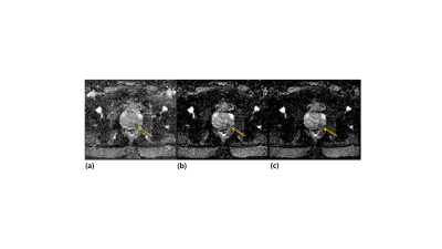 |
Radiomics Analysis of Apparent Diffusion Coefficient Maps with Various b-value Combinations for Differentiation of Prostate Cancer
Eo-Jin Hwang 1 and Moon Hyung Choi2
1Seoul St. Mary's Hospital, College of Medicine, The Catholic University of Korea, Seoul, Republic of Korea, 2Eunpyeong St.Mary's Hospital, College of Medicine, The Catholic University of Korea, Seoul, Republic of Korea
Prostate Imaging-Reporting and Data System (PI-RADS) suggests acquiring multiple apparent diffusion coefficient (ADC) maps including the lowest b-values between 50-100s/mm2 and highest b-values greater than 1400s/mm2. Radiomics is a novel field in medical imaging to advance decision support by utilizing large amount of quantitative features. In this study, we employed radiomics from ADC maps and a linear regression model to differentiate prostate cancer from benign tissues and evaluated the effect of various b-value combinations on ADC maps. We discovered that ADC with the b-values of 100 and 1000s/mm2 was most effective in discriminating prostate cancer with high accuracy.
|
|
4563. |
Two-step approach in IVIM parameter quantification
Xin Li1, Ryan Kopp2,3, William D Rooney1, Fergus Coakley4, and Mark Garzotto2,3
1Advanced Imaging Research Center, Oregon Health & Science University, Portland, OR, United States, 2Portland VA Medical Center, Portland, OR, United States, 3Urology, Oregon Health & Science University, Portland, OR, United States, 4Diagnostic Radiology, Oregon Health & Science University, Portland, OR, United States
The goal of this study is to investigate a new two-step approach in intra-voxel incoherent motion (IVIM) imaging data quantification. This methods first determines the slow diffusion constant (D) then the pseudo-perfusion parameters (D*, f) using the IVIM model fitting but holding D at pre-determined value. Results show that the new approach returns more consistency parametric maps and the new modeling is favored by Akaike information criterion (AIC).
|
|
4564. |
Prostate cancer detection with biophysical modeling of diffusion and relaxometry
Gregory Lemberskiy1,2, Yousef Mazaheri3, Herbert Alberto Vargas3, Ricardo Otazo3, Els Fieremans1, and Dmitry S Novikov1
1Radiology, New York University School of Medicine, New York, NY, United States, 2Microstructure Imaging INC, New York, NY, United States, 3Memorial Sloan Kettering Cancer Center, New York, NY, United States
For prostate cancer imaging, increasing the echo time selectively suppresses the cellular tissue, and emphasizes luminal water. We study the ADC dependence on echo time and diffusion time in 3 patients with PIRADS≥4 lesions, and propose a modeling strategy to interpret the changes in signal, as lumen diameter and volume fraction shrink during cancer progression. We evaluate cellular and luminal diffusivities, volume fractions, and luminal diameters as cancer biomarkers.
|
|
4565. |
Breast MRI combined with clinicopathologic characteristics for Preoperative Prediction of Axillary Lymph Node Metastasis in Breast Cancer
Mei Xue1, Lizhi Xie2, and Jing Li3
1Radiology, Cancer Hospital Chinese Academy of Medical Sciences, Beijing, China, 2GE Healthcare, MR Research China, Beijing, Beijing, China, 3Cancer Hospital Chinese Academy of Medical Sciences, Beijing, China
Accurate identification of axillary lymph node(ALN) involvement in patients with breast cancer is crucial for prognosis and treatment strategy decisions. We developed a clinical model based on breast MR imaging and clinicopathologic characteristics to predict ALN metastasis. Our findings suggest that the clinicopathological features of breast tumor are highly correlated with axillary lymph node metastasis. It can be used to assist clinicians to predict LN metastasis non-invasively.
|
|
4566.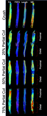 |
Assessment with Partial Peripheral Nerve Transection with Diffusion MRI
Isaac Vicente Manzanera Esteve1, Angel F Farinas1, Alonda C Pollins1, Wesley P Thayer1, Mark Does1, and Richard Dortch1
1VUIIS, Vanderbilt University Medical Center, Nashville, TN, United States
High-resolution DTI of ex vivo rat sciatic nerve yields viable biomarkers of peripheral nerve recovery following partial transection and surgical repair. FA values decreased with increasing cut depth at 4 weeks. By week 12, the three partial cuts showed elongated FA distributions, most likely representing regions with regenerated (high FA values) and degenerated axons (low FA values). FA finding are influenced by pathological features that affect diffusion both perpendicular and parallel to the axons.
|
|
4567.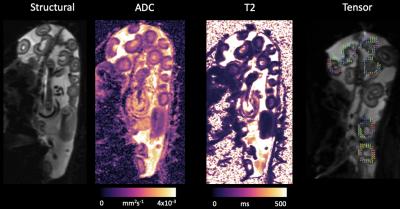 |
Characterisation of placentome function using combined diffusion-relaxometry MRI and flow anisotropy
Dimitra Flouri1,2, Jack RT Darby3, Stacey L Holman3, Sunthara R Perumal4, Anna L David5,6, Andrew Melbourne1,2, and Janna L Morrison3
1School of Biomedical Engineering & Imaging Sciences, King's College London, London, United Kingdom, 2Department of Medical Physics & Biomedical Engineering, University College London, London, United Kingdom, 3Early Origins of Adult Health Research Group, University of South Australia, Adelaide, Australia, 4Preclinical Imaging and Research Laboratories, South Australian Health and Medical Research Institute, Adelaide, Australia, 5Institute for Women's Health, University College London, London, United Kingdom, 6NIHR University College London Hospitals Biomedical Research Center, London, United Kingdom
Abnormal development of the placenta is postulated as the root cause of preeclampsia and fetal growth restriction. Diffusion-Weighted imaging techniques are considered to give additional placental information. Animal models have been important in invasive validation studies for MRI measurements, as they allow for controlled experiments and analysis of multiple time-points during pregnancy. This study characterises diffusion and perfusion properties of the placenta such as the apparent diffusion coefficient, T2 measurements, fractional anisotropy and perfusion fraction derived from intravoxel incoherent motion analysis on sheep placental tissue in order to validate new imaging markers of placental function.
|
|
4568.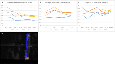 |
Tracing and Therapeutic Evaluation of Transplanted Mesenchymal Stem Cell in The Spinal Cord Injury of Beagles using Diffusion Tensor Imaging
Junting Zou1, Jilei Zhang2, Yuanyuan Xie3, yunpeng Shen4, Jiacheng Du4, Yang Chen4, Yang Chen4, Bing Zhang3, and Xiaoli Mai5
1Nanjing Drum Tower Hospital Clinical College of Nanjing Medical University, Nanjing, China, 2Clinical Science, Philips Healthcare, Shanghai, China, 3Nanjing Drum Tower Hospital, Nanjing, China, 4Southeast University, Nanjing, China, 5Radiology, Nanjing Drum Tower Hospital, Nanjing, China
The labelled SPIO of the transplanted MSCs were found in spinal cord segment of TSCI based on T1w and T2w images. The DTI can be of great value in the dynamic evaluation of morphological and neurological changes in beagles after TSCI. FA values are correlated with the impairment and repair of the fiber tracts of the spinal cord. The FA value can satisfactorily evaluate the completeness of the fiber tracts after SCI and the repair of them by stem cells. The combination of conventional MRI and DTI is of great clinical importance for the diagnosis and therapeutic evaluation of SCI.
|
|
4569. |
Mono-exponential bi-exponential and stretched-exponential diffusion imaging in characterization of nonalcoholic fatty liver disease
Xianfu Luo1, Jing Ye1, Weiqiang Dou2, Jun Sun1, and Wei Xia1
1Radiology, Clinical Medical School of Yangzhou University, Northern Jiangsu People’s Hospital, Yangzhou, China, 2GE Healthcare,MR Research China, Beijing, China
We aimed to compare mono-exponential bi-exponential and stretched-exponential diffusion-weighted imaging(DWI) model in characterizing nonalcoholic liver disease (NAFLD) by providing multiple quantitative parameters. The diagnosis performance for early detection nonalcoholic steatohepatitis (NASH) was compared using characteristic operating curve analysis. Stretched-exponential DWI performed as well as bi-exponential DWI and better than mono-exponential DWI in the noninvasive characterization of NAFLD severity.
|