
Digital Poster Session
fMRI: Contrast & Physiology
fMRI
3815 -3829 fMRI: Contrast & Physiology - fMRI Contrast Mechanisms
3830 -3845 fMRI: Contrast & Physiology - fMRI Physiology 1
3846 -3861 fMRI: Contrast & Physiology - fMRI Physiology 2
Session Topic: fMRI: Contrast & Physiology
Session Sub-Topic: fMRI Contrast Mechanisms
Digital Poster
fMRI
3815.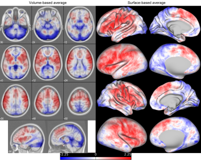 |
Effect of the perfusion lag structure removal on task fMRI
Toshihiko Aso1 and Takuya Hayashi1
1Laboratory for Brain Connectomics Imaging, RIKEN Center for Biosystems Dynamics Research, Kobe, Japan
A lag structure in the fMRI signal, created by a combination of the low-frequency oscillation and perfusion delay, has been suggested as being a noise component, but its removal has not been established as a denoising approach. Using an HCP task-fMRI dataset, we report that the removal of this lag structure, or deperfusioning, has a unique effect on fMRI group analysis both qualitatively and quantitatively. Increased sensitivity of the treatment in some condition was emphasized by switching from volume- to surface-based analysis, suggesting concomitant improvement of specificity. Unfavorable effect was also suggested depending on the task condition, warranting further investigation.
|
|
3816.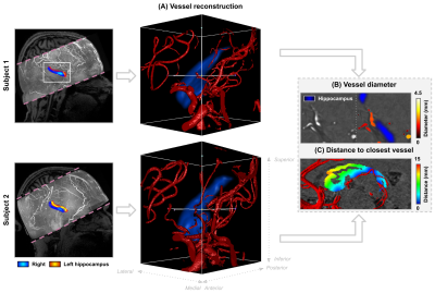 |
Disentangling hippocampal perfusion from the macrovasculature using ASL and TOF at 7T
Roy AM Haast1, Sriranga Kashyap2, Jordan DeKraker1, Dimo Ivanov2, Benedikt A Poser2, and Ali R Khan1,3
1Robarts Research Institute, Western University, London, ON, Canada, 2Department of Cognitive Neuroscience, Maastricht University, Maastricht, Netherlands, 3Department of Medical Biophysics, Schulich School of Medicine and Dentistry, Western University, London, ON, Canada
Arterial spin labeling (ASL) at 7T can be used to map cerebral blood flow (CBF) at a high spatial resolution. Previously, we have shown that different hippocampal subfields have differences in perfusion, owing to potential differences in their vascular density. Hippocampal imaging at 7T can be challenging and requires optimizations of acquisition methods, as well as integration of novel analysis strategies. Here, we map high-resolution quantitative multi-modal MRI data (T1, CBF, time-of-flight angiography) in the human hippocampus to disentangle perfusion and macrovasculature at 7T.
|
|
3817.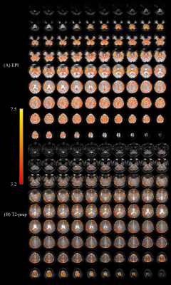 |
Whole-Brain Functional MRI in Human Subjects Wearing Metallic Orthodontic Braces
Xinyuan Miao1,2, Yuankui Wu1,2,3, Dapeng Liu1,2, Qin Qin1,2, Peter C.M van Zijl1,2, Jay J. Pillai4,5, and Jun Hua1,2
1Neurosection, Division of MRI Research, Russell H. Morgan Department of Radiology and Radiological Science, Johns Hopkins University School of Medicine, Baltimore, MD, United States, 2F.M. Kirby Research Center for Functional Brain Imaging, Kennedy Krieger Institute, Baltimore, MD, United States, 3Department of Medical Imaging, Nanfang Hospital, Southern Medical University, Guangzhou, China, 4Johns Hopkins University School of Medicine, Division of Neuroradiology, Russell H. Morgan Department of Radiology and Radiological Science, Baltimore, MD, United States, 5Department of Neurosurgery, Johns Hopkins University School of Medicine, Baltimore, MD, United States
Metallic objects such as dental braces bring substantial susceptibility artifacts in MR images acquired using echo-planar-imaging (EPI) sequences. Gradient-echo EPI is currently the most commonly adopted sequence for blood oxygenation level–dependent (BOLD) functional MRI (fMRI) in humans, which suffers from susceptibility artifacts including signal dropout and geometric distortion in the presence of metallic implants. Here, we demonstrate that T2-prepared (T2prep) BOLD fMRI can significantly reduce susceptibility artifacts that are commonly seen in GRE EPI in the presence of metallic orthodontic braces.
|
|
3818.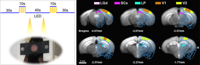 |
Mouse fMRI of visual stimulation in awake vs. anesthetized condition
THI NGOC ANH DINH1 and Seong Gi Kim1
1Department of Biomedical Engineering, Sungkyunkwan University, Center for Neuroscience Imaging Research, Institute for Basic Science (IBS), Suwon-si, Korea, Republic of
Mouse fMRI has been increasingly used in the MRI community. Here, we developed an awake mouse fMRI protocol and compared BOLD fMRI responses responding to visual stimulation of 5 Hz and 10 Hz, obtained during awake and anesthetized conditions. Our 9.4-T BOLD-fMRI showed activities in all visual areas including LGN, superior colliculus, and V1, and can be well-explained by electrophysiology literature.
|
|
3819.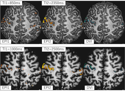 |
Simultaneous functional and anatomical high-resolution imaging using BISEPI at 7T
Guoxiang Liu1,2, Adnan Shah1,2, and Takashi Ueguchi1,2
1Brain Function Analysis and Imaging Lab, CiNet, NICT, Osaka, Japan, 2Graduate School of Frontier Biosciences, Osaka University, Osaka, Japan
A novel method for simultaneous functional and anatomical imaging is presented for high-resolution fMRI at 7T. An RF inversion pulse is used before two EPI acquisition trains where functional scans are acquired alternating between shorter and longer inversion times (TIs). Post-processing division of the shorter TI functional scan with the longer TI functional scan allows improved localization of brain activity in high-resolution fMRI at 7T. The proposed method allows simultaneous imaging of brain function and anatomy as the grey matter tissues can easily be segmented out from the resulting functional images, ensuring their perfect alignment required for detecting functional activity.
|
|
3820.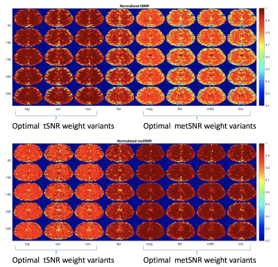 |
Temporal SNR in multiecho fMRI and its dependence on the choice of weights
Thomas Liu1, Bochao Li2, Conan Chen1, Brice Fernandez3, Baolian Yang4, and Suchandrima Banerjee5
1Center for Functional MRI, UC San Diego, La Jolla, CA, United States, 2Biomedical Engineering, University of Southern California, Los Angeles, CA, United States, 3GE Healthcare, Buc, France, 4GE Healthcare, Waukesha, WI, United States, 5GE Healthcare, Menlo Park, CA, United States
In multi-echo fMRI, data from multiple echoes are combined using weights. We use the framework of the generalized Rayleigh quotient (GRQ) to derive the optimal weights for temporal SNR (tSNR) and multi-echo tSNR (metSNR) measures of performance. We also examine the sensitivity of both these measures to the choice of weights. We find that metSNR measures are relatively insensitive to the weights in gray matter regions as compared to tSNR measures, which show greater sensitivity in these regions. We interpret the findings using the form of the GRQ.
|
|
3821.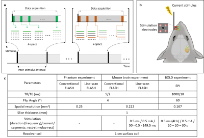 |
In vivo Direct MR Imaging of Mouse Whisker Sensory Responses
Tan Toi Phan1,2 and Jang-Yeon Park1,2
1Department of Biomedical Engineering, Sungkyunkwan University, Suwon, Republic of Korea, 2Center for Neuroscience Imaging Research, Institute for Basic Science, Suwon, Republic of Korea
Although there are debates on direct MR imaging of neuronal activity, the feasibility of this topic has been finding in various different ways. Herein, we propose an effective method of line-scan-based FLASH (LS-FLASH) imaging to directly capture sensory responses of in vivo mouse brain with a 5-ms temporal resolution. The response delay of ~10 - 15 ms between the thalamus and somatosensory barrel cortex (S1BF) was observed when the electrical stimulation was applied to the left mouse whisker area.
|
|
3822.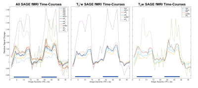 |
Optimization of vascular and functional sensitivity using multi-contrast, multi-echo SAGE-EPI for fMRI
Maurizio Bergamino1, Lori Steffes1, Ashlyn Gonzales2, Leslie Baxter2, and Ashley M. Stokes1
1Neuroimaging Research, Barrow Neurological Institute, Phoenix, AZ, United States, 2Neuropsychology, Mayo Clinic Arizona, Phoenix, AZ, United States
The purpose of this study is to develop a multi-contrast, multi-echo sequence using spin- and gradient-echo (SAGE) for fMRI; furthermore, we sought to assess various analysis schemes for optimal quantification of fMRI. For this purpose, we acquired SAGE-fMRI data with five echoes (2 gradient-echo, 2 asymmetric spin-echoes, and 1 spin-echo) using a visual stimuli task in 8 healthy subjects. Analysis was performed using each echo signal individually, using weighting factors to combine dynamic signals, and by quantifying dynamic R2* and R2 time-courses. These methods are compared to determine the optimal analysis method for SAGE-fMRI.
|
|
3823.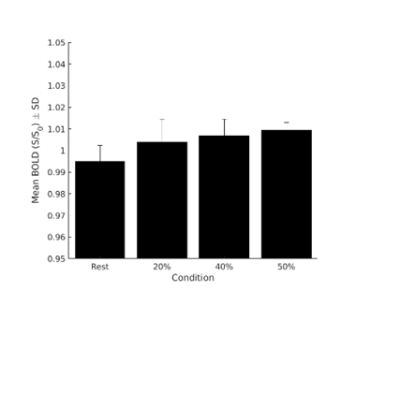 |
Study of the Spinal Cord BOLD functional response
Michela Fratini1,2, Mauro Di Nuzzo2, Marta Moraschi3, Laura Maugeri2,4, fabio mangini2, daniele mascali3, and federico giove3
1CNR-Nanotec, Roma, Italy, 2fondazione Santa Lucia, Roma, Italy, 3centro fermi, Roma, Italy, 4CNR-Nanotec, piazzale Aldo Moro 5, Italy
Spinal Cord Functional Magnetic Resonance Imaging (fMRI) techniques are widely exploited for the study of brain activation. Similar approaches have been repeatedly attempted in the spinal cord, but spinal cord fMRI has not yet affirmed as habitual tool for the assessment of spinal cord function. One of the reasons is that the features of the functional contrast are still partially unknown. In the present work, we study the relationship between the intensity of the motor stimulation (performing a controlled motor task with the dominant hand ) and the amplitude of the functional response in healthy
|
|
3824.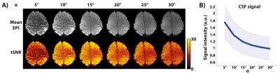 |
Characterising the effect of flip angle in the acquisition of sub-millimetre resolution 3D GE-EPI at 7T
Sriranga Kashyap1, Laurentius Huber1, Dimo Ivanov1, Denizhan Kurban1, David A Feinberg1,2,3, and Benedikt A Poser1
1Maastricht Brain Imaging Centre (MBIC), Faculty of Psychology and Neuroscience, Maastricht University, Maastricht, Netherlands, 2Advanced MRI Technologies, Sebastopol, CA, United States, 3Helen Wills Neuroscience Institute, University of California, Berkeley, CA, United States
Sub-millimetre resolution is desirable for laminar-fMRI to reduce partial-voluming with neighbouring tissues. CSF signal fluctuations can be a confound in voxels that lie at the CSF-GM interface due to physiological processes such as activation. The 3D-EPI readout, used with sub-millimetre fMRI studies, suppresses the CSF signals (long-T1 vs. short-TR). However, the degree of suppression is dependent on the flip angle used. In this study, we investigate the signal contributions of different tissue compartments by systematically varying the flip angle and map the flip angle dependence of tSNR as a function of cortical depth at 0.8 mm isotropic resolution at 7T.
|
|
3825.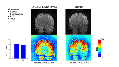 |
Multislab multiband 3D EPI for simultaneous high spatial and temporal resolution at 7T
Luca Vizioli1, Steen Moeller1, Edward Auerbach1, Kamil Ugurbil1, and Essa Yacoub1
1CMRR, University of Minnesota, Minneapolis, MN, United States
Ultra-high field scanners allow recording BOLD images with unprecedented spatio-temporal resolution. These highly precise measurements permit studying the human brain at the mesoscale level, investigating the roles of some of the most fundamental units of neural computations, namely, layers and columns. These submillimeter recordings are however SNR-starved and the ideal choice of sequence remains to be determined. Here, we show that by using a multislab multiband 3D EPI approach we can achieve simultaneously high spatial and temporal resolution for sub-millimeter fMRI applications at 7T. We compare this approach to the more common strategies of 3D EPI and 2D MB EPI.
|
|
3826. |
Apparent attenuation of BOLD macro-vascular contributions with high-frequency stimuli
Daniel E. P. Gomez1,2,3, Nina E. Fultz1,3, Shahin Nasr1,2, Jonathan R. Polimeni1,2,4, and Laura D. Lewis1,3
1Athinoula A. Martinos Center for Biomedical Imaging, Massachusetts General Hospital, Boston, MA, United States, 2Department of Radiology, Harvard Medical School, Boston, MA, United States, 3Department of Biomedical Engineering, Boston University, Boston, MA, United States, 4Harvard-Massachusetts Institute of Technology Division of Health Sciences and Technology, Boston, MA, United States
Fast fMRI signals may reflect an enhanced relative contribution of fast microvascular signals in the BOLD response. In the current work we tested whether using fast oscillatory stimulus frequencies indeed leads to a relative attenuation of the signal from draining veins, as compared to the microvasculature. Our results suggest that the relative contribution of deep BOLD signals is enhanced at high stimulus frequencies.
|
|
3827.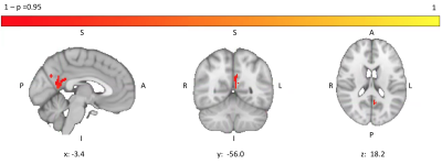 |
Locally reduced cerebral blood flow in patients with Idiopathic Pulmonary Fibrosis
Kirsty Hett1, Eleonora Patitucci1, Hannah Chandler1, Michael Germuska1, Benjamin Hope-Gill2, and Richard Wise1,3
1Cardiff University Brain Research Imaging Centre (CUBRIC), Cardiff University, Cardiff, United Kingdom, 2Respiratory Medicine, Cardiff and Vale University Health Board, Cardiff, United Kingdom, 3Institute for Advanced Biomedical Technologies, Department of Neuroscience, Imaging and Clinical Sciences, "G. D'Annunzio University" of Chieti-Pescara, Chieti, Italy
Idiopathic Pulmonary Fibrosis (IPF) is a palliative lung condition. Neuroimaging has the potential to shed light on the neural pathways of cough, a common, troublesome symptom in IPF. However, given the nature of the condition, it is important to first understand the physiological state of brain tissue before investigating functional networks. No significant differences in brain volume were observed in IPF patients but a localised reduction in resting perfusion was seen. There was no difference in functional response when performing a combined motor and visual task. The results support future neuroimaging investigation into the cough pathway in IPF.
|
|
3828.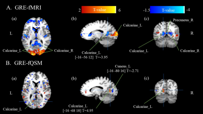 |
Investigating Human Brain Negative Blood Oxygenation Level Dependent (BOLD) with Functional Quantitative Susceptibility Mapping (fQSM)
Wei-Hao Huang1, Hong-Yi Wu1, Yun-An Huang2, Po-Wei Cheng1, Chia-Ming Shih1, and Jyh-Horng Chen1,2
1Graduate Institute of Biomedical Electronics and Bioinformatics, National Taiwan University, Taipei, Taiwan, 2Interdisciplinary MRI/MRS Lab, Department of Electrical Engineering, National Taiwan University, Taipei, Taiwan
We aim to use fQSM to investigate the phenomenon of negative BOLD with a visual stimulation task. fQSM data of 14 subjects was reconstructed with the phase information. The results show the negative BOLD is located in the identical brain region of Calcarine in both fMRI or fQSM analysis. In the future, we will optimize fQSM reconstruction method. A reliable fQSM is able to improve the understanding of the underline mechanism of fMRI BOLD.
|
|
3829.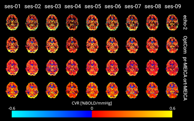 |
Improving breath-hold cerebrovascular reactivity mapping with multi-echo BOLD fMRI
Stefano Moia1, Maite Termenon1, Eneko Uruñuela-Tremiño1, Rachael C. Stickland2, Molly G. Bright2,3, and César Caballero-Gaudes1
1Basque Center on Cognition Brain and Language, San Sebastián, Spain, 2Physical Therapy and Human Movement Sciences, Feinberg School of Medicine, Northwestern University, Chicago, IL, United States, 3Biomedical Engineering, McCormick School of Engineering, Northwestern University, Evanston, IL, United States
Cerebrovascular reactivity (CVR) is an emerging metric for quantifying vascular health. During a BOLD fMRI acquisition, breath-holding is a simple approach for increasing blood CO2 levels, thereby driving the vasodilatory CVR response. However, breath-holding is associated with increased head motion artifacts, which can confound our estimates of CVR. We demonstrate that multi-echo BOLD fMRI acquisitions, in conjunction with denoising with multi-echo independent component analysis approaches, enable better differentiation of true vasodilatory responses and improve the reliability of CVR estimates across multiple sessions in highly sampled individuals.
|
 Watch the Video
 View the Poster
Session Topic: fMRI: Contrast & Physiology
Session Sub-Topic: fMRI Physiology 1
Digital Poster
fMRI
3830.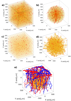 |
A novel statistical 3D cortical vascular network to simulate realistic hemodynamic changes in the human brain
Mario G. Báez-Yáñez1, Jeroen Siero1,2, and Natalia Petridou1
1Department of Radiology, Center for Image Sciences, University Medical Center Utrecht, Utrecht, Netherlands, 2Spinoza Centre for Neuroimaging Amsterdam, Royal Netherlands Academy of Arts and Sciences, Amsterdam, Netherlands
We present a statistical 3D computational model that mimics the human cortical vascular network. This approach allows to study the hemodynamic changes and to simulate biophysical effects across the vascular network, as well as to investigate the dynamic BOLD fMRI signal formation. In order to extract quantitative parameters of the BOLD fMRI signal in humans, it is necessary to adopt a computational model that resembles the human cortical vasculature and mimics realistic hemodynamic fluctuations triggered by neurovascular coupling. Simulating the biophysical effects of tissue related to hemodynamic changes will provide accurate information on the origins of the BOLD signal time-course.
|
|
3831.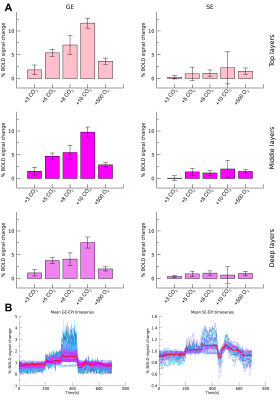 |
Vascular and neurovascular effects on micro- and macro-vascular compartments in human visual cortex.
Wouter Schellekens1, Alex Bhogal1, Jeroen Siero1, and Natalia Petridou1
1Radiology, UMC Utrecht, Utrecht, Netherlands
In the current study, we investigate the contribution of non-neuronal and neuronal-related hemodynamic changes to the BOLD signal for micro- and macro-vascular compartments. Vascular reactivity was assessed using CO2 and O2 gas administration, as well as brief visual stimuli, while we used gradient-echo and spin-echo across different cortical depths to specifically target different vascular compartments. We found that gradient-echo responded clearly to the gas challenges, also affecting the neurovascular hemodynamic response function. These effects were not seen for spin-echo, suggesting that the spatiotemporal characteristics of micro-vessels may not solely be affected by vascular properties.
|
|
3832.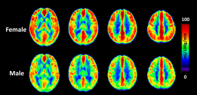 |
Characterization of regional differences in cerebral vascular response to breath holding using BOLD fMRI
Hui-Chieh Yang1, Tsai-Ying Liao2, Guan-Jie Wang3, Chun-Ming Chen4, and Shin-Lei Peng3
1Institute of Brain Science, National Yang-Ming University, Taipei, Taiwan, 2School of Medicine, China Medical University, Taichung, Taiwan, 3Department of Biomedical Imaging and Radiological Science, China Medical University, Taichung, Taiwan, 4Department of Radiology, China Medical University Hospital, Taichung, Taiwan
Previous studies have demonstrated that cerebrovascular reactivity (CVR) depends on the baseline vascular dilation status between groups. Within the brain, there also exist spatial variations in resting cerebral blood flow (CBF) and CVR across different cerebral regions; however, the relationship between regional CBF and CVR remains unclear. We found the frontal lobe had the maximal resting CBF but the minimal vascular response to hypercapnia, whereas the occipital lobe had the lowest rest CBF but the maximal reactivity to hypercapnia, suggesting that there is a small and a large autoregulatory efficiency at high and low resting CBF in the brain, respectively.
|
|
3833.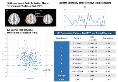 |
30-day Reliability Assessments between Cortisol, Vigilance and Brain Activity
Yu-Lun Su1, Hong-Yi Wu2, Po-Yi Chen2, Chi-Yun Liu1, Ai-Ling Hsu3, Yi-Ping Chao4, and Changwei Wesley Wu5
1Taipei Medical University, Taipei, Taiwan, 2National Taiwan University, Taipei, Taiwan, 3Quanta Computer Inc., Taoyuan, Taiwan, 4Chang Gung University, Taoyuan, Taiwan, 5Graduate Institute of Mind, Brain and Consciousness, Taipei Medical University, Taipei, Taiwan
To estimate the reliability of task-fMRI, we conducted a repeated measure fMRI study of psychomotor vigilance task once per day and 30 days in total. Salivary cortisol and sleep duration were taken into consideration based on previous study. However, we found dramatic within-subject variability in the brain activity of dACC and primary motor cortex. The cortisol showed insignificant association with brain activity, but sleep duration affected the motor and temporal cortices. Such finding suggests that the PVT-based fMRI showed large variability across different days, which might be associated with the deviations of sleep duration.
|
|
3834.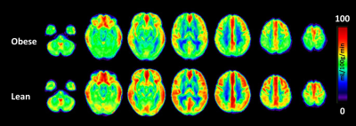 |
CBF alteration in human obesity: An arterial spin labeling study
Guan-Jie Wang1, Chun-Ming Chen2, and Shin-Lei Peng1
1Department of Biomedical Imaging and Radiological Science, China Medical University, Taichung, Taiwan, 2Department of Radiology, China Medical University Hospital, Taichung, Taiwan
Obesity is accompanied with damage to several organs including the brain. Although an extensive body of neuroimaging literature indicates that brain structure deteriorates with obesity, little information related to the relationship between CBF and obesity is available. In this study, we investigated the potential influence of body mass index (BMI) on brain abnormalities in young adults by combining functional and structural MRI studies. Results show CBF measured with the noninvasive MRI technique decreased as the BMI increased, as manifested by altered CBF in thalamus and visual-associated areas, including Brodmann areas (BA)7, BA18, and BA19
|
|
3835.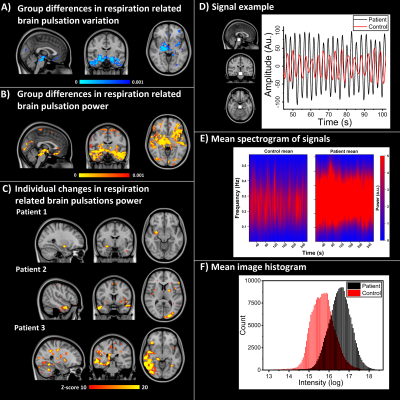 |
Respiratory related brain pulsations are disturbed in epilepsy
Janne Kananen1, Timo Tuovinen2, Vesa Korhonen2, Niko Huotari2, Hanna Ansakorpi3, Pierre LeVan4, and Vesa Kiviniemi2
1Medicine, Radiology, OFNI, University of Oulu, Oulu, Finland, 2University of Oulu, Oulu, Finland, 3Oulu University Hospital, Oulu, Finland, 4University of Freiburg, Freiburg, Germany
Fast-fMRI has become a potent tool in researching brain physiology more accurately than before and it can detect changes in the blood oxygen level‐dependent signal, even if there is no epileptiform activity present. These detectable changes suggest that in epilepsy there are alterations in the brain physiology. With utilization of coefficient of variation (CV) method for patients with epilepsy (PWE), we detected a robust increase of CV in patients with in white matter, brainstem and temporal lobes in PWE at group level. Importantly, individual mapping of possible epileptic abnormality is also possible.
|
|
3836. |
Regional effects of caffeine on BOLD fMRI calibration constant M
Rebecca J Williams1, Jacinta L Specht1, Wen-Ming Luh2, Erin L Mazerolle1, and G. Bruce Pike1
1University of Calgary, Calgary, AB, Canada, 2National Institute on Aging, National Institutes of Health, Baltimore, MD, United States
Estimating M value using a gas challenge is an essential component of calibrated fMRI. M is the maximal possible BOLD response and reflects baseline physiology such as CBF, CBV and CMRO2. Despite its common use, it is unknown how caffeine ingestion affects M. Here it is demonstrated that caffeine increases M regionally, with frontal brain regions including the paracingulate and anterior cingulate gyrus particularly affected. M increases were found in the context of decreased baseline CBF. These results indicate that caffeine may be implicated in regional uncoupling between CBF and CMRO2 which causes an increase in M.
|
|
3837. |
Voluntary action rhythmically modulates 7T BOLD visual responses in primary visual cortex
Maria Concetta Morrone1, Alessandro Benedetto1, Mauro Costagli2, Michela Tosetti2, and Paola Binda1
1Department of Translational Research on New Technologies in Medicine and Surgery, University of Pisa, Pisa, Italy, 2IRCCS Stella Maris, Calambrone, Pisa, Italy
Behavioral visual sensitivity modulates rhythmically in synchrony with the onset of a voluntary action. Using 7T fMRI, we demonstrate similar oscillations of primary visual cortex (V1) BOLD responses to brief visual stimuli presented at different delays from the onset of an action. The oscillations are limited to the stimulated V1 region and not to peripheral V1 (which responds to the motor action). The peak responses of V1 BOLD activity are functionally connected to primary motor cortex, suggesting that motor cortex drives the rhythmic oscillation of V1. These results support the suggestion of a possible role of oscillations for visuo-motor coordination.
|
|
3838. |
Characteristics of temporal dynamics of intrinsic brain activity in unmedicated bipolar II disorder with suicidality
JiaYing Gong1,2, Guanmao Chen1, Yanbin Jia3, Long Qian4, Li Huang1, and Ying Wang1
1Medical Imaging Center, First Affiliated Hospital of Jinan University, Guangzhou, China, 2Department of Radiology, Six Affiliated Hospital of Sun Yat-sen University, Guangzhou, China, 3Department of Psychiatry, First Affiliated Hospital of Jinan University, Guangzhou, China, 4MR Research, GE Healthcare, Beijing, China
The alterations of brain dynamics in bipolar disorder (BD) with suicidality are still unknown. Dynamic amplitude of low-frequency fluctuations (dALFF) was evaluated using sliding window analysis, and the severity of suicidality was predicted using a multivariate regression model between 106 BD II participants. Our findings suggest that alterations of temporal variability in the precuneus/ posterior cingulate cortex (PCC) is a common feature of BD participants, the right temporal lobe involved in impulsivity, social and emotional processing are associated with suicidality in BD II depression
|
|
3839.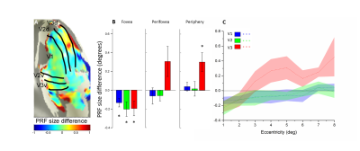 |
Attention field size alters patterns of population receptive field in the early visual cortex
Bo Liu1, Xiaochun Wang1,2, Le Wang2, Qiaojun Qu1, Xiang Feng3, Wei Zhang1, Bin Wang4, and Hui Zhang1,2
1College of Medical Imaging, Shanxi Medical University, Taiyuan 030001, Shanxi Province, China, 2Department of Radiology, First Clinical Medical College, Shanxi Medical University, Taiyuan 030001, Shanxi Province, China, 3MR Scientific Marketing, Diagnosis Imaging, Siemens Healthcare Ltd, Beijing 100102, China, 4College of Information and Computer, Taiyuan University of Technology, Taiyuan 030001, Shanxi Province, China
The effect of attention field (AF) is not only constrained to spatial positions of attention but also to size. However, the direct link of AF size to receptive field (RF) has not been well established. In our study, we demonstrated remarkable modulation of AF in the early visual cortex, with regard to size and location of population receptive field (pRF), emphatically. Specially, the differences in pRF size between focused and distributed attention conditions increase with eccentricity, especially in the V3 area. Considering pRF location, eccentricity is larger under distributed attention conditions than under focused attention conditions at the perifovea in the V3 area.
|
|
3840.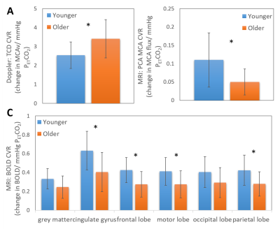 |
Comparing MRI and Doppler measures of resting Cerebral Blood Flow and Cerebrovascular Reactivity in the young and old
Claire V Burley1,2, Susan T Francis3, Samuel JE Lucas1, and Karen Mullinger4
1Centre for Human Brain Health, University of Birmingham, Birmingham, United Kingdom, 23Dementia Centre for Research Collaboration, University of New South Wales, Sydney, Australia, 3Sir Peter Mansfield Imaging Centre, School of Physics and Astronomy, University of Nottingham, Nottingham, United Kingdom, 4School of Physics and Astronomy, University of Nottingham, Nottingham, United Kingdom
Resting cerebral blood flow (CBF) and cerebrovasculature responsiveness (CVR) are becoming key measures of brain health. However, traditionally accepted CBF/CVR changes with age/fitness have been recently questioned using a variety of imaging modalities. Here we investigate the source of these discrepancies, performing Doppler and MRI measures on the same groups. We find similar changes in CBF but opposing changes in CVR measures between groups. There was no significant correlation of CBF MRI and Doppler measures across the cohort. Our work shows the necessity to further understand driving factors of CBF/CVR across modalities before use as a clinical research tool.
|
|
3841.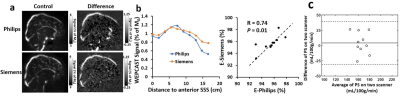 |
Cross vendor harmonization of water-extraction-phase-contrast-arterial-spin-tagging (WEPCAST) MRI for blood brain barrier assessment
Zixuan Lin1, Dengrong Jiang1, Zachary Baker1, Dapeng Liu1, Yang Li1, Xirui Hou1, Jay J. Pillai1, Qin Qin1, Yulin Ge2, and Hanzhang Lu1
1Department of Radiology, Johns Hopkins University, Baltimore, MD, United States, 2Department of Radiology, New York University Langone Medical Center, New York, NY, United States
Water-extraction-with-phase-contrast-arterial-spin-tagging (WEPCAST) MRI was proposed recently as a non-invasive technique to assess blood-brain barrier (BBB) permeability to water. However, the reproducibility of this technique and cross-vendor reliability has not been reported. In this study, we harmonized WEPCAST technique across two major MRI vendors, Philips and Siemens and examined the test-retest reproducibility of the technique. The results showed that WEPCAST MRI can give a reliable assessment of BBB permeability with an excellent reproducibility.
|
|
3842.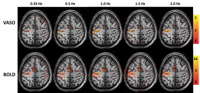 |
BOLD and CBV nonlinear responses during a hand movement task with increasing movement rates using 3D-EPI VASO at 7T
Ícaro A F de Oliveira1,2, Wietske van der Zwaag1, Luisa Raimondo1, Serge O Dumoulin1,2,3, and Jeroen CW Siero1,4
1Spinoza Centre for Neuroimaging, Amsterdam, Netherlands, 2Experimental and Applied Psychology, VU University, Amsterdam, Netherlands, 3Experimental Psychology, Helmholtz Institute, Utrecht University, Utrecht, Netherlands, 4Radiology, University Medical Centre Utrecht, Utrecht, Netherlands
We used 3D-EPI VASO to simultaneously measure CBV and BOLD signal changes in the primary motor cortex. We aimed to investigate differences in neurovascular coupling as measured by BOLD and CBV signal changes during a hand movement task with increasing movement rates. In agreement with previous findings, the BOLD response was found to behave nonlinearly with respect to the movement rate. Remarkably, the CBV response amplitude also shows a similar and strong nonlinear behavior where the amplitude saturates at fast movement rates (≥ 1 Hz).
|
|
3843.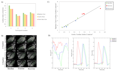 |
Investigating Cerebral and Cardiac Blood Flow and Cerebral Oxygen Extraction Concurrently During and Immediately after Exercise
Rosemary Nicholas1, Paul Greenhaff1, Jordan McGing1, Ayushman Gupta1, and Susan Francis1
1University of Nottingham, Nottingham, United Kingdom
To investigate the integrated impact of exercise on cardiac (CI) and cerebral (CBF) blood flow and oxygenation (OEF), measures were made concurrently in older active males at rest, during steady-state exercise and recovery. There was high association between aortic and biplane CI measures, and CBF increases with CI. Exercise induced increases in CI, CBF and the cerebral metabolic rate of oxygen (CMRO2), which were not seen on recovery. OEF was inversely associated with CI at rest, and increased on recovery. Exercise and recovery changes support the need for and highlight the utility of in-scanner exercise studies to investigate cardiovascular regulation.
|
|
3844. |
Amplitude of slow fluctuations in CSF as a time-resolved marker of sleep states for resting-state fMRI: a validation study.
Javier Gonzalez-Castillo1, Daniel A Handwerker1, and Peter A Bandettini1,2
1Section on Functional Imaging Methods, NIMH, Bethesda, MD, United States, 2FMRI Facility, NIH, Bethesda, MD, United States
Wakefulness fluctuations during rest are a key confound for dynamic functional connectivity. Yet, tracking such fluctuations is not trivial when lacking concurrent EEG and/or eye-tracking. Recent work suggests that ultra-slow CSF fluctuations accompany descent into sleep. Here we evaluate how such fluctuations help track wakefulness in rest scans acquired on non sleep-deprived subjects using sequences not optimized for detecting such inflow-related fluctuations. We conclude that those fluctuations can be easily detected in other samples, and that they may provide valuable time-resolved information about fluctuations in wakefulness, as well as a means to segregate subjects according to their overall wakefulness levels.
|
|
3845.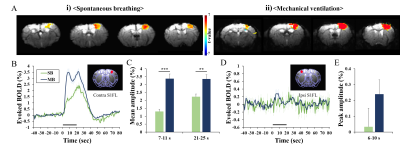 |
Anesthetized Mouse fMRI and optical imaging under spontaneous-breathing vs. mechanical ventilation
Hyun-Ji Shim1,2 and Seong-Gi Kim1,2,3
1Center for Neuroscience Imaging Research (CNIR), Institute for Basic Science (IBS), Suwon-si, Korea, Republic of, 2Department of Health Sciences and Technology, Samsung Advanced Institute for Health Sciences and Technology (SAIHST), Sungkyunkwan University (SKKU), Seoul, Korea, Republic of, 3Department of Biomedical Engineering, Sungkyunkwan University (SKKU), Suwon-si, Korea, Republic of
Evoked fMRI findings of anesthetized mice are inconsistent possibly due to uncontrolled vascular physiology. In our studies under ketamine and xylazine anesthesia, localized BOLD response was observed in the spontaneously breathing condition, which is known to cause hypercapnia. Here we performed blood gas analysis and functional studies of spontaneously breathing vs. mechanical ventilating mice. The mechanical ventilation maintained mice at normal physiology and induced larger hemodynamic and BOLD responses to forepaw stimulation. Spontaneous breathing induced severe hypercapnia and acidosis, but surprisingly showed significant evoked functional responses. These results suggest that both methods can be used for functional experiments.
|
Session Topic: fMRI: Contrast & Physiology
Session Sub-Topic: fMRI Physiology 2
Digital Poster
fMRI
3846.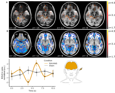 |
Vascular effects in fMRI during respiratory-gated auricular vagal nerve stimulation
Nikos Priovoulos1, Dimo Ivanov1, Benedikt Poser1, Linda Pagen1, Vitaly Napadow2,3, Roberta Sclocco2, Frans Verhey1, and Heidi Jacobs1,2
1Maastricht University, Maastricht, Netherlands, 2Harvard Medical School, Boston, MA, United States, 3Logan University, Chesterfield, MT, United States
Vagus nerve stimulation has been suggested to cause wide-spread changes in the hemodynamic response function in rats. Here, we demonstrate in humans that a non-invasive variant of vagus nerve stimulation is linked with neurovascular and global blood flow changes, and associated with increased salivary alpha-amylase, suggesting potential noradrenaline release. Our results provide support for the role of noradrenaline in modulating BOLD fMRI contrast via blood flow.
|
|
3847.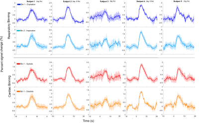 |
Modulation by cardiac and respiratory cycles of single-trial BOLD responses to visual stimulation using ultrafast fMRI at 7T
Francisco Moreira Azevedo1, João Jorge2,3, Wietske van der Zwaag4, and Patrícia Figueiredo1
1ISR-Lisboa/LARSyS and Department of Bioengineering, Instituto Superior Técnico, Universidade de Lisboa, Lisboa, Portugal, Lisbon, Portugal, 2Laboratory for Functional and Metabolic Imaging, École Polytechnique Fédérale de Lausanne, Lausanne, Switzerland, Lausanne, Switzerland, 3Systems Division, Swiss Center for Electronics and Microtechnology (CSEM), Neuchâtel, Switzerland, Lausanne, Switzerland, 4Spinoza Centre for Neuroimaging, Amsterdam, Netherlands, Amsterdam, Netherlands
The haemodynamic response to neuronal activity measured by BOLD-fMRI exhibits substantial trial-by-trial variability, but the underlying sources remain largely unknown. Here, we investigated whether single-trial BOLD responses were modulated by cardiac and respiratory cycles, by recording the BOLD responses to single trials of brief visual stimulation using an ultrafast acquisition (TR=0.4s) at ultrahighfield (7T). We found that, when binning trials according to the respiratory phase, the BOLD responses were more similar to each other when occurring up to 1sec following expiration. Our results suggest that trial-by-trial variability of BOLD responses may partly be explained by the respiratory cycle.
|
|
3848. |
Ultrafast EPI enables event-related fMRI and the mapping of hemodynamic response function in the mouse visual pathway
Xiaoqing Alice Zhou1, Zengmin Li2, Hsu-Lei Lee3, Elizabeth Coulson4, and Kai-Hsiang Chuang3
1QBI/SBMS, The University of Queensland, Brisbane, Australia, 2QBI, The University of Queensland, Brisbane, Australia, 3QBI/CAI, The University of Queensland, Brisbane, Australia, 4SBMS/QBI, The University of Queensland, Brisbane, Australia
|
|
3849.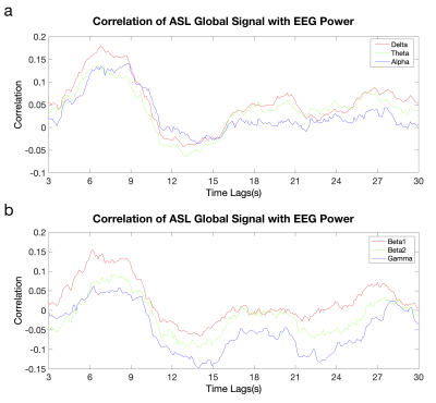 |
Neural Basis of Global Resting-state ASL Perfusion Signals
Shichun Chen1, Wenna Duan1, Li Zhao2, David C. Alsop2, Wenming Luh3, and Weiying Dai1
1Computer Science, State University of New York at Binghamton, Binghamton, NY, United States, 2Radiology, Beth Israel Deaconess Medical Center & Harvard Medical School, Boston, MA, United States, 3National Institute on Aging, National Institite of Health, Baltimore, MD, United States
Global fluctuations of arterial spin labeling (ASL) signals may emerge from global synchronization of resting-state brain networks. We aimed to investigate the synchrony of ASL global signal with brain neural activity using simultaneously recorded ASL perfusion images and EEG. The correlation between the EEG frequency power time series and ASL global time courses was evaluated. The shape for time course of the correlation is very similar to hemodynamic response functional curve. The peak correlation time indicates that the global ASL signals follow the neural activity and the function of human brain is organized by the presence of global functional synchronization.
|
|
3850. |
Mouse BOLD fMRI of innocuous and putative noxious forepaw stimulation without cardiovascular modulation
Won Beom Jung1,2, Hyun-Ji Beom Shim1,3, and Seong-Gi Kim1,2
1Cener for Neuroscience Imaging Research (CNIR), Institute for Basic Science (IBS), Suwon-si, Gyeonggi-do, Korea, Republic of, 2Department of Biomedical Engineering, Sungkyunkwan University, Suwon-si, Gyeonggi-do, Korea, Republic of, 3Department of Health Sciences and Technology, Samsung Advanced Institute for Health Sciences and Technology (SAIHST), Sungkyunkwan University, Suwon-si, Gyeonggi-do, Korea, Republic of
The wide-spread and/or bilateral activity patterns in cortical and thalamic areas were common observation in response to innocuous and noxious electrical stimulation in mouse BOLD fMRI studies. Those findings may be related to the global BOLD effect confounded by stimulus-induced changes in cardiac functions such as heart rate and blood pressure, rather than originated from underlying neural activity. Under the ketamine-xylazine anesthesia, sympathetic activity is known to be suppressed. Here, we measured the response specificity of the BOLD signal responding from innocuous to putative noxious stimuli in mice under ketamine-xylazine anesthesia.
|
|
3851.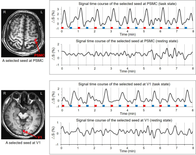 |
The relationship of functional connectivity of the sensorimotor and visual cortical networks between resting and task states
Zhenliang Xiong1, Chong Tian1, Jie Huang2, Xianchun Zeng1, Dongxue Li1, Lisha Nie3, Pu-Yeh Wu3, and Rongpin Wang1
1Department of Radiology, Guizhou Provincial People's Hospital, Guiyang, China, 2Department of Radiology, Michigan State University, East Lansing, MI, United States, 3GE Healthcare, Beijing, China
The current study investigated the relationship of the sensorimotor and visual cortical functional connectivity (FC) networks between the resting and task states. Our study demonstrated a general relationship of the task-evoked FC network with its corresponding intrinsic FC network, regardless of the tasks.
|
|
3852.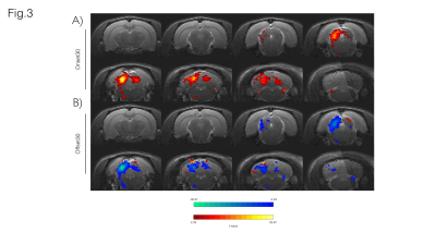 |
BOLD Responses in the Rat Auditory Pathway Upon the Cessation of Sound
Frederico Severo1, Alfonso Renart1, and Noam Shemesh1
1Champalimaud Research, Champalimaud Centre for the Unknown, Lisboa, Portugal
Auditory offset responses due to cessation of auditory stimuli have been previously described, suggesting the existence of a dedicated auditory offset pathway. These responses are of major importance in auditory perception, as temporal cues like sound duration can be crucial for sound identification and interpretation. Still, studies on the subject are scarce compared to classic auditory onset responses. Here, we investigate the BOLD dynamics upon the cessation of sound using BOLD fMRI in the rat. We find differences between Onset and Offset paradigms suggesting negative BOLD responses observed in the latter are likely due to an active process.
|
|
3853.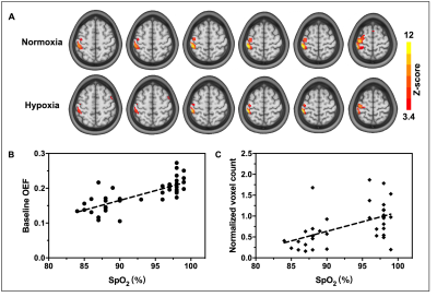 |
Effects of mild hypoxia on oxygen extraction fraction responses to brain stimulation
Yayan Yin1, Lang Qin2, and Jie Lu1
1Radiology, Capital Medical University, Beijing, China, 2Linguistics, the University of Hong Kong, Beijing, China
The oxygen extraction fraction (OEF) is a key physiological marker of cerebral oxygen metabolism. While the availability of oxygen plays a crucial role in oxygen metabolism, the effect of mild hypoxia on OEF during brain activity remains largely unclear. In this study, a novel OEF dynamic measurement technique, multiecho asymmetric spin echo pulse sequence, was used to investigate the OEF response during motor task in both normoxia and mild hypoxia. Results demonstrated that the motor-induced OEF response change was larger in mild hypoxia (-23% ± 6%) than that in normoxia (-12% ± 4%) and was positively correlated with oxygen saturation.
|
|
3854.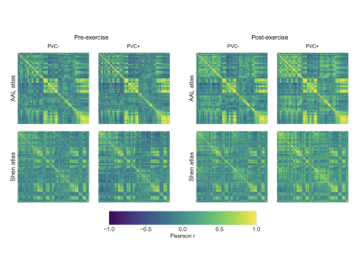 |
Region-to-region covariation of cerebral blood flow in the young brain before and after acute exercise
Nicholas J. Luciw1,2, Benjamin I. Goldstein2,3, and Bradley J. MacIntosh1,2
1Department of Medical Biophysics, University of Toronto, Toronto, ON, Canada, 2Sunnybrook Research Institute, Toronto, ON, Canada, 3Departments of Psychiatry and Pharmacology, University of Toronto, Toronto, ON, Canada
Functional brain networks reflect inherent inter-regional relationships that subserve behaviour and information processing. There is evidence that the correlations of regional brain function across individuals partly reflect functional connectivity. The current study focuses on region-to-region covariation of cerebral blood flow (CBF) across participants using arterial spin-labelled MRI. To investigate the stability of this CBF covariance mapping, we compare maps acquired within the same day but before and after a single exercise session. To refine our CBF covariance mapping methodology, we consider whether choice of anatomical atlas and use of partial volume correction impact the results of our between-session comparison.
|
|
3855.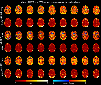 |
Assessment of longitudinal cerebrovascular reactivity measurements based on breath-hold and resting state BOLD multi-echo fMRI
Stefano Moia1, Rachael C. Stickland2, Maite Termenon1, Eneko Uruñuela-Tremiño1, César Caballero-Gaudes1, and Molly G. Bright2,3
1Basque Center on Cognition Brain and Language, San Sebastián, Spain, 2Physical Therapy and Human Movement Sciences, Feinberg School of Medicine, Northwestern University, Chicago, IL, United States, 3Biomedical Engineering, McCormick School of Engineering, Northwestern University, Evanston, IL, United States
Cerebrovascular reactivity (CVR) is an important measure of vascular function, but true variability in CVR can be masked by numerous confounds. We explore the variability of CVR maps, extracted using breath-holding and resting state data, over 9 weekly scanning sessions. Our results show that CVR demonstrates robust spatial patterns that vary across known neural networks.
|
|
3856.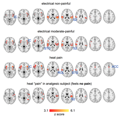 |
Resolving Spatially Distinct Patterns of Nociception with Functional MRI at 7.0 T En Route to Pain-Specific Neurosignatures
Henning Matthias Reimann1, Jurjen Heij1, Joao Periquito1, Antje Els1, Haopeng Han1, and Thoralf Niendorf1,2
1Berlin Ultrahigh Field Facility (B.U.F.F.), Max Delbrück Center for Molecular Medicine in the Helmholtz Association, Berlin, Germany, 2Experimental and Clinical Research Center, a joint cooperation between the Charité Medical Faculty and the Max Delbrück Center for Molecular Medicine in the Helmholtz Association, Berlin, Germany
In recent years, the identification of pain-specific biomarkers has become the holy grail of pain research in the fMRI community. Painful stimuli, salient (attention capturing) non-painful stimuli, and nociceptive stimuli in analgesic subjects (who cannot feel pain) elicit similar BOLD patterns that cannot be resolved at 3T. Recognizing this shortcoming we present preliminary fMRI data at 7T (double spatial resolution), comparing painful stimuli with isosalient non-painful stimuli in healthy volunteers and an analgesic subject who is insensitive to pain. The overall goal of this study is to detail the evoked neurosignatures for pain-specificity and identify diagnostic biomarkers of pain experience.
|
|
3857.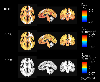 |
Dynamic Brain-Body Coupling of Breath-by-Breath O2-CO2 Exchange Ratio with Resting State Cerebral Hemodynamic Fluctuations
Suk-tak Chan1, Karleyton C Evans2, Tian-yue Song1, Juliette Selb1, Andre van Kouwe1, Bruce R Rosen1, Yong-ping Zheng3, Andrew C Ahn1, and Kenneth K Kwong1
1Athinoula A. Martinos Center for Biomedical Imaging, Massachusetts General Hospital, Boston, MA, United States, 2Biogen Inc., Cambridge, MA, United States, 3Biomedical Engineering, Hong Kong Polytechnic University, Kowloon, Hong Kong
We showed that the oscillations of breath-by-breath O2-CO2 exchange ratio (bER) were superior to those of end-tidal CO2 in correlating with the low frequencies (0.008-0.03Hz) of resting state cerebral hemodynamic fluctuations (CHF). Brain regions showing significant association of ΔBOLD with bER overlapped with many regions of default mode network. Transcranial Doppler sonography and fMRI were used to measure CHF and time series of partial pressure of O2 and CO2 were collected. bER-CHF coupling is a novel metric to measure brain-body interaction that may provide some answers to the physiological contributions to low frequencies of CHF.
|
|
3858.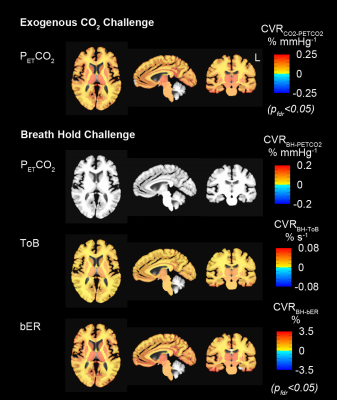 |
Cerebrovascular Reactivity Assessment with O2-CO2 Exchange Ratio Under Brief Breath Hold Challenge
Suk-tak Chan1, Karleyton C Evans2, Tian-yue Song1, Juliette Selb1, Andre van Kouwe1, Bruce R Rosen1, Yong-ping Zheng3, Andrew C Ahn1, and Kenneth K Kwong1
1Athinoula A. Martinos Center for Biomedical Imaging, Massachusetts General Hospital, Boston, MA, United States, 2Biogen Inc., Cambridge, MA, United States, 3Biomedical Engineering, Hong Kong Polytechnic University, Kowloon, Hong Kong
Here we tested the hypothesis that the cerebrovascular responses to brief breath hold epochs were coupled not only with increased partial pressure of carbon dioxide (PCO2), but also with decreased partial pressure of oxygen (PO2). fMRI map of cerebrovascular reactivity (CVR) to breath-by-breath O2-CO2 exchange ratio covers similar regions as map of CVR to exogenous CO2 challenge. Substantially fewer regions in fMRI map of CVR to endogenous end-tidal CO2 satisfied statistical significance. Our results support the hypothesis that hypoxia and hypercapnia work synergistically to enhance cerebrovascular responses to breath hold.
|
|
3859.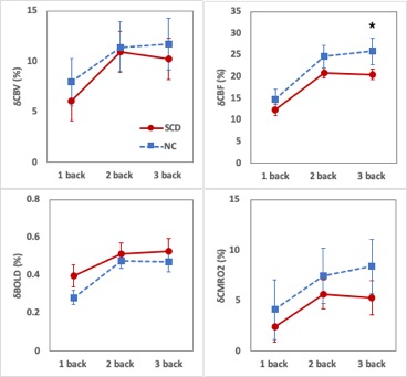 |
Cerebral hemodynamic responses of subjective cognitive decline evoked by loaded N-back tasks
Yaoyu Zhang1, Wenying Du2, Ying Han2,3,4, and Jia-Hong Gao1
1Center for MRI Research, Peking University, Beijing, China, 2Department of Neurology, XuanWu Hospital of Capital Medical University, Beijing, China, 3Center of Alzheimer’s Disease, Beijing Institute for Brain Disorders, Beijing, China, 4National Clinical Research Center for Geriatric Disorders, Beijing, China
Subjective cognitive decline (SCD) is a high risk for Alzheimer’s disease. Most of the existing studies made their observations during the resting state of the brain. This study compared the responses of SCD and normal controls (NC) evoked by loaded N-back tasks. A load-dependent effect is clearly seen in both groups. CBF, as compared to other parameters, shows higher sensitivity in detecting the difference between SCD and NC subjects. Our results may help understand the physiological mechanisms and develop early diagnostic strategies of SCD.
|
|
3860.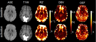 |
R2’-mapping of baseline and visual stimulus states in the human brain using streamlined-qBOLD
Fatemehsadat Arzanforoosh1, Marion Smits1, and Esther AH Warnert 1
1Radiology and Nuclear medicine, Erasmus MC, Rotterdam, Netherlands
Cerebral hypoxia occurs in a plethora of brain diseases, including stroke and brain tumor. This work provides a step towards a rapid, non-invasive imaging protocol for clinically feasible cerebral oxygenation mapping. In this study we use an asymmetric spin echo (ASE)-based streamlined-qBOLD (sq-BOLD) technique to non-invasively monitor hemodynamic properties of the brain in two states (baseline and activation). Our results show that, despite the low signal-to-noise ratios likely due to macroscopic magnetic field gradients (MFGs), sq-BOLD has the potential to measure changes in oxygen extraction fraction in the activated area.
|
|
3861. |
The difference of left and right side after chronic pontine infarction based on resting-state fMRI
Luo bing Wu1, Ying Wei1, Cai hong Wang1, Kai yu Wang1, and Jing liang Cheng1
1GE Healthcare, MR Research China, Beijing, Zheng Zhou, China
The resting state functional images of 53 patients with chronic pontine infarction and 35 healthy controls were collected, and A and B were used to evaluate the difference and sensitivity of functional information integration between single brain regions and hemispheres.It was associated with clinically relevant cognitive function scores.Finally, the contribution of the left and right sides to the difference was evaluated.The results showed that the difference and contribution of the right infarction group were significantly higher than that of the left infarction group.In the future, functional integration between the two hemispheres will be further studied.
|