
Digital Poster Session
fMRI: Acquisition & Applications
fMRI
3862 -3876 fMRI: Acquisition & Applications - fMRI Acquisition 1
3877 -3891 fMRI: Acquisition & Applications - fMRI Acquisition 2
3892 -3906 fMRI: Acquisition & Applications - Non-Connectivity-Based Applications
3907 -3921 fMRI: Acquisition & Applications - fMRI Applications
3862.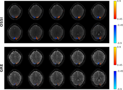 |
High SNR and High-Resolution fMRI using 3D OSSI and Tensor Model Reconstruction
Shouchang Guo1, Jeffrey A. Fessler1, and Douglas C. Noll2
1Electrical Engineering and Computer Science, University of Michigan, Ann Arbor, MI, United States, 2Biomedical Engineering, University of Michigan, Ann Arbor, MI, United States
The goals of fMRI acquisition methods include high spatial and temporal resolutions with high signal to noise ratio (SNR). Oscillating Steady-State Imaging (OSSI) is a new fMRI acquisition method that provides large signals with high SNR, but may result in a slower acquisition of modest spatial resolution. This work improves OSSI spatial and temporal resolutions by exploiting the inherent high-dimensional structure of OSSI data and developing a tensor low-rank model for OSSI prospectively undersampled reconstruction. Compared to GRE imaging with the same spatial-temporal resolution, 3D OSSI demonstrated 2 times higher temporal SNR and 2 times larger activation region.
|
|
3863. |
Detection of Cortical Depth-dependent Functional Activation using Whole-brain, Half-millimetre Resolution EPIK at 7T
Seong Dae Yun1, Patricia Pais-Roldán1, and N. Jon Shah1,2,3,4
1Institute of Neuroscience and Medicine 4, INM-4, Forschungszentrum Juelich, Juelich, Germany, 2Institute of Neuroscience and Medicine 11, INM-11, Forschungszentrum Juelich, Juelich, Germany, 3JARA - BRAIN - Translational Medicine, Aachen, Germany, 4Department of Neurology, RWTH Aachen University, Aachen, Germany
The steady development of fMRI techniques has enabled the use of submillimetre-resolution in recent fMRI studies. The use of submillimetre-resolution allows the detection of cortical, depth-dependent brain activation. Although there have been numerous attempts to perform submillimetre-resolution fMRI, the level of spatial resolution in several recent works is still around 0.7 mm. Moreover, most methods do not provide whole-brain coverage. Therefore, this work aims to develop a novel half-millimetre resolution fMRI technique capable of providing whole-brain coverage. Here, the method was employed for exemplary finger-tapping fMRI at 7T and the identification of cortical-depth dependent brain activation was demonstrated.
|
|
3864. |
Beyond the limits of layer-dependent CBV fMRI in humans: strategies towards whole brain coverage, sub-second TR, and very high 0.5mm resolutions
Laurentius Huber1, Yuhui Chai2, Rüdiger Stirnberg3, Sriranga Kashyap1, Deni Kurban1, Arman Khojandi2, Dimo Ivanov1, Sean Marrett2, Tony Stöcker3,4, Kamil Uludag5, Peter Bandettini2, and Benedikt Poser1
1MR-Methods group, MBIC, Faculty of Psychology and Neuroscience, Maastricht, Netherlands, 2SFIM, NIMH, Bethesda, MD, United States, 3German Center for Neurodegenerative Diseases (DZNE), Bonn, Germany, 4Physics and Astronomy, University of Bonn, Bonn, Germany, 5UHN Toronto, Toronto, ON, Canada Recent developments of ultra-high field (UHF) MRI and high-resolution CBV-sensitive VASO sequences have made it possible to measure activity changes across cortical depths. However, most methods are limited to individual brain areas of large cortical thicknesses (4mm in M1), long repetition times and small matrix sizes. In this study, we developed multiple VASO sequence approaches to achieve: 1.) Whole-brain coverage (104 slices) at sub-millimeter layer resolutions 2.) Sub-second TRs (TR = 650ms) at sub-millimeter layer resolutions 3.) Extra-high 0.5mm isotropic resolutions with matrix sizes of 316 We tested new sequence concepts and validated their applicability for the neuroscientific context. |
|
3865. |
Fingerprinting individual subjects from resting-state functional magnetic resonance imaging head motion
Thomas Bolton1,2, Stefano Moia3, Eneko Urunuela3, Dimitri Van De Ville1,2, and César Caballero-Gaudes3
1Institute of Bioengineering, Ecole Polytechnique Fédérale de Lausanne, Lausanne, Switzerland, 2Department of Radiology and Medical Informatics, University of Geneva, Geneva, Switzerland, 3Basque Center on Cognition, Brain and Language, San Sebastian, Spain
Head motion during resting-state functional magnetic resonance imaging acquisitions is an infamous confound requiring dedicated preprocessing steps. Here, we probed the level of complexity at which motion should be characterised. To do so, we designed a clustering-based approach that determines the spatio-temporal motion patterns that can fingerprint an individual. We found that each subject's motion could be fingerprinted at a different space/time granularity, some more easily than others. Our results call for refined motion descriptions in which space and time should not be over-simplified, and point towards the relevance of motion-related fingerprints as an individual's functional trait.
|
|
3866. |
Transition-Band SSFP and EPI Functional MRI on a High-Performance 0.55 T Scanner
Yicun Wang1, Peter van Gelderen1, Jacco A. de Zwart1, Adrienne E Campbell-Washburn2, and Jeff H. Duyn1
1AMRI, LFMI, NINDS, National Institutes of Health, Bethesda, MD, United States, 2Cardiovascular Branch, Division of Intramural Research, NHLBI, National Institutes of Health, Bethesda, MD, United States
Low magnetic susceptibility contrast and inadequate gradient performance have limited the use of low-field systems for BOLD fMRI. With the recent introduction of low-field scanners equipped with high-performance gradients, we re-evaluated the feasibility of low-field fMRI by comparing transition-band SSFP and GRE-EPI at 0.55 T. With optimized acquisition parameters both techniques proved practical for detection of brain activation in response to visual stimulation. EPI was found to be more robust to system imperfections and physiological noise. In the current study, SSFP does not appear to outperform conventional EPI in the level and spatial extent of activation.
|
|
3867.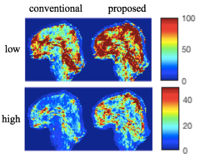 |
Reduced Inter-shot Physiological Variability in 3D Multi-Shot fMRI using Structured Low-Rank Matrix Completion
Xi Chen1, Wenchuan Wu1, and Mark Chiew1
1University of Oxford, Oxford, United Kingdom
Three-dimensional encoding methods like multi-shot 3D-EPI are increasingly being explored as alternatives to multi-slice 2D acquisitions in functional MRI, particularly in cases where high isotropic resolution is needed. However, multi-shot 3D methods can suffer from artifacts and reduction of temporal SNR (tSNR) due to inter-shot variability from motion or physiological fluctuations. Here, we present a method for reconstruction of multi-shot 3D EPI data, that is insensitive to smooth inter-shot phase inconsistencies due to physiologically-induced B0 variations. This approach is based on annihilating filter Hankel structured low-rank matrix completion, illustrating improved tSNR compared to conventional multi-shot reconstruction.
|
|
3868.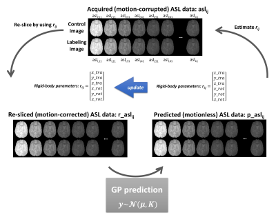 |
A novel motion correction for ASL-fMRI with multi-PLD: non-parametric Gaussian Processes prediction of background suppression
Yuriko Suzuki1,2, Thomas Okell2, Joseph G. Woods2,3, and Michael Chappell1,2
1Institute of Biomedical Engineering, Department of Engineering Science, University of Oxford, Oxford, United Kingdom, 2Wellcome Centre for Integrative Neuroimaging, FMRIB, Nuffield Department of Clinical Neurosciences, University of Oxford, Oxford, United Kingdom, 3Department of Radiology, University of California, La Jolla, CA, United States
For fMRI using ASL, multi-PLD acquisitions may have advantages, improving reliability and specificity. However, the varying static-tissue signal in multi-PLD ASL can confound motion estimation when conventional motion correction is applied. In this study, we propose a novel framework using Gaussian processes to address this problem, in which motionless ASL images are predicted, so that they can be used as a reference for motion correction for each ASL volume. Simulation and in-vivo studies show the new motion correction framework using Gaussian Processes eliminates the influence of multi-PLD and provides a suitable reference for each volume.
|
|
3869.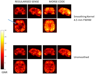 |
Multiple Orthogonal Reference Sensitivity Encoding Combined Over Dominant Eigencoils (MORSE CODE): Motion robust accelerated fMRI
Oliver Josephs1, Nadege Corbin1, Isobel Green2, Jonathan Roiser2, and Martina Callaghan1
1Wellcome Centre for Human Neuroimaging, UCL Queen Square Institute of Neurology, UCL, London, United Kingdom, 2Institute of Cognitive Neuroscience, UCL Queen Square Institute of Neurology, UCL, London, United Kingdom
Motion-robust reconstruction using a regularised SENSE-based unfolding of 3D-EPI time series for fMRI.
|
|
3870.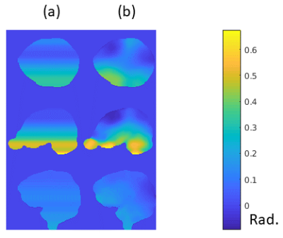 |
7T EPI Nyquist Ghost Reduction by Combining Trajectory Measurement, Navigator Echoes and an Inverted Readout Polarity Calibration Scan
Oliver Josephs1, Nadege Corbin1, and Martina Callaghan1
1Wellcome Centre for Human Neuroimaging, UCL Queen Square Institute of Neurology, UCL, London, United Kingdom
A combination and development of several existing calibration and reference techniques for Nyquist ghost reduction allowing 2D EPI fMRI images to be reconstructed with very low ghost level at 7T in demanding gradient regimes.
|
|
3871.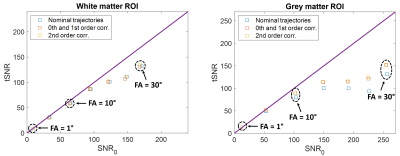 |
tSNR increase with field monitoring for correction of scanner instabilities and physiological noise at 7T in single-shot 2D-EPI sequences
Caroline Le Ster1, Alexandre Vignaud1, and Nicolas Boulant1
1Université Paris-Saclay, Neurospin, CEA, Gif-sur-Yvette, France
Signal-dependent noise contributions in a voxel limit the temporal SNR (tSNR) gain brought by increasing the image SNR. The effect of retrospectively correcting for field fluctuations on the tSNR for different SNR regimes at 7T in single-shot 2D-EPI sequences was assessed using field sensor measurements. For the slice located at isocenter, results of the study show an increase of up to 20% in vivo on the grey matter region and for high SNR regimes.
|
|
3872.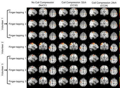 |
Effect of Coil Compression on fMRI Time Courses: Preliminary Analysis
Brice Fernandez1, Baolian Yang2, Gaohong Wu3, Jeff McGovern2, and Suchandrima Banerjee4
1Applications and Workflow, GE Healthcare, Buc, France, 2Applications and Workflow, GE Healthcare, Waukesha, WI, United States, 3Engineering, GE Healthcare, Waukesha, WI, United States, 4Applications and Workflow, GE Healthcare, Menlo Park, CA, United States
Coil compression was introduced as a mean to reduce image reconstruction computational complexity of data acquired with large coil arrays with negligible SNR penalty. Previous work using a different acquisition strategy (not a standard multiband EPI) suggests that coil compression has a negligible effect on fMRI time courses. In this preliminary study, the effects of our coil compression implementation on fMRI time courses is evaluated using different level of compression and a standard multiband EPI. The results suggest a negligible effects of coil compression on fMRI time courses.
|
|
3873.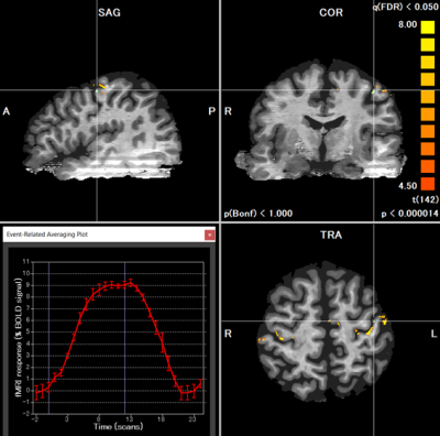 |
Distortion- and Resolution-Matched T1w-Like Anatomy for Investigating Depth-Dependent Activity in Submillimeter-Resolution fMRI at 7T
Adnan Shah1,2, Guoxiang Liu1,2, and Takashi Ueguchi1,2
1Brain Function Analysis and Imaging Lab, CiNet, NICT, Osaka, Japan, 2Graduate School of Frontier Biosciences, Osaka University, Osaka, Japan
We demonstrate the reconstruction of isotropic submillimeter-resolution distortion- and resolution-matched (DRM) T1w-Like anatomy from the image inversion of T1-maps obtained as five slice-shifted anatomical volumes covering the whole brain. The proposed anatomy avoids the distortion-mismatch between function and anatomy resulting in a high alignment with the functional data, necessary for activity localization in submillimeter-resolution fMRI. We use the same pulse sequence for acquiring both functional and DRM anatomical images with aligned slice acquisitions except differing in parameters unrelated to distortions. Moreover, the proposed anatomy allows the generation of cortical surface for investigating depth-dependent activity in isotropic submillimeter-resolution fMRI analysis.
|
|
3874.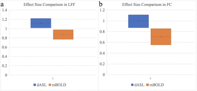 |
Sensitivity of Dynamic ASL and Resting-state BOLD in Patients with Bipolar Disorder
Zongpai Zhang1, Wenna Duan1, Nicolas R. Bolo2, Carol Tamminga3, Brett A. Clementz4, Godfrey D. Pearlson5, Matcheri Keshavan2, David C. Alsop6, and Weiying Dai1
1Computer Science, State University of New York at Binghamton, Binghamton, NY, United States, 2Psychiatry, Beth Israel Deaconess Medical Center & Harvard Medical School, Boston, ME, United States, 3Psychiatry, University of Texas Southwestern Medical Center, Dallas, TX, United States, 4Psychology and Neuroscience, University of Georgia, Athens, GA, United States, 5Psychiatry, Yale University, New Haven, CT, United States, 6Radiology, Beth Israel Deaconess Medical Center & Harvard Medical School, Boston, ME, United States
The effect sizes of bipolar disorder (BD) on low frequency fluctuation (LFF) and functional connectivity (FC) using dASL and rsBOLD imaging were evaluated in forty-five subjects (19 BD patients, 26 control). dASL showed significant increase of LFF and FC in BD, while rsBOLD did not show any difference. dASL demonstrated significantly higher effect sizes compared to rsBOLD, which lead to decreases of 39% and 49% in sample size for LFF and FC measures respectively. These findings support that dASL is more sensitive to BD than rsBOLD and therefore may offer advantages in reducing costs for clinical trials of BD therapies.
|
|
3875.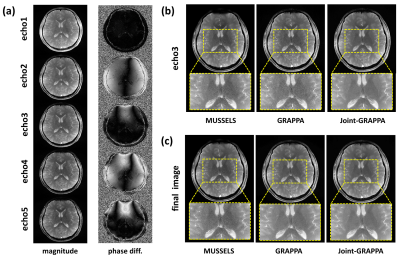 |
Robust Nyquist Ghost Correction for Multi-echo Balanced SSFP at 7T Using Joint-GRAPPA
Ziyi Pan1, Huilou Liang2,3, Kaibao Sun2, Danny J.J. Wang4, Rong Xue2,3,5, and Hua Guo1
1Center for Biomedical Imaging Research, Department of Biomedical Engineering, School of Medicine, Tsinghua University, Beijing, China, 2State Key Laboratory of Brain and Cognitive Science, Beijing MRI Center for Brain Research, Institute of Biophysics, Chinese Academy of Sciences, Beijing, China, 3University of Chinese Academy of Sciences, Beijing, China, 4Laboratory of FMRI Technology (LOFT), Stevens Neuroimaging and Informatics Institute, University of Southern California, Los Angeles, CA, United States, 5Beijing Institute for Brain Disorders, Beijing, China
Multiline bSSFP is an acceleration technique that has the potential to approach the speed of EPI while overcoming inherent EPI artifacts in fMRI. However, the highly segmented EPI readout in multiline bSSFP can cause severe Nyquist artifacts. Additionally, extra magnitude and phase differences among the echoes also exist within the echo train, making it more complicated. In this study, a joint-GRAPPA based 2D phase correction method is proposed, which can robustly correct artifacts in multi-echo bSSFP with different ETLs while maintaining the image SNR. To evaluate its performance, this approach is also compared to other parallel imaging based correction methods.
|
|
3876. |
Functional line-scanning in humans with ultra-high spatiotemporal resolution: reconstruction and BOLD sensitivity assessment
Luisa Raimondo1, Tomas Knapen1,2, ĺcaro A.F. de Oliveira1, Xin Yu3,4, Serge O. Dumoulin1,5, Wietske van der Zwaag1, and Jeroen C.W Siero1,6
1Spinoza Centre for Neuroimaging, Amsterdam, Netherlands, 2VU University, Amsterdam, Netherlands, 3Max Planck Institute for Biological Cybernetics, Tuebingen, Germany, 4Athinoula A. Martinos Center for Biomedical Imaging, Massachusetts General Hospital and Harvard Medical School, Charlestown, SC, United States, 5Experimental and Applied Psychology, VU University, Amsterdam, Netherlands, 6Radiology, University Medical Centre Utrecht, Utrecht, Netherlands
We present initial results of line-scanning fMRI in humans. The potential of this technique lies in the combination of both high spatial and temporal resolution while sacrificing spatial coverage outside the region of interest. We reached a 250 μm resolution along the line direction with a temporal resolution of 200 ms. Coil sensitivity profiles and the average tSNR per channel were used to optimize the line reconstructions. We obtained similar BOLD sensitivity compared to standard 2D GE-EPI BOLD and high spatial specificity for a visual task. Hence, we demonstrate the feasibility of ultra-high spatiotemporal resolution in humans using line-scanning.
|
 View the Poster
 Watch the Video
3877.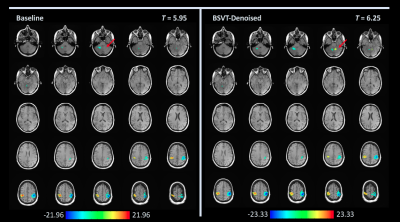 |
Locally low-rank denoising of complex-valued EPI reconstructions preceding task fMRI analysis
Nolan K Meyer1, Norbert G Campeau2, David F Black2, Kirk M Welker2, Jeffrey L Gunter2, Uten Yarach2, Daehun Kang2, MyungHo In2, John Huston III2, Yunhong Shu2, Matt A Bernstein2, and Joshua D Trzasko2
1Biomedical Engineering and Physiology, Mayo Clinic Graduate School of Biomedical Sciences, Rochester, MN, United States, 2Radiology, Mayo Clinic, Rochester, MN, United States
This work examines the removal of physiologic and measurement noise (i.e. "denoising") of complex-valued EPI timecourse data preceding task-based fMRI analysis. The locally low-rank properties of the EPI data are leveraged with a blockwise singular value thresholding (BSVT) algorithm applied as a preprocessing step. Two EPI datasets (single-band and SMS multi-band) concomitant with task-based finger tapping fMRI exams were preprocessed with BSVT; activation maps were then compared by board-certified neuroradiologists. BSVT denoising of complex-valued fMRI time-course data prior to task analysis improves statistical confidence in activation areas identified by conventional processing or reveals new activation regions under fixed confidence levels.
|
|
3878. |
Distortion-free, radial under-sampled fMRI with GRASP reconstruction
Chantelle Y Lim1, Yang Li1, Xirui Hou1, and Hanzhang Lu1
1Department of Radiology, Johns Hopkins University School of Medicine, Baltimore, MD, United States
Golden-angle RAdial Sparse Parallel MRI (GRASP) is a fast field echo radial acquisition with golden-angle rotation sequence used in dynamic imaging due to its motion robustness and high temporal and spatial resolution. In this study, we implemented GRASP for BOLD fMRI during visual stimulation, which yielded distortion-free images in addition to reliable activation maps and signal time-courses as compared to EPI-based fMRI. This initial feasibility study suggests that GRASP could be extended towards other parts of the brain to serve as an alternative when EPI suffers from signal distortion.
|
|
3879.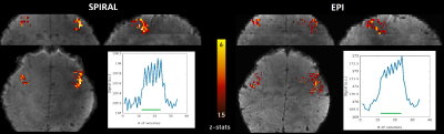 |
Simultaneous multi-slice spiral-out acquisitions for high resolution perfusion fMRI at 7T
Denizhan Kurban1, Gilad Liberman1,2, Christian Mirkes3, Sriranga Kashyap1, Laurentius Huber1, Debora Niekämper 1, Dimo Ivanov1, and Benedikt A Poser 1
1Maastricht Brain Imaging Centre (MBIC), Faculty of Psychology and Neuroscience, Maastricht University, Maastricht, Netherlands, 2A. A. Martinos Center for Biomedical Imaging, Massachusetts General Hospital, Charlestown, MA, United States, 3Skope MRT, Zürich, Switzerland
At ultra-high fields, high-resolution and large coverage functional perfusion mapping with Arterial Spin Labeling (ASL) becomes challenging due to shorter T2* requiring shorter read-out times and B0 inhomogeneity causing geometric distortions. We address these challenges with a single-shot, highly undersampled, short-TE spiral-out trajectory, and simultaneous multi-slice sampling with incoherent CAIPIRINHA for increased slice coverage. Spiral ASL acquisitions at short TE allows functional perfusion mapping with higher SNR compared to conventional EPI acquisitions, showing potential for high-resolution perfusion fMRI applications at ultra-high field.
|
|
3880.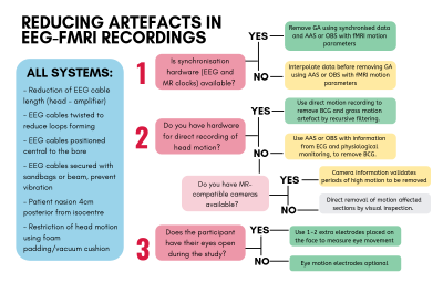 |
Artefact reduction in simultaneous EEG-fMRI: improving current practice.
Madeleine Bullock1,2, David F Abbott1,2, and Graeme Jackson1,2
1Faculty of Medicine, Dentistry and Health Sciences, University of Melbourne, Melbourne, Australia, 2Florey Institute of Neuroscience and Mental Health, Melbourne, Australia EEG recorded during fMRI is subject to artefact many times greater than neuronal events of interest, therefore, artefact removal methods are crucial for accurate EEG-fMRI studies. This work systematically reviews all novel artefact reduction methods (1998-2018), as well as the use of artefact reduction methods (2016-2018). Results show that whilst there are many published artefact reduction methods, contemporary studies overwhelmingly use only a few established methods. It is recommended that: 1. Artefact reduction techniques are adequately reported, 2. Novel software is robust to help adoption by others, and 3. Commercial EEG-fMRI vendors consider including additional hardware for recording artefact. |
|
3881.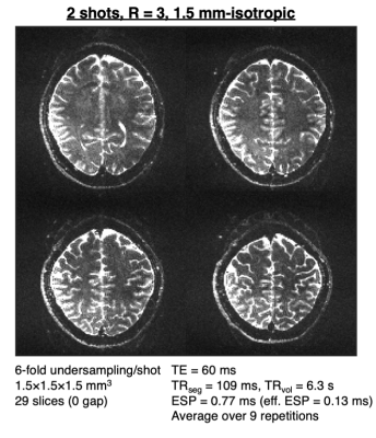 |
Segmented spin-echo BOLD fMRI using a variable flip angle FLEET acquisition with recursive RF pulse design for high spatial resolution fMRI
Avery JL Berman1,2, William A Grissom3, Thomas Witzel1,2, Daniel J Park1, Olivia Viessmann1,2, Kawin Setsompop1,2,4, and Jonathan R Polimeni1,2,4
1Athinoula A. Martinos Center for Biomedical Imaging, Massachusetts General Hospital, Charlestown, MA, United States, 2Department of Radiology, Harvard Medical School, Boston, MA, United States, 3Vanderbilt University Institute of Imaging Science, Nashville, TN, United States, 4Harvard-MIT Division of Health Sciences and Technology, Massachusetts Institute of Technology, Cambridge, MA, United States
Spin-echo (SE) EPI has long been desired for fMRI acquisitions with reduced macrovascular sensitivity; however, to achieve a purely T2-weighted signal, requires sampling a short window around the spin-echo—generally achieved by a segmented readout. Segmented EPI is well-known to be temporally unstable due to sensitivity to subject motion between segments. Here we propose a reordering of the EPI segments, known as FLEET, combined with a variable flip angle excitation to maximize the image signal level and a recursive RF pulse design to maintain consistent slice profiles at the spin-echo. We demonstrate the feasibility of this approach at 3T.
|
|
3882.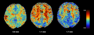 |
Comparison of 2D and 3D EPI for high-resolution functional imaging at 7T MRI
Hankyeol Lee1, Rüdiger Stirnberg2, Tony Stöcker2,3, and Kâmil Uludağ1,4,5
1Center for Neuroscience Imaging Research, Institute for Basic Science, Suwon, Republic of Korea, 2German Centre for Neurodegenerative Diseases (DZNE), Bonn, Germany, 3Department of Physics and Astronomy, University of Bonn, Bonn, Germany, 4Department of Biomedical Engineering, Sungkyunkwan University, Suwon, Korea, Republic of, 5Techna Institute & Koerner Scientist in MR Imaging, University Health Network, Toronto, ON, Canada
This study presents the performance of 2D and 3D EPI sequences at 7T for functional imaging. Eleven subjects were scanned with individually optimized 2D and 3D EPI sequences with TR and total parallel imaging acceleration that were matched. Three spatial resolutions: 0.8 mm, 1.1 mm, and 1.7 mm isotropic, were used. Resulting temporal signal-to-noise ratios (tSNR) were compared between 2D and 3D EPI sequences, and their potential functional sensitivities are discussed. The results show images acquired with 3D EPI have higher tSNR at 0.8 mm and 1.1 mm resolutions, while 2D EPI images have higher tSNR at 1.7 mm resolution.
|
|
3883.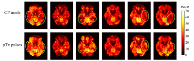 |
Parallel transmit (pTx) with online pulse design for task-based fMRI at 7T
Belinda Ding1, Catarina Rua1, Johan D Carlin2, Marta M Correia2, Ajay D Halai2, Patrick Liebig3, Robin Heidemann3, Iulius Dragonu4, and Christopher T Rodgers1
1Wolfson Brain Imaging Centre, University of Cambridge, Cambridge, United Kingdom, 2MRC Cognition and Brain Science Unit, Cambridge, United Kingdom, 3Siemens Healthcare Limited, Erlangen, Germany, 4Siemens Healthcare Limited, Firmley, United Kingdom
Parallel transmit (pTx) has developed in recent years to show promising reductions in signal dropouts and imaging artifacts from B1+ field inhomogeneities in 7T MRI. However, pTx methods have rarely been applied for functional MRI. To our knowledge, no published task fMRI study has used pTx with online pulse calculation. We therefore implemented pTx spokes excitation in our vendor’s product EPI sequence, and tested it in volunteers using two different task-based fMRI paradigms. Comparing with CP+ (or “TrueForm”) mode, using pTx spokes pulses significantly improved both the tSNR and fCNR in task-based fMRI acquisitions at 7T.
|
|
3884.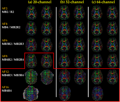 |
Evaluation of multiband EPI for T2*-weighted and diffusion-weighted imaging
Manoj Shrestha1, Daniela van Hinsberg2, H. Sean Lee2, Ulrike Nöth1, David Poeppel2, and Ralf Deichmann1
1Brain Imaging Center (BIC), Goethe University Frankfurt, Frankfurt am Main, Germany, 2Max Planck Institute for Empirical Aesthetics, Frankfurt am Main, Germany
The combination of multiband EPI with in-plane parallel imaging yields a total acceleration factor AF=MB*R, MB and R representing the slice and in-plane acceleration factors, respectively. The data quality of T2*-weighted and diffusion-weighted imaging was compared for different AF, using different combinations of MB and R, and for different head coils, providing advice on the optimum experimental set-up. Results suggest that severe artifacts may occur for large MB and R, a low number of slices (SL) and head coils with a reduced number of elements (NE). Suggestions comprise SL≥48, NE≥32, MB≤8 and R≤4.
|
|
3885.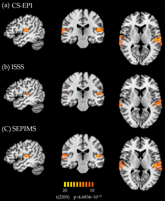 |
Functional MRI of the Auditory Cortex: Comparison of Different Sequences to investigate Speech and Amplitude Modulated Sounds
Manoj Shrestha1, Xiangbin Teng2, H. Sean Lee2, Ulrike Nöth1, David Poeppel2, and Ralf Deichmann1
1Brain Imaging Center (BIC), Goethe University Frankfurt, Frankfurt am Main, Germany, 2Max Planck Institute for Empirical Aesthetics, Frankfurt am Main, Germany
This study compares three fMRI sequences with silent breaks for acoustic stimulation: conventional sparse EPI (CS-EPI), interleaved silent steady-state (ISSS) sampling and the proposed SEPIMS scheme. CS-EPI and SEPIMS yield stronger activations than ISSS for simple artificial amplitude modulated sound stimuli and complex speech-based stimuli. Constant TR in CS-EPI and ISSS creates a regular auditory environment which participants may recognize, thus anticipating the timing of stimuli and scanner noise. In contrast, SEPIMS allows for interleaved TR values of different lengths, creating irregular temporal patterns and avoiding correlations between data acquisition and a regular heart beat or breathing rhythm.
|
|
3886.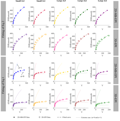 |
The SNR, tSNR, physiological noise and spatial correlation characteristics of 2D-MB-EPI and 3D-EPI.
Nadège Corbin1, Yael Balbastre 1, Oliver Josephs1, and Martina F Callaghan1
1Wellcome Centre for Human Neuroimaging, UCL Institute of Neurology, London, United Kingdom, London, United Kingdom
Here we use an extended model describing the impact of smoothing on tSNR to characterize 2D-Multiband-EPI and 3D-EPI time-series, in vivo, in terms of their SNR, tSNR, intrinsic smoothness, as well as the level of physiological noise (λ) and its degree of spatial correlation. The model fitting suggested that higher intrinsic smoothness and physiological noise levels in 3D-EPI can explain why spatial smoothing is less beneficial than for 2D-MB-EPI, as previously observed. Furthermore, the model captured the facts that the level of physiological noise is higher for 3D-EPI than 2D-MB-EPI and that λ increases with the number of acquired segments.
|
|
3887.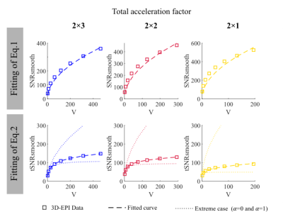 |
To smooth or not to smooth? Modelling the impact of spatially correlated physiological noise in fMRI
Nadège Corbin1, Oliver Josephs1, and Martina F Callaghan1
1Wellcome Centre for Human Neuroimaging, UCL Institute of Neurology, London, United Kingdom
Spatial smoothing is common in fMRI analyses but the benefit to functional sensitivity can vary depending on baseline signal-to-noise, inherent smoothness and physiological noise characteristics. Here we propose an extended model that can quantify each of these properties in addition to parameterising the degree of spatial correlation in the physiological noise. The model is validated through simulation and in vivo experiment. This new model allows the complete characterisation of the impact of spatial smoothing from a single fMRI time series enabling researchers to efficiently gain insight into their data and to optimise processing pipelines.
|
|
3888.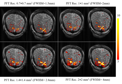 |
How far PFT reconstruction technique can improve spatial resolution in radial fMRI acquisition: An experimental study
Vahid Malekian1, Fatemeh Rastegar2, and Abbas Nasiraei Moghaddam2
1School of Cognitive Science, Institute for Research in Fundamental Sciences (IPM), Tehran, Iran (Islamic Republic of), 2Biomedical Engineering, Amirkabir University of Technology, Tehran, Iran (Islamic Republic of)
Radial acquisition along with Polar Fourier Transform (PFT) reconstruction allows to retrospectively choose the image pixel-size. We experimentally investigated how this selected pixel-size was related to spatial resolution in fMRI studies. The functional contrast to noise ratio (CNR) was considered as a measure to assess whether the improvement in apparent spatial resolution is real or just resulted from interpolation. In an fMRI study on 9 subjects, SSFP raw data was reconstructed by PFT technique with four pixel areas. Results showed that the CNR improvement stopped at the boundaries of reduced-FOV, where the spacing in azimuthal and radial directions are equal.
|
|
3889. |
3D Inner Volume bSSFP Functional MRI: Preliminary results
Tianrui Luo1, Douglas Noll1, and Jon-Fredrik Nielsen1
1fMRI Lab, Univ. of Michigan., Ann Arbor, MI, United States
3D inner-volume (IV) imaging enables increased spatiotemporal resolution within a reduced field-of-view (FOV), but is not directly compatible with steady-state sequences such as balanced SSFP. We present an IV bSSFP sequence for transition-band functional MRI, a technique that can produce high functional signal within a relatively narrow off-resonance frequency (B0) range. We use spatially tailored 3D radiofrequency (RF) pulses for IV excitation and outer-volume (OV) saturation, and a pair of gradient spoilers that make the overall sequence balanced (unbalanced) for IV (OV) spins. This approach produces good OV suppression in most regions, and high functional signal near a transition band.
|
|
3890.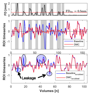 |
Dynamic Missing-data Completion Removes Additional Effect of Motion Contamination Caused by Temporal Filtering during fMRI Preprocessing
Seyhmus Guler1, Burak Erem2, Onur Afacan1, Alexander L. Cohen1, and Simon K. Warfield1
1Boston Children's Hospital and Harvard Medical School, Boston, MA, United States, 2TrueMotion, Inc., Boston, MA, United States
Subject movement during fMRI acquisition creates motion artifact. “Scrubbing” removes motion-corrupted volumes and is performed after temporal filtering since it creates temporal discontinuities. Thus, it does not prevent the spread of corrupted time samples from high motion volumes to their neighbors during temporal filtering. To mitigate this “leakage”, we propose a novel method, Dynamic Missing-data Completion (DMC), that replaces motion corrupted volumes with synthetic data matching the temporal dynamics of the uncorrupted no-motion volumes. We analyzed six rsfMRI scans with different motion levels and found that DMC provides added benefit in further reduction of motion contamination that remains after scrubbing.
|
|
3891. |
Connectivity analyses of accelerated 3D resting-state ASL
Fanny Munsch1, Manuel Taso1, John A. Detre2, and David C. Alsop1
1Radiology, Division of MRI Research, Beth Israel Deaconess Medical Center, Harvard Medical School, Boston, MA, United States, 2Neurology and Radiology, University of Pennsylvania, Philadelphia, PA, United States
We used a single-shot accelerated volumetric Arterial Spin Labeling (ASL) sequence to study brain connectivity in regions where resting-state (rs) BOLD analyses can be challenging due to low signal to noise ratio (SNR). Thus, we chose to study the medial prefrontal cortex, part of the default-mode network (DMN), and the subcallosal cortex for which RS-BOLD is impacted by susceptibility artifacts. We also studied the connectivity of the intracalcarine cortex because it belongs to a network involving deep nuclei, for which BOLD signal is reduced due to low blood volume.
|
3892.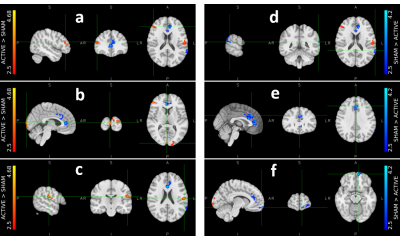 |
Paired Sham and Active Groups Activate Different Brain Regions to the Same Feedback Signal during fMRI Neurofeedback
Seyhmus Guler1, Onur Afacan1, Alexander L. Cohen1, and Simon K. Warfield1
1Boston Children's Hospital and Harvard Medical School, Boston, MA, United States
There is growing interest in fMRI neurofeedback (fMRI-nf) to facilitate therapeutic reorganization of brain function. However, the mechanisms underlying self-regulation processes are incompletely understood. Here we interrogate the mechanisms of fMRI-nf using an experimental protocol designed to increase lateralized motor activity. Twelve right-handed healthy adults were assigned into age- and sex-matched active and sham study arms. Each participant received active or sham feedback during one scanning session. We constructed group-averaged activation maps and lateralization index. During neurofeedback, active and sham groups demonstrated different brain activation. We did not observe any long-lasting functional reorganization and improvement in lateralization index.
|
|
3893.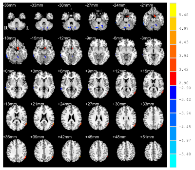 |
Aberrant amplitude of low frequency fluctuations of spontaneous brain activity in patients with Parkinson’s disease
Yanjun Liu1, Mengyan Li2, Xinhua Wei3, Xiuhang Ruan3, Guihe Hu2, Haobo Chen2, and Yaoqin Xie1
1Institute of Biomedical and Health Engineering, Shenzhen Institutes of Advanced Technology, Chinese Academy of Sciences, Shenzhen, China, 2Department of Neurology, Guangzhou First People’s Hospital, School of Medicine, South China University of Technology, Guangzhou, China, 3Department of Radiology, Guangzhou First People’s Hospital, School of Medicine, South China University of Technology, Guangzhou, China
Parkinson’s disease (PD) patients are widely reported with abnormalities in motor and cognitive features. Early diagnosis can be benefit if neuroimaging markers are well developed. This study investigated the altered amplitude of low frequency fluctuations (ALFF) of brain activity by functional MRI and explored the neural correlates of motor and cognitive symptoms of PD. Compared to normal controls, PD patients exhibited increased ALFF in default mode network and decreased in cerebellum. The ALFF was negatively correlated with the motor performances of PD in cerebellum. The findings suggest the cerebellum as critical area associated with motor and cognitive performances in PD.
|
|
3894.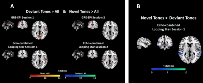 |
Silent functional MRI for novel sound discrimination using the auditory oddball paradigm
Nikou Louise Damestani1, David John Lythgoe1, Owen O'Daly1, Florian Wiesinger1,2, Ana Beatriz Solana2, Steven Charles Rees Williams1, and Fernando Zelaya1
1Department of Neuroimaging, King's College London, London, United Kingdom, 2GE Healthcare, Munich, Germany
We present the first demonstration that a multi-echo variant of a silent fMRI pulse sequence (Looping Star) is sensitive to the BOLD response elicited by the processing of novel auditory stimuli. We employed an established event-related paradigm known as the ‘oddball’ task. Our results show remarkable consistency with a previous investigation using conventional loud fMRI with the same paradigm. We also demonstrate that Looping Star reveals activation differences between auditory challenges not visible using conventional fMRI. Additionally, between-subject correlations and differences in activation between sessions were evaluated. This study supports the use of Looping Star for studies of sound-averse populations.
|
|
3895.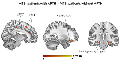 |
Serum levels of inflammatory markers modulates brain structural changes and post-traumatic headache trajectory in mild traumatic brain injury
Xuan Niu1,2, Yingxiang Sun2, Shuoqiu Gan2, Ming Zhang2, and Lijun Bai3
1Department of Radiology, Washington University School of Medicine, St. Louis, MO 63110, St.Louis, MO, United States, 2Department of Medical Imaging, the First Affiliated Hospital of Xi’an Jiaotong University, Xi’an, China, Xi'an, China, 3The Key Laboratory of Biomedical Information Engineering, Ministry of Education, Department of Biomedical Engineering, School of Life Science and Technology, Xi’an Jiaotong University, Xi’an 710049, China, Xi'an, China
Post-traumatic headache (PTH) is one of the most frequent and persistent physical symptoms following mild traumatic brain injury (mTBI). However, the underlying neurobiological basis and modulatory component remained unclear. Evidence indicated that neuroinflammation is a major contributor in the pathogenesis of PTH. We hypothesized that the effect of peripheral inflammatory signaling on PTH could be produced by influencing brain structure that subserve pain modulatory function. Our findings demonstrated that neuroinflammation following mTBI is a potential process affecting structural changes in cognitive component of pain modulation, which may serve as a potential neurobiological mechanism underlying the emergence and persistence of PTH.
|
|
3896.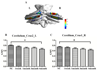 |
Progressive gray matter atrophy and altered structural covariance network in pontine infarction
Ying Wei1, Caihong Wang1, Peifang Miao1, Luobing Wu1, Yingying Wang1, Kaiyu Wang2, and Jingliang Cheng1
1The First Affiliated Hospital of Zhengzhou University, Zhengzhou, China, 2GE Healthcare, MR Research China, GE Healthcare, MR Research, Beijing, China
In order to identify longitudinal changes in gray matter volumes (GMVs) and structural covariance network after pontine infarction (PI), eleven patients and twenty normal underwent MRI scans and neurological examinations during six-month period. Changes of GMV were evaluated by using the voxel-based morphometry (VBM), and structural covariance networks were constructed. In general, the patients exhibited significant decreased GMV in bilateral cerebellum posterior lobe and extended disordered structural covariance network. In addition, the GMVs were correlated with behavior scores showing the abnormal results may be the mechanism of some impaired neurological function.
|
|
3897.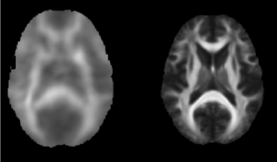 |
Use of functional correlation tensors for correlating white matter fMRI and brain structure
Tory Frizzell1,2, Lukas Algis Grajauskas2,3, Careesa Chang Liu1,2, Sujoy Ghosh Hajra1,2, Xiaowei Song2,4, and Ryan C.N. D'Arcy2,5,6
1Engineering Science, Simon Fraser University, Burnaby, BC, Canada, 2SFU ImageTech Lab, Health Science and Innovation, Surrey Memorial Hospital, Fraser Health, Surrey, BC, Canada, 3Cummings School of Medicine, University of Calgary, Calgary, AB, Canada, 4Biomedical Physiology and Kinesiology, Simon Fraser University, Burnaby, BC, Canada, 5Faculty of Applied Science, Simon Fraser University, Burnaby, BC, Canada, 6Djavad Mowafaghian Centre for Brain Health, University of British Columbia, Vancouver, BC, Canada
White matter functional activity is a neglected area of research and key component for understanding the brain’s ability to adapt and learn. Participants completed a fine motor task during functional scans. DTI images were also collected for structural comparison. Functional correlation tensors were computed to examine local functional signal synchronicity. Strong agreement was found between the functional anisotropy maps and the structural anisotropy maps. Functional correlation tensors substantiate white matter functional response and identify a novel link between structure and function.
|
|
3898. |
Improved Accuracy of Resting State Fluctuation Amplitude for Cerebrovascular Reactivity Estimation Using a Multiband Multi-Echo Acquisition
Alexander D. Cohen1, Baolian Yang2, Brice Fernandez3, Suchandrima Banerjee4, and Yang Wang1
1Medical College of Wisconsin, Milwaukee, WI, United States, 2GE Healthcare, Waukesha, WI, United States, 3GE Healthcare, Buc, France, 4GE Healthcare, Menlo Park, CA, United States
Resting state fMRI (rs-fMRI) can potentially be used to measure cerebrovascular reactivity (CVR). Specifically, resting state fluctuation amplitude (RSFA) is related to the breath hold response. In this study a multiband (MB), ME sequence was compared to a MB, single echo (SE) sequence in terms of their ability to estimate CVR using rs-fMRI-derived RSFA by comparing to BH-CVR. The MBME scan showed higher voxelwise across-subject correlation between RSFA and CVR. The spatial correlation between mean RSFA and CVR maps was also higher for MBME scans, indicating improved accuracy of RSFA as an alternative to CVR using an MBME sequence.
|
|
3899.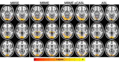 |
Comparing Multiband Single Echo and Multi-echo Sequences With and Without Arterial Spin Labeling for Task Activation
Alexander D. Cohen1, Baolian Yang2, Brice Fernandez3, Suchandrima Banerjee4, and Yang Wang1
1Medical College of Wisconsin, Milwaukee, WI, United States, 2GE Healthcare, Waukesha, WI, United States, 3GE Healthcare, Buc, France, 4GE Healthcare, Menlo Park, CA, United States
Multi-echo functional MRI has shown increased BOLD sensitivity compared to single echo acquisitions; however, it suffers from increased TR, especially when combined with arterial spin labeling (ASL). This study compared multiband multi-echo scans with and without pseudocontinuous ASL to a multiband single echo acquisition during a visual task. Temporal signal to noise and group task activation was higher for the multi-echo scans. Furthermore, the multi-echo PCASL sequence showed similar activation to multi-echo alone despite significantly higher TR. This study showed the benefits of ME acquisitions for task fMRI, despite higher TR. The addition of ASL did not reduce task activation.
|
|
3900.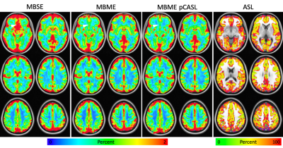 |
Evaluating Multiband Single Echo and Multi-Echo Sequences With and Without Arterial Spin Labeling for Cerebrovascular Reactivity Calculations
Alexander D. Cohen1, Baolian Yang2, Brice Fernandez3, Suchandrima Banerjee4, and Yang Wang1
1Medical College of Wisconsin, Milwaukee, WI, United States, 2GE Healthcare, Waukesha, WI, United States, 3GE Healthcare, Buc, France, 4GE Healthcare, Menlo Park, CA, United States
A multiband (MB) multi-echo (ME) pseudocontinuous arterial spin labeling (pCASL) sequence has shown improved cerebrovascular reactivity (CVR) calculations compared to a single echo; however, pCASL significantly increases TR. This study compared a MBME pCASL sequence to MBME and MB single echo acquisitions for the calculation of CVR. TSNR was higher for the MBME pCASL sequence compared to the MBME and MBMS sequences, and mean CVR was comparable between all sequences. The ME sequences had improved quality in areas with high susceptibility. Despite significantly longer TR, the MBME pCASL sequence performed comparably to the MBME sequence, while simultaneously providing ASL data.
|
|
3901.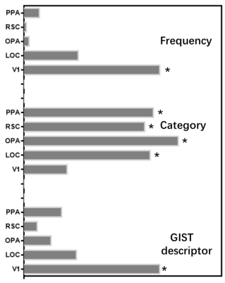 |
FMRI response representation associated with lower space structure of scene perception
Yun Qin1, Xiaole Zhao1, and Tao Zhang1
1University of electronic science and technology of China, Chengdu, China
This study identified the similarity of fMRI-based brain activity in certain scene-selective regions associated with scene categories and low-level spatial structure. In addition, similar hierarchy was found between fMRI response and convolution neural network features of different layers. The results may provide new insights to the processing of lower and high-level knowledge, as well as the performance-optimized hierarchical models.
|
|
3902.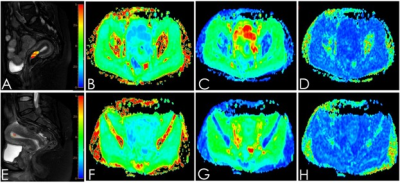 |
APT combined DKI for differential diagnosing endometrial carcinoma from benign endometrial lesions
Ye Ju1, Xing Meng1, Shifeng Tian1, Liangjie Lin2, Jiazheng Wang2, Zhiwei Shen2, Yishi Wang2, Yaxin Niu1, Wan Dong1, and Ailian Liu1
1First affiliated hospital of dalian medical university, Dalian, China, 2Philips Healthcare, Beijing, China
Amide proton transfer (APT) combined diffusion kurtosis imaging (DKI) imaging technology have been preliminarily applied in the diagnosis of cervical diseases. However, there is no study on the differentiation of endometrial carcinoma and endometrial benign lesions with APT combined DKI. In this examine, we used APT combined DKI to identify endometrial carcinoma and endometrial benign lesions.
|
|
3903.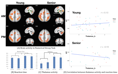 |
Compensatory Effect of Thalamus on Numerical Stroop Task in Senior Adults
Chu-Shin Peng1, Shang-Cheng Chiu2, Fan-Chi Hsiao2, Chih-Mao Huang3, Chi-Yun Liu1, Chien-Ming Yang2, and Changwei Wesley Wu4
1Taipei Medical University, Taipei, Taiwan, 2Department of Psychology, National Chengchi University, Taipei, Taiwan, 3National Chiao Tung University, Hsinchu, Taiwan, 4Graduate Institute of Mind, Brain and Consciousness, Taipei Medical University, Taipei, Taiwan
To unveil the time-of-day effect in the aging process, we recruited both young and senior adults to participate fMRI experiments with the numerical Stroop task before and after nap. Beyond the dorsal attention network, we found the thalamus activity showed facilitating effect in the senior adults but the opposite way in the young adults. Such finding supports the compensation-related utilization of neural circuits hypothesis across age groups, but the time-of-day effect was relatively minor compared to the age effect.
|
|
3904. |
Neurobiology of Financial Decision-Making: An fMRI Based Study Using IGT
Mrinalini Srivastava1, Pankaj Pankaj2, S Senthil Kumaran2, Gagan Sharma1, and Achal Srivastava3
1University School of Management Studies, Guru Gobind Singh Indraprastha University, New Delhi, India, 2Department of NMR & MRI Facility, All India Institute of Medical Sciences, New Delhi, India, 3Department of Neurology, All India Institute of Medical Sciences, New Delhi, India
The neural underpinnings of financial behaviour using modified Iowa Gambling Task (IGT) was assessed through fMRI technique. Data was processed using SPM 12. The results reveal differential brain activation in fronto-parietal lesion patients (n=3, mean age: 47 ±12 years) with respect to controls (n=6, mean age:42±17years). IGT results demonstrated activations during experience of majorly four conditions viz. gains, losses, draws and penalty. The decision-making and associated tasks invoked memory, attention and execution networks providing the insights about the underlying structures of financial investment behaviour.
|
|
3905. |
Negative correlations of brain activation between daily recognition and trauma memory remembering in PTSD
Kayako Matsuo1,2, Jun Inoue3, Toshiki Iwabuchi4, and Hidenori Yamasue5
1Center for Research Collaboration and Support, Dokkyo Medical University, Tochigi, Japan, 2Department of Psychiatry, Hamamatsu University School of Medicine (former), Hamamatsu, Japan, 3Department of Child and Adolescent Psychiatry, Hamamatsu University School of Medicine, Hamamatsu, Japan, 4Research Center for Child Mental Development, Hamamatsu University School of Medicine, Hamamatsu, Japan, 5Department of Psychiatry, Hamamatsu University School of Medicine, Hamamatsu, Japan
We found a negative correlation between brain activity estimates of two conditions, daily recognition and trauma memory remembering, that reflected altered responses in PTSD. We conducted two task-fMRI runs for 9 patients with PTSD and the matched controls employing a script-driven imagery task. A region-of-interest analysis revealed a negative correlation between a hyperarousal subscale of psychological assessment and the activity estimate in the hippocampus in the daily recognition whereas a positive correlation in the trauma memory remembering. When computing voxel-based correlations between the activity estimates of the two conditions, extensive negative correlations emerged around the hippocampus in patients.
|
|
3906.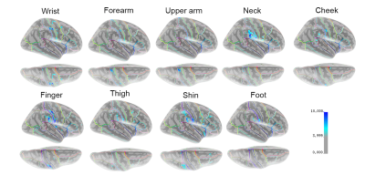 |
An automated pneumatic tactile stimulator for somatotopic mapping
Ryan Willoughby1 and Mark Bolding1
1Radiology, University of Alabama at Birmingham, Birmingham, AL, United States
An MR-safe pneumatic tactile stimulator was developed and tested for automated somatotopic mapping using functional MRI. The device was used in a non-invasive fMRI experiment to perform basic somatotopic mapping on three healthy volunteers. Results from fMRI are consistent with maps of S1 obtained from cortical stimulation studies done in surgical patients, and the technique shows promise for future studies.
|
 |
3907.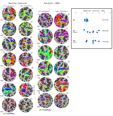 |
Assessing somatotopic and mototopic organisation in Focal Hand Dystonia using high-resolution 7T-fMRI
Michael Asghar1, Daisie Pakenham1, Rosa Sanchez-Panchuelo1, Denis Schluppeck2, Paul Glover1, Miles Humberstone3, George O'Neill4, and Susan Francis1
1Sir Peter Mansfield Imaging Centre, University of Nottingham, Nottingham, United Kingdom, 2Psychology, University of Nottingham, Nottingham, United Kingdom, 3Nottingham Trust University Hospitals, University of Nottingham, Nottingham, United Kingdom, 4Wellcome Centre for Human Neuroimaging, UCL, London, United Kingdom
Six patients with Focal Hand Dystonia (FHD) were scanned at 7T both 4 weeks and 3 months after Botox treatment, along with age-matched healthy controls (HCs). Behaviourally, spatial acuity was raised in the FHD patients compared to HCs. Travelling wave fMRI data were collected to assess somatotopy and mototopy. Compared to age-matched healthy controls and a healthy probabilistic atlas, FHD patients show little difference in somatotopy. Spatial overlap of digits was not found to be significantly different between FHD patients and HCs. In both groups there was high spatial overlap of somatosensory and motor responses in the post-central gyrus.
|
3908.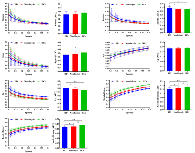 |
Hemodialysis is associated with more severe disrupted brain functional networks in patients with end-stage renal disease
Baolin Wu1, Lei Li1, Zhiyun Jia1,2, and Qiyong Gong1,3
1Huaxi MR Research Center (HMRRC), Department of Radiology, West China Hospital of Sichuan University, Chengdu, China, 2Department of Nuclear Medicine, West China Hospital of Sichuan University, Chengdu, China, 3Department of Psychology, School of Public Administration, Sichuan University, Chengdu, China
Although patients with end-stage renal disease (ESRD) have shown brain structural and functional alterations, the change patterns of the whole-brain functional networks remain largely unknown. We aimed to explore the brain functional topologic organization differences among ESRD patients with hemodialysis (HD) and without dialysis and healthy controls (HCs) using graph-based network analysis. Compared with HCs, ESRD patients showed aberrant global and local topologic organizations, which is more obvious in HD patients. Furthermore, some topologic parameters were associated with cognitive performances and clinical markers.
|
|
3909.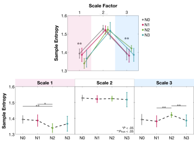 |
Local and Distant Communications in Sleeping Brain Modeled by Multiscale Entropy
Yi-Chia Kung1, ChangWei Wesley Wu2,3, Pei-Jung Tsai4, Chia-Wei Lee5, Chun-Yi Zac Lo6, and Ching-Po Lin1
1Institute of Neuroscience, National Yang-Ming University, Taipei, Taiwan, 2Graduate Institute of Mind, Brain and Consciousness, Taipei Medical University, Taipei, Taiwan, 3Research Center of Brain and Consciousness, Shuang-Ho Hospital, New Taipei, Taiwan, 4Institute on Drug Abuse, National Institutes of Health, Baltimore, MD, United States, 5Department of Radiology, Wan Fang Hospital, Taipei Medical University, Taipei, Taiwan, 6Institute of Science and Technology for Brain-inspired Intelligence (ISTBI), Shanghai, China
Multiscale entropy (MSE) was used to disclose the mixture between functional integration and segregation of brain circuits across NREM sleep. MSE showed N0>N2 and N1>N2 in Scale 1, accompanied with N2>N1 and N2>N3 in Scale 3. The scale-dependent entropy reflects distinct aspects of information processing in the sleeping brain: brain tends to distribute information distantly during the N2 stage and disintegrate both locally and distantly at the N3 stage.
|
|
3910. |
Application of machine learning multivariate pattern analysis for type 2 diabetes: A resting-state fMRI study
Jingge Lian1, Jilei Zhang2, and Kangan Li1
1Shanghai General Hospital, Shanghai Jiaotong University School of Medicine, Shanghai, China, 2Philips Healthcare, Shanghai, China
Type 2 diabetes (T2DM) mellitus can increase risk of cognition impairment and dementia. Recently, machine learning, espicailly support vector machine, were introduced to functional MRI studies in individual classification of diseases. In current study, we used support vector machine to perform individual classification of T2DM and healthy controls (HC) using ALFF features based on rs-fMRI data. The selected features were determined to be key features for classification between groups using recursive feature elimination and may be associated with abnormalities of the spontaneous brain activity
|
|
3911.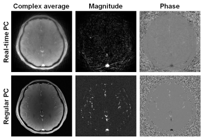 |
Towards accurate quantification of cerebrovascular reactivity using real-time phase-contrast MRI
Wenqi Zhou1, Kristyna Herman1, Dengrong Jiang1, Hanzhang Lu1, and Peiying Liu1
1Johns Hopkins University School of Medicine, Baltimore, MD, United States
Cerebrovascular reactivity (CVR) is typically measured from changes in cerebral perfusion responsive to a hypercapnic gas challenge. Recently, a real-time PC MRI technique using highly undersampled radial FLASH acquisitions with regularized nonlinear inversion reconstruction has been developed and showed great promise in quantifying CBF-based CVR during resting-state without hypercapnic gas challenge. However, quantification of CVR using this method requires optimization. In the present work, using the regular PC MRI as the gold standard, we compared four different analysis methods of the real-time PC MRI results, in order to identify the optimal approach for accurate CVR quantification using real-time PC MRI.
|
|
3912.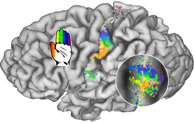 |
Modeling human primary somatosensory cortex using population Receptive Fields.
Wouter Schellekens1, Martijn Thio1, Nick Ramsey2, and Natalia Petridou1
1Radiology, UMC Utrecht, Utrecht, Netherlands, 2Neurosurgery, UMC Utrecht, Utrecht, Netherlands
In the current study, we apply population Receptive Field modeling to somatosensory vibrotactile stimulation, using 7 Tesla functional MRI. We find somatotopic structures within primary somatosensory cortical areas BA3, BA1, and BA2. Furthermore, the receptive field sizes describe tactile information integration and allows for the direct assessment of processing hierarchy within primary somatosensory cortex. This finding is further supported by the estimated HRFs, showing that the BOLD response is quickest to emerge in BA3 which is known to receive primary input from the thalamus.
|
|
3913.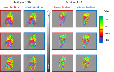 |
A cortical-depth-dependent analysis of fingertip maps in human somatosensory cortex measured at ultra-high field (7T).
Ashley York1, Saskia Bollmann2, Clinton Condon1, Markus Barth2, Ross Cunnington1, and Alexander Puckett1
1School of Psychology, University of Queensland, St Lucia, Australia, 2Centre for Advanced Imaging, University of Queensland, St Lucia, Australia
While cortical-depth-dependent fMRI studies are becoming increasingly common, these studies often focus on comparing average responses at different depths. Here we aimed, instead, to explore the integrity of a spatial response pattern across depth. For this, we analysed sub-millimeter fingertip mapping data from human somatosensory cortex. Data were acquired using fMRI at 7T and was comprised of two sets of somatotopic maps (bottom-up or top-down driven). Both sets of somatotopic maps were found to vary across depth and were marked by a banded pattern at superficial layers; however, this pattern dissipated in deeper layers – being dominated by noise.
|
|
3914.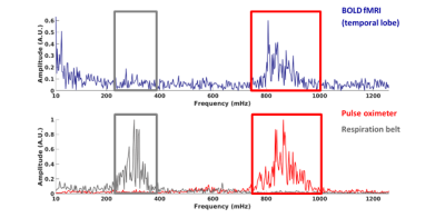 |
The amplitude of spontaneous fluctuations with high-frequency resting-state fMRI at 7T: influence of age and cardiorespiratory pulsations
Marieke van den Kerkhof1,2, Jacobus F.A. Jansen1,2,3, Lisanne P.W. Canjels1,2,3, Robert J. van Oostenbrugge2,4,5, Benedikt A. Poser6, and Walter H. Backes1,2
1Department of Radiology and Nuclear Medicine, Maastricht University Medical Center, Maastricht, Netherlands, 2School for Mental Health and Neuroscience, Maastricht University, Maastricht, Netherlands, 3Department of Electrical Engineering, Eindhoven University of Technology, Eindhoven, Netherlands, 4Department of Neurology, Maastricht University Medical Center, Maastricht, Netherlands, 5Cardiovascular Research Institute Maastricht, Maastricht University, Maastricht, Netherlands, 6Maastricht Brain Imaging Center, Faculty of Psychology and Neuroscience, Maastricht University, Maastricht, Netherlands
Resting-state fMRI with a short TR enables unaliased sampling of the BOLD-signal, by disentangling cardiorespiratory pulsation signals. In this study, we aimed to explore the feasibility of obtaining a high-frequency spectrum and determined the influence of aging. Structural and high-frequency resting-state fMRI was performed using 7T MRI on 5 young and 5 elderly subjects. The power spectra, calculated for different brain regions, showed a clear separation of spontaneous BOLD fluctuations and the respiratory and cardiac pulsations. This pilot study demonstrated the feasibility of acquiring high-frequency spectra using fMRI. Furthermore, initial results confirm that the BOLD effect attenuates with aging.
|
|
3915.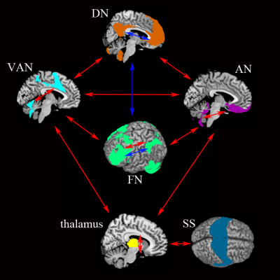 |
Large-scale network abnormality in bipolar disorder: A multimodal meta-analysis of resting-state functional connectivity and VBM
JiaYing Gong1,2, Junjing Wang3, Shaojuan Qiu1, Long Qian4, Li Huang1, and Ying Wang1
1Medical Imaging Center, First Affiliated Hospital of Jinan University, Guangzhou, China, 2Department of Radiology, Six Affiliated Hospital of Sun Yat-sen University, Guangzhou, China, 3Department of Applied Psychology, Guangdong University of Foreign Studies, Guangzhou, China, 4MR Research, GE Healthcare, Beijing, China
The present study is the first meta-analysis to integrate these diverse seed-based whole-brain resting-state functional connectivity (rs-FC) and voxel-based morphometry (VBM) results in bipolar disorder (BD) that exhibits abnormal connectivity within and between brain networks involved in internally oriented attention, processing of emotion or salience, goal-directed regulation of these functions, gating information, and sensorimotor processing. These findings motivate a large-scale neurocognitive model in which network abnormality is tightly linked to deficits in maintaining the integrated self (DN), processing of emotion (AN) or salience (VAN), and goal-directed regulation (FN) in BD.
|
|
3916.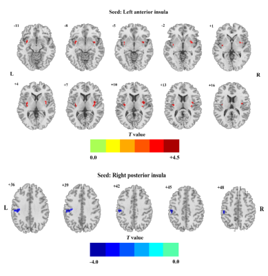 |
Insular subdivisions functional connectivity and immune dysregulation in patients with bipolar disorder: a resting‑state fMRI study
Pan Chen1, Feng Chen1, Guanmao Chen1, Long Qian2, and Ying Wang1
1Medical Imaging Center, First Affiliated Hospital of Jinan University, Guangzhou, China, 2MR Research, GE Healthcare, Beijing, China
Systemic inflammation and immune dysregulation have been considered as risk factors in the pathophysiology of mood disorders including bipolar disorder (BD). Previous metabolism, structural and functional neuroimaging studies have reported the specific regional brain volumetric alteration and dysfunction of the insula in BD. Taken together, in current study, the associations between the whole-brain dFC of each insular subdivision in unmedicated patients with BD and the level of pro-inflammatory cytokines were analyzed. Our results demonstrated that the clinical risk factors might relate to the aberrant dFC in specific insular subdivision.
|
|
3917.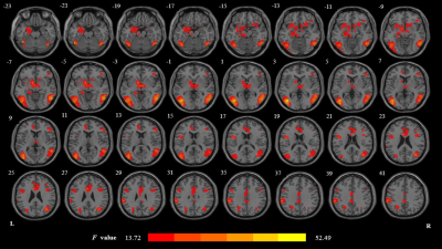 |
Assessing drug cue-induced brain response in heroin dependents treated by methadone maintenance and protracted abstinence measures one year
Xuan Wei1
1radiology department, Tangdu Hospital, the Fourth Military Medical University, Xi'an, China
Our research aims to compare PA with MMT to reveal which abstinence way is better to recover the brain function in heroin-dependent individuals. Twenty-four heroin-dependent male males with about 12 months of PA, 21 heroin-dependent male patients stabilized on MMT for about 12 months and 20 demographically HC completed an event-related fMRI task including heroin-related and neutral cues. In the last part of this study, we proved PA is closer to HC group. This study showed PA is more beneficial for the heroin-dependent patients to lower the salience value of drug related cues, in turn to reduce relapse risks.
|
|
3918.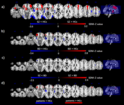 |
Aberrant Regional Homogeneity in Bipolar disorder and schizophrenia: a meta-analysis of resting-state functional MRI
Zhangzhang Qi1, JiaYing Gong1,2, Long Qian3, and Ying Wang1
1Medical Imaging Center, First Affiliated Hospital of Jinan University, Guangzhou, China, 2Department of Radiology, Six Affiliated Hospital of Sun Yat-sen University, Guangzhou, China, 3MR Research, GE Healthcare, Beijing, China
Resting-state functional MRI (RS-fMRI) studies have provided evidences for abnormal intrinsic brain activity in both schizophrenia and Bipolar disorder, but results are inconsistent. We conducted a meta-analysis of whole-brain, RS-fMRI studies to explore the Regional Homogeneity (ReHo) differences between patients with bipolar disorder (BD) and schizophrenia(SCZ). Our results suggested that the brain regions with convergent changes of ReHo in SCZ and BD included the Insular and prefrontalis lobe. While, the occipital lobe showed divergent change, where the ReHo value in SCZ decreases more. Patients with SCZ demonstrated much more widespread brain functional damage, including the decreased ReHo in sensorimotor area.
|
|
3919.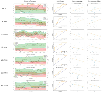 |
Abnormal Stationary and Dynamic Functional Connectivity in Multiple System Atrophy
Weimin Zheng1, Xiang Feng2, and Zhiqun Wang1
1Aerospace Center Hospital, Beijing, China, 2MR Scientific Marketing, Diagnosis Imaging, Siemens Healthcare Ltd, Beijing, China
This study aims to explore whether the differences in static and dynamic functional connectivity (s-FC and d-FC) can serve as potential biomarkers of multiple system atrophy (MSA). 24 MSA patients and 20 normal controls (NCs) were enrolled. We applied both s-FC and d-FC to evaluate functional changes in MSA patients and calculated the graph theory attributes based on s-FC and d-FC. We found that there were significant correlations between indicators of s-FC and d-FC and clinical performance. Furthermore, the receiver operating characteristic (ROC) analysis revealed that the substantially different FC features can serve as predictors to distinguish MSA from NC.
|
|
3920.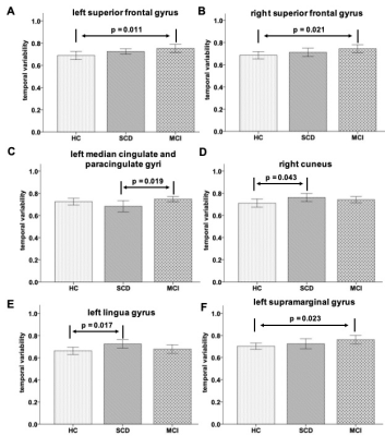 |
Evidences of Disrupted Temporal Variabilities in patients with subjective cognitive decline: a resting-state fMRI study
Ling Zhang 1, Yi Zhu 2, Han Wu 3, Hongyuan Ding 1, Yaxin Gao 4, Qian Zhong 4, Qiumin Zhou 2, Ming Qi 1, Long Qian 5, Weiqiang Dou5, and Tong Wang2
1Radiology department, The First Affiliated Hospital of Nanjing Medical University, Nanjing, China, 2Rehabilitation Department, The First Affiliated Hospital of Nanjing Medical University, Nanjing, China, 3Rehabilitation Department, Nanjing Drum Tower Hospital, The Affiliated Hospital of the Medical School at Nanjing University, Nanjing, China, 4Nanjing Medical University, Nanjing, China, 5MR Research China, GE Healthcare, Beijing, China
In this study, the whole brain temporal variability (TV) changes have been respectively investigated for patients with subjective cognitive decline (SCD) and mild cognitive impairment (MCI) and healthy controls (HCs). Significantly different TVs have been separately found between SCD, MCI patients and HCs in the regions involved in executive function, episodic memory, visual processing and visual memory and language perception and processing. Additionally, the TVs at these regions also showed strong correlations with multiple clinical scales. Therefore, TV method can be considered an effective tool in the early detection of SCD patients.
|
|
3921.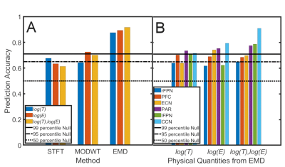 |
EMD-derived Energy-Period Profiles of Brain Networks in fMRI Resting-State Data: An Application to Parkinson’s Disease
Dietmar Cordes1,2, Muhammad Kaleem3, Zhengshi Yang1, Xiaowei Zhuang1, Tim Curran2, Karthik Sreenivasan1, Virendra Mishra1, Rajesh Nandy4, and Ryan Walsh5
1Cleveland Clinic Lou Ruvo Center for Brain Health, Las Vegas, NV, United States, 2University of Colorado, Boulder, CO, United States, 3University of Management & Technology, Lahore, Pakistan, 4University of North Texas Health Science Center, Fort Worth, TX, United States, 5Muhammad Ali Parkinson Center at Barrow Neurological Institute, Phoenix, AZ, United States
Traditionally, functional networks in resting-state data were investigated with Fourier and wavelet-related methods to characterize their frequency content. In this study, Empirical Mode Decomposition (EMD), a nonlinear method, is used to determine energy-period profiles of Intrinsic Mode Functions (IMFs) for different resting-state networks. In an application to early Parkinson’s disease (PD) vs. normal controls (NC), energy and period content were computed with EMD and compared with results using short-time Fourier transform (STFT) and maximal overlap discrete wavelet transform (MODWT) methods. Using a support vector machine, EMD achieved highest prediction accuracy in classifying NC and PD subjects among the three methods.
|