
Digital Poster Session
fMRI: Multimodal & Preclinical
fMRI
3922 -3933 fMRI: Multimodal & Preclinical - fMRI Multimodal 1
3934 -3945 fMRI: Multimodal & Preclinical - fMRI Multimodal 2
3946 -3958 fMRI: Multimodal & Preclinical - fMRI Preclinical
3922.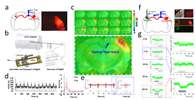 |
Mapping the brain-wide network effects by optogenetic activation of the corpus callosum with an MRI-guided robotic arm
Yi Chen1,2, Filip Sobczak1,2, Patricia Roldán Pais1,2, Cornelius Schwarz3, Alan P. Koretsky4, and Xin Yu1,5
1Research Group of Translational Neuroimaging and Neural Control, High-field Magnetic Resonance, Max Planck Institute for Biological Cybernetics, Tübingen, Germany, 2Graduate School of Neural Information Processing, University of Tübingen, Tübingen, Germany, 3Werner Reichardt Center for Integrative Neuroscience, Tübingen, Germany, 4Laboratory of Functional and Molecular Imaging, National Institute of Neurological Disorders and Stroke, National Institutes of Health, Bethesda, MD, United States, 5Athinoula A. Martinos Center for Biomedical Imaging, Massachusetts General Hospital and Harvard Medical School, Charlestown, MA, United States
This study not only specifies the optogenetically driven corpus callosum-mediated regulation of the local excitation/inhibition balance in the local barrel cortex, but also depicts the power of the multi-modal fMRI to characterize the brain-wide network activity associated with circuit-specific optogenetic activations. It highlights a vital aspect of the brain-wide activation for circuit-specific causality studies with optogenetic tools.
|
|
3923.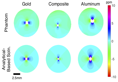 |
Gold-Aluminum Composite Electrodes for Brain Stimulation and LFP Recording with Reduced Image Artifacts at 16.4 Tesla
Corey Cruttenden1, Mahdi Ahmadi1, Yi Zhang2, Wei Zhu2, Rajesh Rajamani1, Xiao-Hong Zhu2, and Wei Chen2
1Mechanical Engineering, University of Minnesota, Minneapolis, MN, United States, 2Center for Magnetic Resonance Research, University of Minnesota, Minneapolis, MN, United States Poster Permission Withheld
Understanding the mechanisms of deep brain stimulation (DBS) would benefit from the ability to perform DBS simultaneously with functional magnetic resonance imaging (fMRI), but DBS electrodes introduce severe artifacts in fMRI. We constructed a novel gold-aluminum composite wire bundle and tested it for reducing B0 field distortion and image artifacts in phantom and in vivo at ultrahigh field (UHF) of 16.4T. The composite wire bundle exhibited superior performance compared to controls made of only gold or only aluminum. The new composite material structure can be applied to make multi-channel DBS electrodes with significantly reduced image artifacts even in UHF fMRI.
|
|
3924.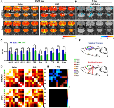 |
How brain reacts to attack at a hub region
Wenyu Tu1, Zilu Ma2, Yuncong Ma2, and Nanyin Zhang2
1The Huck Institutes of the Life Sciences, Penn State University, University Park, PA, United States, 2Biomedical Engineering, Penn State University, University Park, PA, United States
The brain function is a network phenomenon. However, exactly how brain network reconfigures when a brain region stops functioning is virtually unknown. By combining chemogenetic and resting-state fMRI methods in an awake rats, we investigated the causal impact of inactivating a hub region, dorsal anterior cingulate cortex on brain network properties. We found that disrupting hub activity changed organization of the default-mode network (DMN) and DMN-related behavior. It also altered topological architecture of the whole-brain network. Our study established a system that allows for mechanistically dissecting the relationship between local regions and the whole brain network organization.
|
|
3925.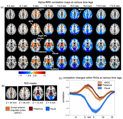 |
Resting-state fMRI and EEG alpha power show distinct correlations at different time lags
Yameng Gu1, Lucas E Sainburg1, Feng Han1, Jack W Williams1, Xiaoxiao Bai1, and Xiao Liu1
1Pennsylvania State University, State College, PA, United States
Alpha rhythm in EEG is prominent in the occipital and parietal cortex under an awake brain state. Previous studies have reported strong correlations between the resting-state fMRI signals and EEG alpha power. Here, we re-examined their correlations at different time delays. We found strong positive fMRI correlations at the zero lag, which is distinct from those observed at the typical hemodynamic delay. The finding suggests that the resting-state EEG alpha modulation is likely associated with a sequential brain process starting much earlier.
|
|
3926.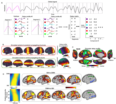 |
Electrophysiological and fMRI signals show consistent global propagations across the cortical hierarchy
Yameng Gu1 and Xiao Liu1
1Pennsylvania State University, State College, PA, United States
Infra-slow (<0.1 Hz) propagating structures have been found in resting-state fMRI. Nevertheless, the neural origin of the infra-slow rsfMRI propagations remains unclear. Besides, the study of propagating activity using fMRI cannot ignore a series issue that the brain has spatial heterogeneity of hemodynamic delays. Here, to address the questions, we combined human rsfMRI and monkey electrophysiology data and found similar propagations swiping the cortex in two opposite directions along an axis of
|
|
3927.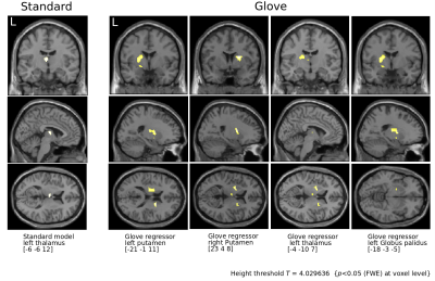 |
Improving brain imaging in Parkinson's disease by accounting for simultaneous motor output.
Renzo Torrecuso1, Karsten Mueller1, Stefan Holiga2, Thomas Sieger3, Jan Vymazal4, Růžička Jan5, Evzem Ruzicka6, Matthias Schroeter7, Robert Jech8, and Harald E. Möller1
1Nuclear Magnetic Resonance, Max Planck Institute for Human Brain and Cognitive Sciences, Leipzig, Germany, 2Roche Pharma Research and Early Development, Roche Innovation Center Basel, Basel, Switzerland, 3Department of Neurology and Center of Clinical Neuroscience, , Charles University in Prague, Prague, Czech Republic, 4| CULS · Faculty of Environmental Science, Czech University of Life Sciences Prague, Prague, Czech Republic, 5Department of Environmental Engineering, Faculty of TechnologyTomas Bata University in Zlín, Zlin, Czech Republic, 6Neurology, General University Hospital in Prague, Prague, Czech Republic, 7Clinic for Cognitive Neurology, University Hospital Leipzig, Leipzig, Germany, 8Department of Neurology, Charles University in Prague | CUNI ·, Prague, Czech Republic
Parkinson's disease leads to a variety of movement impairments. While studying the disease with fMRI, the main motivation for the research becomes one of its major obstacles: the motor output is unpredictable. Therefore it is troublesome to access, inside the scanner, performances of motor tasks and reliably relate them to brain measurements. We proposed to overcome this by expanding the patients’ number and restricting statistical criteria from a previous study which used a glove with non-magnetic sensors during scanning. Our results revealed basal ganglia not observed in the previous study confirming the usefulness of the device in fMRI studies.
|
|
3928.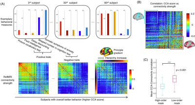 |
Behavior-related resting-state MEG/fMRI connectivity changes are related to the hierarchical organization of the neocortex
Feng Han1 and Xiao Liu1,2
1Department of Biomedical Engineering, Pennsylvania State University, State College, PA, United States, 2Institute for Cyber Science, Pennsylvania State University, State College, PA, United States
It has been shown that the maximal correlation between rsfMRI connectivity and behavioral measures occurs along a positive-negative mode direction characterizing the change of the overall goodness of behavior. Here, we had a thorough examination of rsfMRI/MEG connectivity along this positive-negative mode direction. We found that behavioral changes are associated with significant connectivity modulations that are however distinct at the lower-order sensory/motor areas and higher-order cognitive regions. Moreover, this hierarchy-dependent connectivity modulation is similar for rsfMRI and middle-frequency MEG signals, but reversed for gamma-band MEG signals. The findings may provide novel insight into the neural basis of inter-subject behavioral variability.
|
|
3929.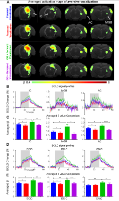 |
The role of hippocampus in central auditory processing – An optogenetic auditory fMRI study
Eddie C. Wong1,2, Alex T. L. Leong1,2, Russell W. Chan1,2,3, Celia M. Dong1,2, Anthea To1,2, Vick Lau1,2, and Ed X. Wu1,2
xxyy-video
1Laboratory of Biomedical Imaging and Signal Processing, The University of Hong Kong, Hong Kong, China, 2Department of Electrical and Electronic Engineering, The University of Hong Kong, Hong Kong, China, 3Neurology and Neurological Sciences, Stanford University, Stanford, CA, United States
Hearing is not just a sensory process as it plays pivotal roles in enabling communication, and memory and learning functions. This indicates that auditory processing is not confined within the central auditory pathways and likely involves higher-order cognitive structures such as hippocampus, which at present remains poorly understood. Here, we employed combined optogenetic and auditory fMRI in rats to investigate whether auditory processing can be influenced by hippocampal inputs. We reveal the specific role of ventral hippocampus in influencing auditory processing of behaviorally relevant sound across the entire central auditory pathways.
|
|
3930.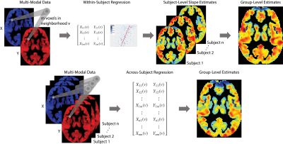 |
Approaches for Modeling Spatially Varying Associations Between Multi-Modal Images
Alessandra Michelle Valcarcel1, Simon N Vandekar2, Tinashe Tapera3, Azeez Adebimpe3, David Roalf3, Armin Raznahan4, Theodore Satterthwaite3, Russell T Shinohara1, and Kristin A Linn1
1Penn Statistics in Imaging and Visualization Center, Department of Biostatistics, Epidemiology, and Informatics, University of Pennsylvania, Philadelphia, PA, United States, 2Department of Biostatistics, Vanderbilt University, Nashville, TN, United States, 3Department of Psychiatry, University of Pennsylvania, Philadelphia, PA, United States, 4Developmental Neurogenomics Unit, National Institute of Mental Health, Bethesda, MD, United States
Multi-modal MRI modalities quantify different, yet complimentary, properties of the brain and its activity. When studied jointly, multi-modal imaging data may improve our understanding of the brain. We aim to study the complex relationships between multiple imaging modalities and map how these relationships vary spatially across different anatomical brain regions. Given a particular location in the brain, we regress an outcome image modality on one or more other modalities using all voxels in a local neighborhood of a target voxel. We apply our method to study how the relationship between local functional connectivity and cerebral blood flow varies spatially.
|
|
3931.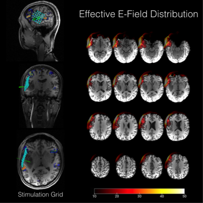 |
Improved pre-surgical causal language mapping combining 3T and 7T fMRI with TMS and E-field modelling in a young tumour patient
Anna-Lisa Schuler1, Georg Widhalm1, Michael Woletz1, Martin Tik1, Roland Fischer1, Karl Rössler1, and Christian Windischberger1
1Medical University of Vienna, Vienna, Austria
Here, we have optimised pre-surgical language mapping using navigated TMS with advanced E-field modelling and precise stimulation pulse timing. We employed a multi-modal approach incorporating ultra-high field (3 and 7 Tesla) functional magnetic resonance imaging, functional causal mapping including neuronavigated repetitive TMS, a software that allows for well-defined targeting and visual stimulus delay timings, as well as an advanced E-field/behavioural analysis. With this approach we could show that ‘virtual lesions’ close to tumour tissue yields clear spatial maps of functional impairment in language production.
|
|
3932. |
Estimating Cerebral Blood Flow from BOLD Signal Using Deep Dilated Wide Activation Networks
Danfeng Xie1, Danfeng Xie1, Yiran Li1, Hanlu Yang1, Li Bai1, Donghui Song 2, Yuanqi Shang2, Qiu Ge2, and Ze Wang3
1Temple University, Philadelphia, PA, United States, 2Hangzhou Normal University, Hangzhou, China, 3University of Maryland School of Medicine, Philadelphia, MD, United States
The purpose of this study was to synthesize Arterial Spin Labeling (ASL) cerebral blood flow (CBF) signal from blood-oxygen-level-dependent (BOLD) fMRI signal using deep machine learning (DL). Experimental results in the dual-echo Arterial Spin Labeling sequence show that the BOLD-to-ASL synthesize networks, the BOA-Net will yield similar cerebral blood flow value to that measured by ASL MRI and the cerebral blood flow maps produced by BOA-Net will show higher Signal-to-Noise Ratio (SNR) than that from ASL MRI.
|
|
3933.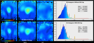 |
EEG-fMRI at 9.4T: Safety assessment and effect on B0, B1 and fMRI scans in a phantom
Vinod Jangir Kumar1, Kai Buckenmaier1, Tracy Warbrick2, Renate Wehrle3, Rolf Pohmann1, and Klaus Scheffler1
1Department for High-field Magnetic Resonance, Max Planck Institute for Biological Cybernetics, Tuebingen, Germany, 2Brain Products GmbH, Gilching, Germany, 3Easycap GmbH, Herrsching, Germany
Simultaneous EEG-fMRI has been attracting significant attention from neuroscientists for the last 20 years. During these years, EEG-fMRI has been in use to investigate brain at different field strengths. However, so far, there has been no study examining the potential use of EEG during MRI at 9.4T. Therefore, in this study, we used a customized EEG and acquired data with RF heating, gradient heating, B0, B1, and fMRI using a phantom. The results revealed no critical increase in temperature in the EEG. However, there is an observable decrease in B1, which indicates the need for further research in this direction.
|
 View the Poster
3934.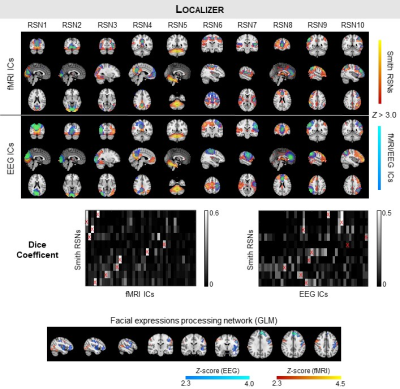 |
Pushing the limits of EEG: estimation of large-scale functional networks and their dynamics validated by simultaneous fMRI
Rodolfo Abreu1, Marco Simões1,2, and Miguel Castelo-Branco1
1Coimbra Institute for Biomedical Imaging and Translational Research (CIBIT), University of Coimbra, Coimbra, Portugal, 2Center for Informatics and Systems (CISUC), University of Coimbra, Coimbra, Portugal
Resting-state networks (RSNs) have been identified on continuous source-reconstructed EEG data (electrical source imaging; EEG-ESI data), but their validation with simultaneous fMRI data is missing. Here, we found a comparable overlap with previous literature between EEG-ESI-derived RSNs and simultaneous fMRI-derived RSNs from 10 subjects. We showed the ability of EEG-ESI to map the task-specific facial expression processing network, and extracted dynamic functional connectivity (dFC) states from EEG-ESI and fMRI, founding a significant match between them. Our results push the limits of EEG towards being used as an imaging tool and support the existence of EEG correlates of fMRI-derived (d)FC.
|
|
3935.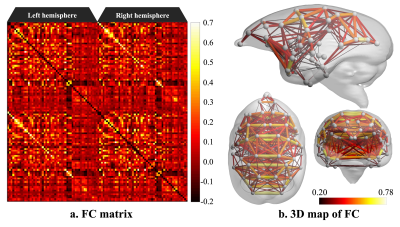 |
Resting-State Functional Connectome Analysis of Awake Common Marmoset with Functional MRI and Electrocorticographic
Yawara Haga1,2,3, Junichi Hata2,3,4, Takaaki Kaneko2,5, Tatsuhiko Yamada6, Yuji Komaki3, Fumiko Seki2,3,5, Hideyuki Okano2,5, Hirotaka James Okano4, Tetsuo Yamamori2, Noritaka Ichinohe2,7, Yuichi Yamashita7, Akira Furukawa1, and Misako Komatsu2,7
1Department of Radiological Sciences, Human Health Sciences, Tokyo Metropolitan University Graduate School, Tokyo, Japan, 2RIKEN Center for Brain Science, Saitama, Japan, 3Live Imaging Center, Central Institute for Experimental Animals, Kanagawa, Japan, 4Division of Regenerative Medicine, The Jikei University School of Medicine, Tokyo, Japan, 5Department of Physiology, Keio University School of Medicine, Tokyo, Japan, 6Graduate School of Computer Science and Systems Engineering, Kyushu Institute of Technology, Fukuoka, Japan, 7National Center of Neurology and Psychiatry, Tokyo, Japan Poster Permission Withheld
We explored the resting-state functional network of awake common marmoset by functional MRI (fMRI) and electrocorticographic (ECoG). As a result, a visual cortex network, a somatomotor network, a default mode network and a striatum network were detected in functional connectome analysis. In addition, some resting-state networks that were observed in the previous studies were detected in ICA. Those resting-state networks were also observed in ECoG data. Therefore, we conclude that fMRI and ECoG data were almost consistent.
|
|
3936.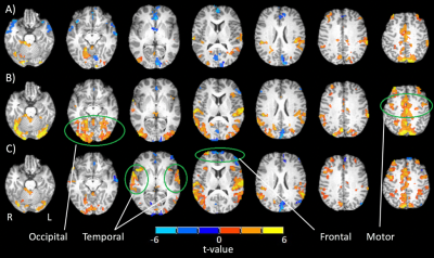 |
Driving with distraction: brain activity and oculomotor behaviour using fMRI and eye-tracking
Nicole Yuen1,2, Fred Tam2, Nathan Churchill3, Tom Schweizer3, and Simon Graham1,2
1Department of Medical Biophysics, University of Toronto, Toronto, ON, Canada, 2Physical Sciences, Sunnybrook Research Institute, Toronto, ON, Canada, 3St. Michael's Hospital, Toronto, ON, Canada
This study sheds further light on the neural correlates of driving behaviour and distracted driving, an important road-safety issue. Functional MRI and simultaneous eye-tracking measurements are performed during simulated driving tasks with and without auditory distraction. Initial results are consistent with previously published fMRI findings, showing changes in the occipital lobes, temporal lobes and frontal regions that are associated with increasing cognitive demand and distraction. These observations, and their interpretation, are consistent with reductions in the gaze field-of-view and increases in pupil diameter that reflect how the brain deals with cognitive challenges during realistic driving scenarios.
|
|
3937.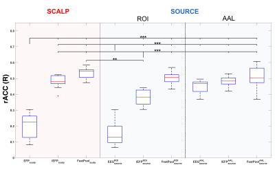 |
Reconstructing the BOLD-fMRI signal at the facial expression processing network from simultaneous EEG-derived predictors
MARCO SIMOES1, Rodolfo Abreu2, Bruno Direito2, Alexandre Sayal2, Joao Castelhano2, Paulo Carvalho3, and Miguel Castelo-Branco2
1CIBIT, Coimbra Institute for Biomedical Imaging and Translational Research, Faculty of Medicine, University of Coimbra, COIMBRA, Portugal, 2CIBIT, University of Coimbra, Coimbra, Portugal, 3CISUC, University of Coimbra, Coimbra, Portugal
fMRI is the neuroimage modality of choice when considering localized neurofeedback applications. However, the high costs and inflexibility of MRI setups limit their widespread application, motivating their transfer to EEG setups by reconstructing the BOLD-fMRI signal at the target regions using EEG only. Here, we systematically investigated the extent at which the BOLD-fMRI signal at the facial expressions processing network could be reconstructed from simultaneously recorded EEG signals. Features from both scalp and source spaces were extracted and used as predictors in a regression problem using random forests. We improved the accuracy of the state-of-the-art method from 20% to 53%.
|
|
3938.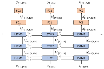 |
Predicting Blood-oxygenation-level Dependent Signal from Local Field Potentials Using Recurrent Neural Networks
Xiaodi Zhang1, Wen-Ju Pan1, and Shella Dawn Keilholz1
1Biomedical Engineering, Emory University/Georgia Institute of Technology, Atlanta, GA, United States
We implemented a stacked long short-term memory neural network to predict the blood-oxygenation-level dependent signal from the band-limited power of local field potentials in a variety of frequencies. The model was trained with simultaneously acquired resting state fMRI and LFP data from rats under Isoflurane anesthesia. The results show that the model prediction has a higher Pearson correlation with the ground truth of BOLD signal than the LFP band-limited power in any frequency bands.
|
|
3939.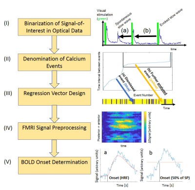 |
Applying Neurophysiologically-Informed Calcium Photometry Data as Regressor for Task-Free fMRI: An Analysis Pipeline.
Dirk Ernst Cleppien1, Felipe Aedo-Jury2, Miriam Schwalm3, and Albrecht Stroh2
1MAIC, German Resilience Center, Mainz, Germany, 2German Resilience Center, Mainz, Germany, 3Massachusetts Institute of Technology, Cambridge, MA, United States
We recently employed optically recorded slow oscillation-associated calcium waves as a linear regressor for simultaneously acquired task-free fMRI revealing a pan-cortical fMRI signature related to the active periods of the slow wave rhythm. Here, we provide a novel analysis pipeline for the synchronization of single events, as obtained by calcium photometry recordings, with fMRI scans usually requiring averaging over multiple trials. This synchronization poses unique problems, such as dealing with the inherent variability of the neurophysiological signal. The pipeline introduced here provides a step-by-step-approach within the framework of fast line scanning fMRI.
|
|
3940.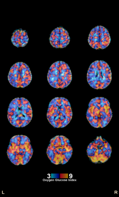 |
Measuring Aerobic Glycolysis with T2-relaxation-under-spin-tagging MRI and 18FDG PET: Preliminary Comparison with 15O PET
Shengwen Deng1, Dengrong Jiang2, Crystal G. Franklin1, Michael O'Boyle1, Wei Zhang1, Betty L. Heyl1, Paul A. Jerabek1, Hanzhang Lu2, and Peter T. Fox1
1Research Imaging Institute, UT Health at San Antonio, San Antonio, TX, United States, 2Department of Radiology, Johns Hopkins University, Baltimore, MD, United States
Preliminary test of using T2 based CMRO2 measurement to measure aerobic glycolysis in human brain, with combination of TRUST MRI, phase contrast MRI and quantitative FDG PET. Preliminary quantative comparison and modeling of MR/PET (TRUST-PC-FDG) and PET/PET (O-15/FDG) aerobic glycolysis measurement.
|
|
3941.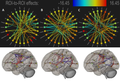 |
Understanding the functional connectivity of neural engagement of motor imagery and task through EEG engagement index informed fMRI
Deepanshi Dabas1,2, Srishti Keshari1,3, Pawan Kumar1, Ardaman Kaur1, Swati Agrawal1, Prabhjot Kaur1, Maria M D'souza1, and Vijayakumar C1
1NMR Research centre, Institute of Nuclear Medicine and Allied Sciences, DRDO, New Delhi, India, 2Maharaja Surajmal Institute of Technology, New Delhi, India, 3Banasthali Vidyapith, Rajasthan, India
Understanding neural engagement of Motor imagery facilitates development of various Brain-Computer Interface systems for rehabilitation purposes effectively. This study brings more insights on the neural underpinnings and associated functional connectivity of basal-ganglia, temporal and frontal-parietal regions during both Motor Imagery (MI) and Motor Action (MA) gripping tasks of randomised left, right and both hands movement with fifteen right-handed volunteers, using EEG-engagement index informed fMRI and functional connectivity approach. Functional connectivity analysis of the neural correlates reveal statistically elevated engagement of basal-ganglia, superior temporal gyrus and frontal-parietal regions in imagery, left handed imagery/action and both hand gripping tasks respectively.
|
|
3942.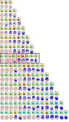 |
fMRI dynamic functional connectivity states associated with EEG alpha power
Afonso Aires1, Rodolfo Abreu1,2, João Jorge3,4, Joana Cabral5, and Patrícia Figueiredo1
1ISR-Lisboa/LARSyS and Department of Bioengineering, Instituto Superior Técnico – Universidade de Lisboa, Lisboa, Portugal, 2Coimbra Institute for Biomedical Imaging and Translational Research (CIBIT), University of Coimbra, Coimbra, Portugal, 3Laboratory for Functional and Metabolic Imaging, École Polytechnique Fédérale de Lausanne, Lausanne, Switzerland, 4Systems Division, Swiss Center for Electronics and Microtechnology (CSEM), Neuchâtel, Switzerland, 5Life and Health Sciences Research Institute (ICVS), School of Medicine, University of Minho, Minho, Portugal
Functional connectivity has been shown to change over short time scales of seconds to minutes, giving rise to the so-called dynamic functional connectivity (dFC). However, the electrophysiological underpinnings of dFC states remain unclear. We investigate EEG spectral correlates of dFC states using simultaneous EEG-fMRI data, by using a high temporal resolution fMRI acquisition combined with a phase coherence approach for dFC estimation and by computing k-means clustering with a varying number of dFC states. We found an association between high alpha power topographies and specific dFC states, which included regions of the frontoparietal network and the default mode network.
|
|
3943.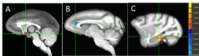 |
Chemically-Induced Hypothermia Altered Hypothalamic Functional Connectivity with Limbic System in the Monkey Brain
Chun-Xia Li1, Xiaohuan Gu2, Doty Kempf1, Ling Wei2, Shanping Yu3,4, and Xiaodong Zhang1,5
1Yerkes Imaging Center, Yerkes National Primate Research Center, Emory University, Atlanta, GA, United States, 2Department of Anesthesiology, Emory University School of Medicine, Atlanta, GA, United States, 32Department of Anesthesiology, Emory University School of Medicine, Atlanta, GA, United States, 4Center for Visual and Neurocognitive Rehabilitation, , Atlanta Veterans Affairs Medical Center, Decatur, GA, United States, 5Division of Neuropharmacology and Neurologic Diseases, Yerkes National Primate Research Center, Emory University, Atlanta, GA, United States
Rhesus monkeys were used to investigate whether ABS-201 induces similar hypothermia effect in large animals as seen in rodents and how this drug influences the hypothalamus centered limbic functional network. Functional connectivity (FC) between anterior hypothalamus and limbic system was examined by using resting state fMRI (rsfMRI). It is found that ABS-201 caused significant hypothermia effects in monkeys and significantly decreased hypothalamic FC with limbic regions as well. The findings suggest ABS-201 may have profound effects on the neurobehavior of subjects mediated by the hypothermia and antipsychotic effect.
|
|
3944.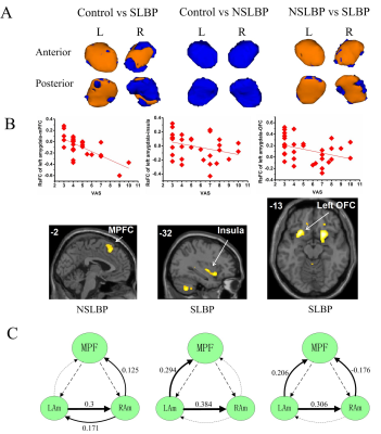 |
Characterizing the morphology and resting-state functional connectivity in chronic specific and nonspecific low back pain
Cui Ping Mao1, Quan Xin Yang1, Qiu Juan Zhang1, Hong Hong Sun1, Hua Juan Yang1, Xiao Qian Zhou1, and Gui Rong Zhang1
1Second Affiliated Hospital of Xi'an Jiaotong University, Xi'an, China
It is important to determine and distinguish the mechanisms underlying different types of pain because different drug targets are useful in pain of different origins. Specific low back pain (SLBP) with a recognizable pathology and nonspecific low back pain (NSLBP) are different pain conditions. The amygdala has been linked with the pathophysiology of chronic LBP. However, it is not known whether the amygdala is differentially affected in both conditions. Our study suggested that the amygdala morphology, resting-state functional connectivity and effective connectivity are differentially affected in subjects with SLBP and NSLBP, indicating different brain mechanisms in SLBP and NSLBP.
|
|
3945.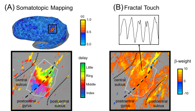 |
Probing human somatosensory cortex using ultra-high field (7T) fMRI and 3D-printed fractal textures
Alexander M Puckett1, Zoey Isherwood2, Ashley York1, Catherine Viengkham3, and Branka Spehar3
1University of Queensland, Brisbane, Australia, 2University of Wollongong, Wollongong, Australia, 3University of New South Wales, Sydney, Australia
Recent advances in fMRI, particularly accelerated image acquisition and ultra-high field (7T), have enabled high-resolution measurements and thus have provided the capability to non-invasively study the organization and function of smaller cortical areas in humans. One such area is primary somatosensory cortex, S1, in which the neuronal processing of tactile information largely occurs. Here we used 7T fMRI to (1) locate and map the fingertip representations in human S1 and then (2) to examine the activity at these locations in response to touching 3D-printed fractal textures. Robust and overlapping activation was seen across both conditions throughout primary somatosensory cortex.
|
 Watch the Video
3946.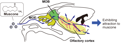 |
Muscone-evoked activations in the whole brains of mice, studied by the BOLD method using periodic stimulation and independent component analysis
Fuyu Hayashi1, Mitsuhiro Takeda1, Naoya Yuzuriha1, Sosuke Yoshinaga1, and Hiroaki Terasawa1
1Faculty of Life Sciences, Kumamoto University, Kumamoto, Japan Poster Permission Withheld
Muscone is a compound that contributes to the smell of musk and attracts male mice. When mice are stimulated with muscone, their main olfactory bulb and olfactory cortex are activated, as revealed by immunohistochemistry. It is conceivable that the signals are further transduced in the cerebrum, resulting in the attraction behavior in male mice. It is important to identify the muscone-evoked activated regions in the whole brain and explore their biological significance. We previously developed a functional MRI method that uses repetitive odor stimulation and independent component analysis. We applied this method to identify the muscone-evoked activated regions.
|
|
3947.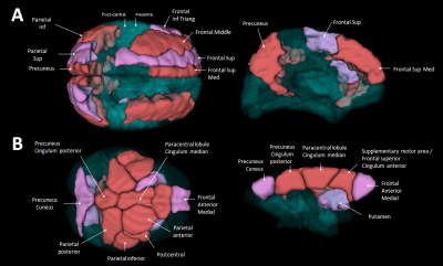 |
Resting state cerebral networks in mouse lemur primates: comparison with humans
Clément M. Garin1, Nachiket N. Nadkarni2, Brigitte Landeau3,4, Jean-Luc Picq2,5, Gaël Chételat3,4, Salma Bougacha3,4, and Dhenain Marc2,6
1Wake Forest University, Winston Salem, NC, United States, 2Commissariat à l’Energie Atomique et aux Energies Alternatives (CEA), Fontenay aux Roses, France, 3INSERM, U1077, CHU de Caen, Neuropsychologie et Imagerie de la Mémoire Humaine, Normandie University, UNICAEN, EPHE, Caen, France, 4Normandie Univ, UNICAEN, GIP Cyceron, Inserm, Inserm UMR-S U1237, Caen, France, 5EA 2027, Université Paris 8, 5 Laboratoire de Psychopathologie et de Neuropsychologie, Saint Denis, France, 6UMR 9199, Neurodegenerative Diseases Laboratory, 1 Centre National de la Recherche Scientifique (CNRS), Fontenay-aux-Roses, France, France
Characterizing neuronal networks in animals is critical to further address their evolutions. Here we compared brain networks in humans and in mouse lemurs (Microcebus murinus), one of the more phylogenetically distant primates as compared to humans. Network hubs were split into parietal and frontal clusters in humans, while they were grouped in lemurs. Human’s default mode network (DMN) embedded more hubs than lemur's DMN. Mouse lemur's motor network embedded more hubs than human motor networks. Hubness properties could constitute a lever of evolution to adapt information flux to brain size and/or cerebral function.
|
|
3948.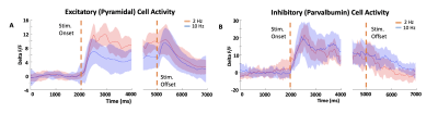 |
Shaping the BOLD signal through Excitatory and Inhibitory Interaction.
Rik Lodevicus Elizabeth Maria Ubaghs1, Harald Dermutz1, Roman Böhringer1, Markus Marks1, Mehmet Fatih Yanik1, and Benjamin Friedrich Grewe1
1Institute of Neuroinformatics University and ETH Zurich, ETH Zurich, Zurich, Switzerland
Characteristics of neural processing such as recurrent signalling, make the definition of functional neural involvement notoriously difficult. This has implications for fMRI interpretation, and especially the relationship between excitatory/inhibitory networks and the BOLD response has not been well studied. Here we provide evidence that there are stimulus-dependent effects on neural processing in the VISp that could explain previously observed differentiation in BOLD polarity. We hypothesize that this is due to local excitatory/inhibitory interactions, and present a novel fiber-photometry setup that allows us to investigate the neural underpinnings of this effect by measuring excitatory and inhibitory activity during fMRI.
|
|
3949.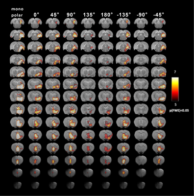 |
Orientation Selective Deep Brain Stimulation of Entorhinal Cortex and Medial Septal Nucleus in rats - functional MRI study at 9.4T.
Lin Wu1, Sheng Sang1, Lauri Lehto1, Antonietta Canna1,2, Jun Ma3, Clairice Pearce3, Zihad Nahas4, Walter Low3, Silvia Mangia1, and Shalom Michaeli1
1Center for Magnetic Resonance Research, University of Minnesota, Minneapolis, MN, United States, 2Department of Medicine, Surgery and Dentistry, Scuola Medica Salernitana, University of Salerno, Salerno, Italy, 3Department of Neurosurgery, University of Minnesota, Minneapolis, MN, United States, 4Department of Psychiatry, University of Minnesota, Minneapolis, MN, United States
Orientation selective deep brain stimulation (OS-DBS) is a flexible strategy to achieve selective stimulation of brain pathways. OS-DBS was used here in rats for stimulating the entorhinal cortex (EC) and the medial septal nucleus (MSN). Stimulating such areas induce neurogenesis in dental gyrus of the hippocampal formation, and can thus be regarded a potential therapy for Alzheimer’s disease (AD). Brain responses during OS-DBS were monitored by MB-SWIFT fMRI at 9.4T. Results demonstrate that different orientations of OS-DBS can modulate and selectively maximize activation of EC/MSN- hippocampal formation networks, and thus hold promise for maximizing neurogenesis and therapy efficacy for AD.
|
|
3950.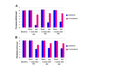 |
fMRI investigation of brain plasticity after unilateral cervical spinal cord injury
Basavaraju G Sanganahalli1, Jyothsna Chitturi2, Stella Elkabes3, Peter Herman1, Fahmeed Hyder1, and Sridhar S kannurpatti2
1Radiology and Biomedical Imaging, Yale University School of Medicine, New haven, CT, United States, 2Radiology, RUTGERS-New Jersey Medical School, Newark, NJ, United States, 3Department of Neurological Surgery, RUTGERS-New Jersey Medical School, Newark, NJ, United States
To assess brain plasticity after a unilateral incomplete cervical spinal cord injury (SCI), a complete neural axis assessment was performed using the adult rat model of cervical SCI at the C2/C3 level. Structural T1 and T2-spinal cord anatomical imaging and resting state BOLD-fMRI (9.4T) of the brain was performed 1 month after SCI in sham and SCI animals. The unilateral SCI induced lesion was discernable from anatomical images. Intrinsic brain activity assessed using functional connectivity density (FCD) mapping of global, short-range and long-range connectivity revealed a significant increase in FCD across SCI animals compared to sham.
|
|
3951.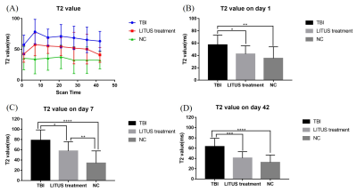 |
Role of the combination of T2 values and edema volume as a biomarker in evaluating the neuroprotective effect of LITUS to moderate TBI with rat model
Du Juan1, Zheng Tao1, Yuan Yi2, Wang Zhanqiu1, Liu Defeng1, Shi Qinglei3, Wu Shuo1, Wang Xiaohan1, and Liu Lanxiang1
1MRI, Qinhuangdao Municipal No. 1 Hospital, Qinhuangdao, China, 2Institute of Electrical Engineering, Yanshan University, Qinhuangdao, China, 3Siemens Healthcare, MR Scientific Marketing, Beijing, China Poster Permission Withheld
In this study, we verified the feasibility of T2 values combined with edema volume changes in evaluating the neuro therapeutic effect of low-intensity transcranial ultrasound stimulation (LITUS) on post-traumatic brain edema with rat model. The neuro therapeutic effect of LITUS was attributed to inhibiting the expression of AQP4 and promoting the removal of β-APP.
|
|
3952.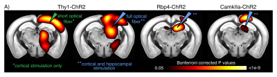 |
Network scale optogenetic fMRI with tapered optical fibers
Elizabeth de Guzman1, Alberto Galbusera1, Filippo Pisano2, Elisa Bellistri2, Alexia Stuefer1, Dania Vecchia3, Massimo De Vittorio2,4, Ferruccio Pisanello2, Tommaso Fellin3, and Alessandro Gozzi1
1Functional Neuroimaging Laboratory, Istituto Italiano di Tecnologia, Rovereto, Italy, 2Center for Biomolecular Nanotechnologies, Istituto Italiano di Tecnologia, Arnesano, Italy, 3Optical approaches to brain function, Istituto Italiano di Tecnologia, Genova, Italy, 4Dip. di Ingegneria dell’Innovazione, Università del Salento, Lecce, Italy
Optogenetic fMRI is an attractive tool for studying neuronal manipulation dynamics at multiple scales, but currently lacks techniques for network (i.e. mesoscale) level investigations due to the small illumination volume of traditional flat-faced optical fibers. Here we show that a recently developed tapered fiber design allowed simultaneous, artefact-free illumination of large cortico-hippocampal areas. We also demonstrated that low-frequency cortico-hippocampal entrainment resulted in a prominent functional desynchronization of the mouse default mode network. These results support the use of this technique for deconstructing network-scale substrates and probing brain-wide functional network dynamics.
|
|
3953.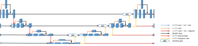 |
Automatic Mouse Skull-Stripping in fMRI Research via Deep Learning Using 3D Attention U-net
Guohui Ruan1, Jiaming Liu1, Ziqi An1, Kaibin Wu2, Wufan Chen1, Ed X. Wu3, and Yanqiu Feng1
1Guangdong Provincial Key Laboratory of Medical Image Processing & Key Laboratory of Mental Health of the Ministry of Education, School of Biomedical Engineering, Southern Medical University, Guangzhou, China, 2School of Biomedical Engineering, Southern Medical University, Guangzhou, China, Guangzhou, China, 3Department of Electrical and Electronic Engineering, Laboratory of Biomedical Imaging and Signal Processing, The University of Hong Kong, Hong Kong SAR, China, Hong Kong, China Skull-stripping is an important preprocessing step in functional magnetic resonance imaging (fMRI) research. In fMRI research using rodents, skull-stripping is still manually implemented, and it is very time-consuming. To address this problem, a 3D Attention U-net was trained for automatically extracting mouse brain from fMRI time series. The experimental results demonstrate that the mouse brain can be effectively extracted by the proposed method, and the corresponding fMRI results agrees well with the results with manual skull-stripping. |
|
3954.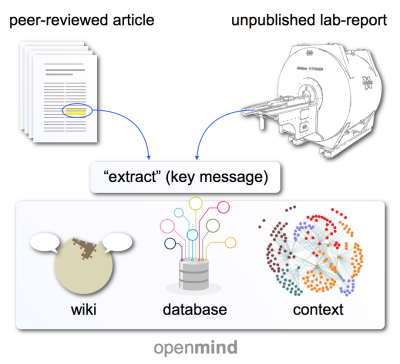 |
OpenMind – An Open Database of Brain Functions under Anesthesia for Preclinical MRI Research (and Beyond)
Henning Matthias Reimann1, Thomas Getgood2, and Thoralf Niendorf1,3
1Berlin Ultrahigh Field Facility (B.U.F.F.), Max Delbrück Center for Molecular Medicine in the Helmholtz Association, Berlin, Germany, 2Unaffiliated, Montreal, QC, Canada, 3Experimental and Clinical Research Center, a joint cooperation between the Charité Medical Faculty and the Max Delbrück Center for Molecular Medicine in the Helmholtz Association, Berlin, Germany
Understanding anesthetic effects in preclinical fMRI is key to produce significant findings. Over the last decades massive amounts of data have been produced on anesthesia, yet major portions lie idle to the community. Here we introduce an open, community driven infrastructure that combines the wiki principle (dynamic creating, correcting, commenting) with a powerful dynamic databank approach (fast and easy search, and network visualization of related findings) on anesthetic effects. OpenMind provides (1) convenient data upload, (2) fast and efficient search and filter functions, (3) embedding of findings in a larger dynamic context, and (4) low maintenance.
|
|
3955.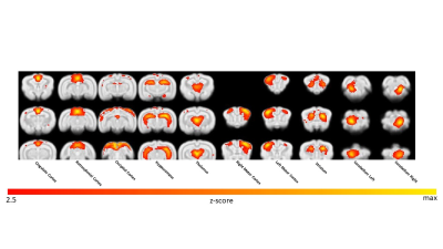 |
Effect of Eyeblink Conditioning on Intrinsic Brain Connectivity in Awake Rabbits: a resting-state fMRI study
Nicola Bertolino1, Daniele Procissi1, John F Disterhoft2, and Craig Weiss2
1Radiology, Northwestern University, Chicago, IL, United States, 2Physiology, Northwestern University, Chicago, IL, United States
Functional brain plasticity is an important characteristic of the brain and enables an individual to learn and adapt as a result of life experiences. In this study we investigated the reorganization of functional brain networks after eyeblink conditioning in awake rabbits by resting state fMRI. This work builds the foundation for studying pre-clinically how cognitive impairment affects learning processes, its implication on brain capability to adapt and learn, and the possible impact of new therapies.
|
|
3956.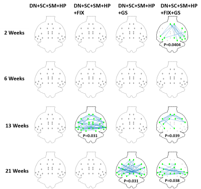 |
An optimized pipeline for functional network identification in rat brain with resting-state fMRI
Yujian Diao1, Rolf Gruetter1, and Ileana Ozana Jelescu1
1Ecole Polytechnique Fédérale de Lausanne, Lausanne, Switzerland
Resting state fMRI (rs-fMRI) is a widely used technique for identifying resting state networks (RSNs) and investigating brain disorders. However, the characterization of RSNs can be seriously hindered by the presence of random and structural noise in the measured fMRI signal. Most tools that correct for these effects are tailored for human brain and are not readily transposable to rat data. Here we propose a data processing pipeline for rat rs-fMRI data which can robustly remove artefacts and clean the rs-fMRI data. We report the performance of the pipeline for analyzing rat RSNs and discriminating between control and disease groups.
|
|
3957.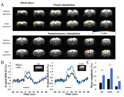 |
Sensory-evoked fMRI of transgenic mouse is enhanced by deafness
Hyun-Ji Shim1,2, Yoonsun Yang1, Gunsoo Kim1, and Seong-Gi Kim1,2,3
1Center for Neuroscience Imaging Research (CNIR), Institute for Basic Science (IBS), Suwon-si, Korea, Republic of, 2Department of Health Science and Technology, Samsung Advanced Institute for Health Sciences and Technology (SAIHST), Sungkyunkwan University (SKKU), Seoul, Korea, Republic of, 3Department of Biomedical Engineering, Sungkyunkwan University (SKKU), Suwon-si, Korea, Republic of
BOLD fMRI is ideal for investigating neural plasticity of transgenic mouse lines. Visual and somatosensory functions can be reorganized following deafness in the early stage and adult in humans and animals. However, the extent of the neural plasticity remains unclear. To determine how deafness affects other sensory areas, 9.4T BOLD fMRI of sensory stimulation was measured before and after deafening in transgenic adult mice. The impact of scanner noise to evoked responses was also investigated in wild-type mice. Our results suggest that the sensory loss in one modality affects other sensory activities.
|
|
3958.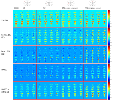 |
Brain mechanism of anesthesia and sedation: fMRI functional connectivity study with minimized impact of physiological background noise in rats
Wen-Ju Pan1, Vahid Khalilzad Sharghi1, Xiaodi Zhang1, and Shella Keilholz1
1Emory University/Georgia Institute of Technology, Atlanta, GA, United States
Different anesthetic states and sedation may have varying contributions to BOLD from physiological background noise that may challenge a comparison across brain states. Little is known on brain functional organization between anesthesia and sedation. We used a scan strategy optimized to minimize impact from physiological background noise to examine the effects of different anesthetics on functional connectivity (FC). Our results demonstrate distinct FC patterns across anesthesia depth transition and sedation, providing insight into potential brain mechanisms of unconsciousness that include global modulation of functional connectivity by isoflurane, and distant functional connectivity impairment in dexmedetomidine.
|