
Digital Poster Session
fMRI: Connectivity
fMRI
3959 -3973 fMRI: Connectivity - fMRI Connectivity Methods
3974 -3988 fMRI: Connectivity - Connectivity-Based Applications 1
3989 -4003 fMRI: Connectivity - Connectivity-Based Applications 2
4004 -4017 fMRI: Connectivity - fMRI Analysis Methods & Software Tools
3959.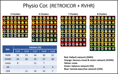 |
Physiologic noise contributes to large scale switching in PCC-seeded spontaneous co-activation
Wanyong Shin1 and Mark J. Lowe1
1Radiology, Cleveland Clinic, Cleveland, OH, United States
We investigated the impact of physiologic noise correction on PCC seeded co-activation pattern (CAP) analysis varying the cluster number. We found that patterns from PCC seeded CAP analysis were best classified as 5 sub-patterns of default mode, sensory visual and motor, salience, central executive networks and noise. Also we observed that physiologic noise correction resulted in less frequent salience and central executive networks from PCC-related co-activations than uncorrected data.
|
|
3960. |
Cohesive parcellation of rsfMRI using constrained hierarchical clustering
Ajay Nemani1 and Mark Lowe1
1Imaging Sciences, Cleveland Clinic, Cleveland, OH, United States Most connectivity-based parcellations of rsfMRI are synthesized from the dense connectome. However, the mean parcel signal is typically used for data reduction prior to network analysis. This results in representative parcel time series that are poorly correlated to their member voxels (poor parcel cohesion). We propose an adjacency-constrained, agglomerative hierarchical clustering framework that uses parcel cohesion as a linkage criteria. This results in parcels with mean time series that are significantly more representative of their member voxels. This parcellation is easily interpretable and well-suited for downstream analyses. |
|
3961.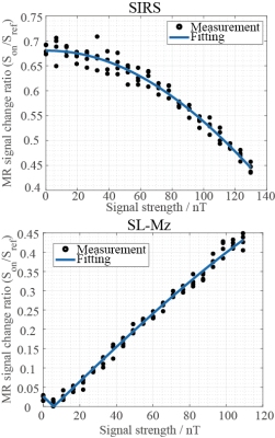 |
Detection of tiny oscillatory magnetic fields toward low-field fMRI: A phantom and simulation study
Hiroyuki Ueda1, Yosuke Ito1, Takenori Oida1, Yo Taniguchi2, and Tetsuo Kobayashi1
1Electrical Engineering, Kyoto University, Kyoto, Japan, 2Hitachi, Ltd., Kokubunji, Japan
To detect neural activities using a low-field MRI, we focused on spin-lock techniques, which are potentially effective tools for low-field functional MRI (fMRI). First, we estimated the magnetic field strength generated from neural activities using an equivalent current dipole model. Subsequently, we compared two spin-lock techniques by simulation and phantom experiments. It was found that the lowest detectable field strength was several tens pT in the simulation, whereas the experimental one was 2.34 nT. In future work, we plan to improve SNR using noise-reduction processing to realize the low-field fMRI.
|
|
3962.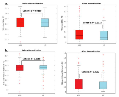 |
Improved Between-group Effect Size via Multisite Functional Connectivity Normalization
Alexandra Reardon1,2, Kaiming Li1,2, Jason Langley2, and Xiaoping Hu1,2
1Bioengineering, University of California, Riverside, Riverside, CA, United States, 2Center for Advanced Neuroimaging, University of California, Riverside, Riverside, CA, United States
Multisite databases allow for increased statistical power and enhanced reproducibility, however data pooled across sites are often acquired with different scanners and/or protocols, leading to significant site-dependent variances and thereby reduced effects in group analyses. We quantified the functional connectivity variance associated with the Autism Brain Imaging Data Exchange, a multisite resting state functional MRI database, and demonstrate how normalization functional connectivity results led to increased effect size when measuring established functional connectivity changes in autistic subjects as compared to healthy controls.
|
|
3963.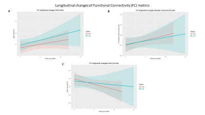 |
Post-surgical human brain functional reorganization in gliomas: a longitudinal study
Francesca Saviola1, Lisa Novello1, Domenico Zacà1,2, Luciano Annicchiarico3,4, Luca Zigiotto3,4, Silvio Sarubbo3,4, and Jorge Jovicich1
1CIMeC, Center for Mind/Brain Sciences, University of Trento, Rovereto, Italy, 2Siemens Healthcare, Milano, Italy, 3Department of Neuroscience,, Division of Neurosurgery, S.Chiara Hospital, Trento, Italy, 4Structural and Functional Connectivity Lab, S.Chiara Hospital, Trento, Italy
The recent brain connectomic framework has introduced a shift in the study of brain tumors. Previously considered as a focal brain disease, brain tumors are nowadays also studied in the context of structural and functional neural networking disruptions and their cognitive consequences. In this longitudinal behavior and resting state functional MRI study, we investigate how tumor grade, behavior and functional connectivity in brain network hubs and their connections to non-hubs are affected following surgical tumor resection.
|
|
3964.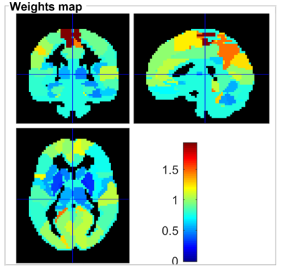 |
A machine learning investigation of resting-state fMRI abnormalities in HIV-associated neurocognitive disorder
Qianlin Ding1, Guanqiao Jin1, Lidong Liu1, Danhui Fu1, Wenjuan Deng1, Yang Zhao 1, Long Qian2, Weiyin Vivian Liu2, and Danke Su1
1GuangXi Medical University Cancer Hospital, Nanning, China, 2MR Research, GE Healthcare, Beijin, China
Frascati criteria for diagnosis of HIV-associated neurocognitive disorder lacks sensitivity, specificity, susceptible to learning effect, socioeconomic position and other confounding factors. fMRI can sensitively discover pathophysiological changes in HIV-infected central nervous system. In our study, ALFF and fALFF separately well classified HIV-positive and HIV-negative groups with AUC = 0.65 and 0.62, respectively. Our findings of injured frontal,parietal and occipital cortex associated with execution and attention function were consistent with previous studies, indicating activations reduced in the frontal lobe but increased in occipital lobe among patients with HAND. Both ALFF and fALFF could be biomarkers for objectively classifying HIV individuals.
|
|
3965.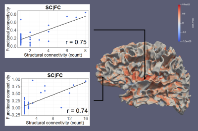 |
Surface Based Connectivity Integration – A Processing Pipeline for High Resolution Integration of Structural and Functional Connectivity
Kyle Murray1, Martin Cole2, Etienne St-Onge3, Maxime Descouteaux3, Jianhui Zhong1,4, Giovanni Schifitto4,5, and Zhengwu Zhang2
1Physics and Astronomy, University of Rochester, Webster, NY, United States, 2Biostatistics and Computational Biology, University of Rochester, Rochester, NY, United States, 3Computer Science, University of Sherbrooke, Sherbrooke, QC, Canada, 4Imaging Sciences, University of Rochester, Rochester, NY, United States, 5Neurology, University of Rochester, Rochester, NY, United States
The integration of structural and functional connectivity thus far has been limited to atlas-based parcellation studies. We present a novel atlas-free processing pipeline to explore the integration of structural and functional connectivity at high spatial resolution. This pipeline has been reliably replicated in other research subjects. We also introduce a dual-modality imaging feature that can be mapped to the gray-white matter surfaces. Early results demonstrate that structural and functional connectivity are likely strongly linked. This pipeline allows for the first time to perform connectivity analyses on individual white surfaces, opening up many more possibilities to future connectivity studies.
|
|
3966. |
Investigating the human brain information flow pattern based on resting-state functional magnetic resonance imaging
Zhizheng Zhuo1, Haiyun Li2, Yingjie Mei3, and Yaou Liu1
1Department of Radiology, Beijing Tiantan Hospital, Beijing, China, 2School of Biomedical Engineering, Capital Medical University, Beijing, China, 3Clinical Science, Philips Healthcare, Guangzhou, China
Different information flow patterns were found within low and high signal frequency bands based on resting-state fMRI.
|
|
3967.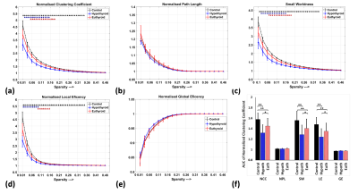 |
Alteration resting state networks functional connectivity associated with Hypothyroidism using Graph theory
Mukesh Kumar1, Poonam Rana1, Tarun Sekhri2, Subash Khushu1, Rajanikant Panda3, Ratnesh Kanwar2, Shilpi Modi1, and Maria Dsouza1
1NMR Research Center, INMAS, Delhi, India, 2Thyroid research Center, INMAS, Delhi, India, 3Cognitive Neuroscience Center, NIMHANS, Bangalore, India
The aim of this study is to explore the alteration RSNs connectivity and its association with underlying neurobehavioral symptoms in hypothyroid as compared to controls and consequences of therapeutic intervention using graph theory-based network analysis. Our finding revealed significantly reduced clustering coefficient, small world connectivity and local network efficiency in hypothyroid as compared to controls in sub-regions FPN, DMN, cingulo-opercular network, SMN and got reversed back in euthyroid patients in the network sub-regions of frontoparietal network, cingulo-opercular network, SMN after thyroxin therapy. These findings of reduced clustering coefficient suggest abated activities of motor, attention, working memory and cognition in hypothyroid.
|
|
| 3968. | Functional Connectivity in Limbic System May Determine the Laterality in Temporal Lobe Epilepsy- A Graph-Theory Approach
Saba Amiri1, Jafar Mehvari Habibabadi2, Seyed Sohrab Hashemi-Fesharaki3, Neda Mohammadi Mobarakeh1, Mehdi Mirbagheri1, and Mohammad-Reza Nazem-Zadeh1,4
1Physics and Biomedical Engineering, Tehran University of Medical Sciences, Tehran, Iran (Islamic Republic of), 2Isfahan Neuroscience Research Center, Isfahan University of Medical Sciences, Isfahan, Iran (Islamic Republic of), 3Pars Advanced Medical Research Center, Pars Hospital, Tehran, Iran (Islamic Republic of), 4Research Center for Molecular and Cellular Imaging, Advanced Medical Technologies and Equipment Inst, Tehran University of Medical Sciences, Tehran, Iran (Islamic Republic of)
Temporal lobe epilepsy (TLE) is a disorder of altered brain networks. We evaluated the functional connectivity of the brain limbic system believed to be affected by TLE, based on resting state functional MRI and the graph network analysis. Our results showed that insula, posterior cingulate gyrus, and thalamus may undergo abnormal functional connectivity in terms of local degree in both right and left TLE compared to the healthy subjects. Furthermore, our results suggest that functional connectivity, as evaluated by local degree in insula and thalamus may have a potential application to help determine the laterality in cases of TLE.
|
|
3969.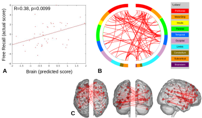 |
Connectome predictive modeling of face-name associations in older adults with mild cognitive impairment
Michelle Karker1, Scott Peltier2, Allison Moll3, Julia Laing3, and Benjamin M. Hampstead3,4
1Biomedical Engineering, University of Michigan, Ann Arbor, MI, United States, 2Functional MRI Laboratory, University of Michigan, Ann Arbor, MI, United States, 3Psychiatry, University of Michigan, Ann Arbor, MI, United States, 4Mental Health Service, VA Ann Arbor Healthcare System, Ann Arbor, MI, United States
Connectome predictive modeling was applied to task data. Leveraging a MCI-relevant face-name task illuminates relationships with measures of total cognition (RBANS) and memory (Free Recall). This provides support for the use of clinically-relevant tasks in cases where a “driven” connectivity network may elucidate pathological changes.
|
|
3970.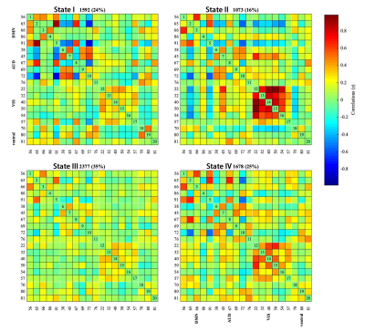 |
The Study of Regional Homogeneity and dynamic functional Connectivity of Trigeminal Neuralgia at Resting-state fMRI
pengfei zhang1, jun wang1, shaoyu wang2, and jing zhang1
1LanZhou University Second Hospital, LanZhou, China, 2MR Scientific Marketing, ShangHai, China
Based on rest-state fMRI, we investigated the changes of regional neural activity in trigeminal Neuralgia (TN) patients via analysis of regional homogeneity (ReHo). Furthermore, the alterations of dynamic functional connectivity was explored. Compared with healthy controls , brain regions with significant ReHo value changes in TN patients are mainly concentrated in default mode network (DMN) and Ventral attention Network(VAN). Dynamic analysis suggested four distinct connectivity ‘States’ across the entire group. Our results demonstrated that TN is associated with abnormal neuronal activities in different areas of the brain and dynamic functional connectivity between functional networks.
|
|
3971.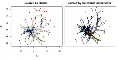 |
A Hierarchical Latent Space Network Model for Population Studies of Functional Connectivity
James David Wilson1, Skyler Cranmer2, and Zhong-Lin Lu3
1Mathematics and Statistics, University of San Francisco, San Anselmo, CA, United States, 2Political Science, The Ohio State University, Columbus, OH, United States, 3Center for Neuroscience and Department of Psychology, New York University, New York, NY, United States
Functional connectivity scans are hierarchical – heterogeneity differentiates people according to clinical diagnosis and stage of disease. Hierarchy furthermore dictates connectivity of an individual - functional networks manifest as a hierarchy of subnetworks, each with their own unique biological function. To model functional connectivity across populations of individuals, we develop the hierarchical latent space model (HLSM), a statistical model that accounts for hierarchy as a function of clinical and connectivity features describing functional images. The HLSM reveals differences in the connectivity patterns between healthy and schizophrenia groups when applied to data from the Center for Biomedical Research Excellence (COBRE) project.
|
|
3972.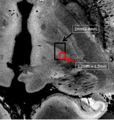 |
Improved Specificity of Functional Mapping of Thalamocortical Connections at Ultra High Field
Mark J Lowe1, Anna Crawford1, and Stephen E Jones1
1Imaging Institute, Cleveland Clinic, Cleveland, OH, United States
We compare whole brain functional connectivity to the Vim thalamic nucleus at 3T and 7T, and demonstrate that the higher spatial resolution and higher contrast-to-noise ratio at 7T results in maps that are more specific to the function of that thalamic region and the connectivity maps from the 3T data are likely lacking in specificity due to partial volume of signal from neighboring thalamic regions.
|
|
3973.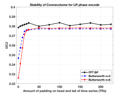 |
Mitigating effects of temporal filter of time-series for reliability of connectome calculations.
Robert Cary Welsh1 and Martin A Lindquist2
1Department of Psychiatry, University of Utah, Salt Lake City, UT, United States, 2Biostatistics, The Johns Hopkins University, Baltimore, MD, United States
Temporal filtering is a typical step in preprocessing of BOLD time-series for calculation of resting-state correlations, which are the basis of a connectome calculation. Bandpass filtering is a usual step in preprocessing of resting-state BOLD time-series data. Temporal filtering can introduce bias in the correlation calculations resulting in artifact in the connectome affecting the reliability in a connectome. We demonstrate this effect in simulations and then show this in data from the Human Connectome Project. We also show how to mitigate this issue for high reliability of the connectome as measured by image intraclass correlation coefficient (I2C2).
|
 View the Poster
 Watch the Video
3974. |
Resting-state Functional Connectivity during Voluntary Mouth Breathing
Chan-A Park1, Ju-Yeon Jung2, Yeong-Bae Lee3,4, and Chang-Ki Kang2,4,5
1Biomedical Engineering Research Center, Gachon University, Incheon, Korea, Republic of, 2Department of Healthy Science, Gachon University Graduate School, Incheon, Korea, Republic of, 3Department of Neurology, Gachon University Gil Medical Center, Incheon, Korea, Republic of, 4Neuroscience Research Institute, Gachon University, Incheon, Korea, Republic of, 5Department of Radiological Science, Gachon University, Incheon, Korea, Republic of Poster Permission Withheld
The purpose of the study is to examine the difference of the functional connectivity between the nasal and mouth breathing conditions in healthy subjects using resting-state functional magnetic resonance imaging via seed-based correlation analysis. “Mouth>Nose” contrast had 5 seeds and 23 connecting pairs, however, 6 seeds and 14 pairs in the “Mouth<Nose” contrast. Especially, caudate had the most number of connections of salience networks, supramarginal gyrus, insular cortex, central opercular cortex, supramarginal gyrus, and parietal operculum in “Mouth>Nose” contrast. These indicated that the limbic system regulates the resting-state functional connectivity during the voluntary mouth breathing compared the nasal breathing.
|
|
3975.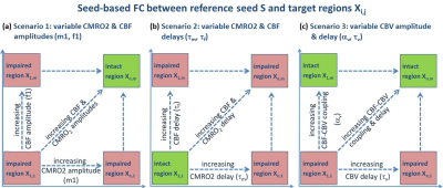 |
Studying the impact of impaired perfusion and oxygen metabolism on resting-state fMRI-based functional connectivity by simulating blood oxygenation fluctuations
Christine Preibisch1,2,3, Mario E. Archila-Melendez1,3, Stephan Kaczmarz1,3, and Christian Sorg1,3
1Department of Neuroradiology, Technische Universität München, School of Medicine, Munich, Germany, 2Clinic of Neurology, Technische Universität München, School of Medicine, Munich, Germany, 3TUM NIC, Technische Universität München, School of Medicine, Munich, Germany
Functional magnetic resonance imaging (fMRI) of blood oxygenation level dependent (BOLD) signals in the resting state is widely used to study functional connectivity of slowly fluctuating ongoing brain activity (BOLD-FC) in humans, particularly also in patients. While physiological impairments, e.g. aberrant perfusion, are common in neurological and psychiatric disorders, their impact on measured BOLD-FC is widely unknown and ignored. The aim of our simulation study, therefore, was to investigate how alterations in neurovascular coupling influence resting-state BOLD-FC measures. Our results demonstrate crucial impact of neurovascular coupling on BOLD-FC due to changes in CMRO2, CBF, CBV, in both amplitudes and delays.
|
|
3976.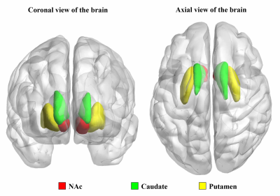 |
Increased Striatal Functional Connectivity is Associated with Improved Smoking Cessation Outcomes
Chao Wang1, Peiyu Huang1, Zhujing Shen1, and Minming Zhang1
1Department of Radiology, the Second Affiliated Hospital, Zhejiang University School of Medicine, Hangzhou, China
Most smoking cessation attempts result in failure. The striatum is critical area of reward processing, and have been repeatedly linked to nicotine addiction. Neuroimaging studies have shown that chronic smokers had altered resting-state functional connectivity (rsFC) of striatum. Here, we further investigated the different rsFC changes of striatum subsets between smokers who relapsed and those who not relapsed after smoking cessation treatment. We found that smokers who relapsed had decreased rsFC of striatum subsets, while those who not relapse had increased rsFC of striatum subsets. These novel findings suggest that increased connectivity of striatum subsets could imporve likelihood of cessation.
|
|
3977.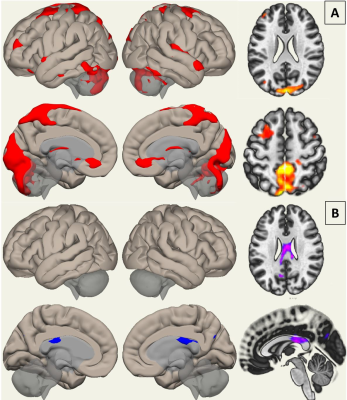 |
Functional Connectivity in Glioma patients using Resting State fMRI: Effect on Default Mode, Fronto-Parietal and Dorsal Attention Networks
Mickael Tordjman1, Pradeep Kumar Gupta1, Guillaume Madelin1, Timothy Shepherd1, Mariana Lazar1, and Rajan Jain1,2
1Department of Radiology, New York University School of Medicine, New York, NY, United States, 2Department of Neurosurgery, New York University School of Medicine, New York, NY, United States
Exploration of functional connectivity in patients with brain tumors with Resting state functional MRI (rsfMRI) is expanding, but methodology used and results in previous studies are heterogenous. We explored the disruption of the functional connectivity of the Default Mode, Dorsal Attention and Fronto-Parietal Networks in 35 glioma patients, with a standardized method, using both Seed-Based Connectivity Analysis and Independent Component Analysis. The Default Mode was the most affected network, with global increased connectivity contrasting with a decreased connectivity in the corpus callosum. No difference in connectivity was found between IDH mutant or wildtype tumors.
|
|
3978.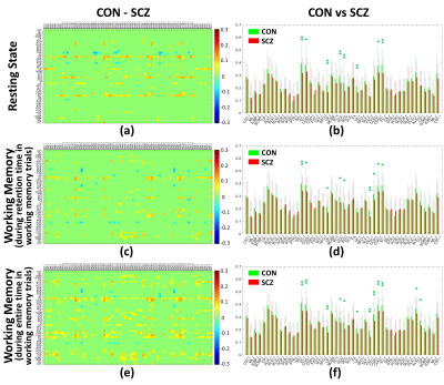 |
Decreases in functional connectivity of white matter in a resting state and during a working memory task in schizophrenia
Yurui Gao1,2, Muwei Li1,3, Anna S Huang4, Adam W Anderson1,2,3, Zhaohua Ding1,5, Stephan H Heckers3,4, Neil D Woodward4, and John C Gore1,2,3
1Vanderbilt University Institute of Imaging Science, Vanderbilt University Medical Center, Nashville, TN, United States, 2Biomedical Engineering, Vanderbilt University, Nashville, TN, United States, 3Radiology and Radiological Sciences, Vanderbilt University Medical Center, Nashville, TN, United States, 4Psychiatry and Behavioral Sciences, Vanderbilt University Medical Center, Nashville, TN, United States, 5Electrical Engineering and Computer Science, Vanderbilt University, Nashville, TN, United States
White matter (WM) pathological changes play a role in disturbing neural connectivity of schizophrenic subjects. We extended our previous analyses of WM-GM connectivity to quantify WM functional differences during a resting state and a working memory task between schizophrenic patients and healthy controls. Significant deficits of functional connectivity were found in several WM tracts in schizophrenic patients relative to healthy controls. These findings add further evidence of the presence of WM changes in schizophrenia subjects compared to controls and further illustrate the potential relevance of functional signals arising from WM in a task and at rest.
|
|
3979.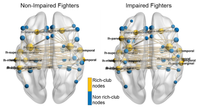 |
Changes of functional brain topology in cognitively impaired professional fighters
Xiaowei Zhuang1, Zhengshi Yang1, Virendra Mishra1, Karthik Sreenivasan1, Sarah J Banks2, Bernick Charles1, and Dietmar Cordes1,3
1Lou Ruvo Center for Brain Health, Cleveland Clinic, Las Vegas, NV, United States, 2Department of Neuroscience, University of California, San Diego, La Jolla, CA, United States, 3University of Colorado, Boulder, Boulder, CO, United States
In this study, we utilized graph-theory to investigate functional brain topological alternations in cognitively impaired active professional fighters, as compared to cognitive non-impaired fighters. We observed reduced global and local efficiency across multiple sparsity levels, suggesting that both functional integration and segregation were affected in cognitively impaired fighters. A different functional topological organization was further observed in impaired fighters, shown by increased number of rich clubs and reduced rich-club edge strength and density. Rich club edge strength and density were further significantly correlated with fighters’ psychomotor speed.
|
|
3980.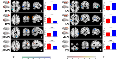 |
The mechanisms of cognitive impairment in patients with pontine infarction:a resting-state fMRI study.
Yingying Wang1, Caihong Wang1, Peifang Miao1, Ying Wei1, Luobing Wu1, Kaiyu Wang2, and Jingliang Cheng1
1Department of MRI, The First Affiliated Hospital of Zhengzhou University, Zhengzhou, China, 2GE Healthcare MR Research, Beijing, China, Beijing, China
In order to explore the mechanisms of cognitive impairment in patients with pontine infarction, we recruited 45 patients with chronic pontine stroke and 48 normal controls. The independent component analysis (ICA) and seed-based analysis were combined to explore altered patterns of functional connections in patients based on resting-state fMRI. The different FCs in the stroke patients and the healthy controls, as well as the positive correlation between the FC within the cerebellar network and cognitive function scores, indicate that the disconnection of the cerebellar network may be related to the cognitive impairment in patients with pons stroke.
|
|
3981.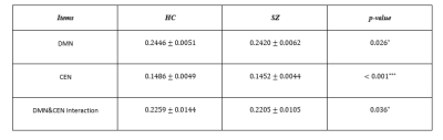 |
Schizophrenia patients exhibit altered network dynamics
Zhenhai Zhang1, Kaiming Li2, and Xiaoping Hu2
1Department of Electrical and Computer Engineering, University of California, Riverside, Riverside, CA, United States, 2Department of Bioengineering, University of California, Riverside, Riverside, CA, United States
The work aims to investigate the nonlinear dynamics in CEN/DMN networks. To this end, we applied phase-space embedding and multivariate autoregressive modeling of the phase-space trajectory. Furthermore, the AR coefficients were analyzed with a linear discriminant analysis to identify principle features that distinguish between patients and controls. The method was able to reveal differences in nonlinear dynamics in CEN and DMN networks respectively and jointly.
|
|
3982.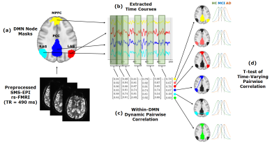 |
Dynamic Changes of the Functional Connectivity in the Default Mode Network across the Spectrum of Alzheimer’s Disease
Hyae Won K Redden1, Daniel Zhu2, Thomas J Grabowski3, and Hesamoddin Jahanian3
1Bioengineering, University of Washington, Seattle, WA, United States, 2Computer Science, University of Washington, Seattle, WA, United States, 3Radiology, University of Washington, Seattle, WA, United States
Default Mode Network functional connectivity (DMN FC) has been proposed as a non-invasive biomarker for Alzheimer’s Disease (AD). However, the dynamic relationships within the DMN over the course of the disease have not been established. We explored the dynamic FC between the DMN nodes in healthy control, mild cognitive impairment (MCI), and AD subjects using a sliding window analysis of ultrafast resting state fMRI (rs-fMRI) data. Group comparisons revealed significant trends in the dynamic measures of functional connectivity within the DMN across the spectrum of AD, suggesting compensatory systems at work within the DMN as AD progresses.
|
|
3983.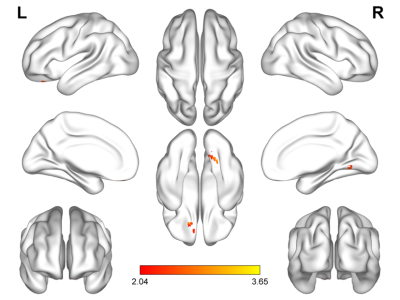 |
Study on brain function changes in patients with neurological depression based on resting state functional magnetic resonance imaging(rs-fMRI)
Hongwei Zhou1, Dezheng Kong1, Tianjing Zhang2, and Yanling Wu2
1Jilin University First Hospital, Changchun, China, 2Philips healthcare, Guangzhou, China
The global incidence of depression is gradually increasing nowadays. However, the exact pathogenesis mechanism is not very clear. Traditional diagnosis approaches to depression usually rely on clinical scales, which is sometimes even subjective. It is essential to make up objective and repetitive methods to diagnose the depression. Resting-state functional magnetic resonance imaging (rs-fMRI) offers a noninvasive technique for detecting and measuring subtle functional brain activities of specific regions. This study aims to explore the change of brain function area in patients with depression and assess the role of these altered brain regions in the pathogenesis.
|
|
3984.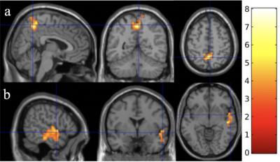 |
The effect of meditation on brain functional connectivity using dynamic ASL
Zongpai Zhang1, Steffi Chettiar1, Swanand A Wagh1, Wenna Duan1, George Weinschenk1, Wen-Ming Luh2, and Weiying Dai1
1Computer Science, State University of New York at Binghamton, Binghamton, NY, United States, 2National Institute on Aging, National Institutes of Health, Baltimore, MD, United States
We applied dynamic arterial spin labeling (ASL) in detecting the changes of brain functional connectivity (FC) in ten college students after 2-months of meditation practice. FC maps before and after meditation practice were compared, and the changes of FC were correlated to practice time. We observed that the increased practice time was associated with increased FC between long-distance nodes within default mode network (DMN) and within dorsal attention network (DAN), between DMN and salience network (SN), with decreased FC between short-distance nodes within DMN and DAN. The findings support efficient global communication even after a short-term meditation for two months.
|
|
3985.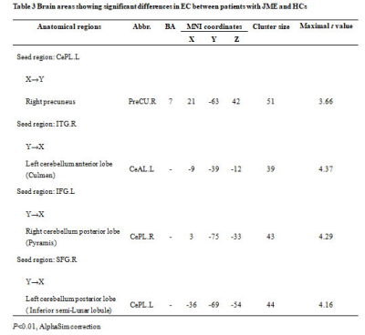 |
Altered cerebro-cerebellar effective connectivity in new-onset juvenile myoclonic epilepsy
Guangyao Liu1, Hong Liu1, Jing Zhang1, and Laiyan Ma1
1Lanzhou University Second Hospital, Lanzhou, China
Resting-state fMRI studies have indicated that juvenile myoclonic epilepsy (JME) could cause widely functional connectivity disruptions between cerebrum and cerebellum. However, the directed influences or effective connectivities (ECs) bewteen these brain regions are poorly understood. In the current study, we aim to evaluate the ECs between cerebrum and cerebellum in new-onset patients with JME.
|
|
3986.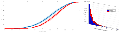 |
Functional brain networks in schizophrenia: chain-like organization goes wild
Tommaso Gili1, Rossana Mastrandrea1, Fabrizio Piras2, Andrea Gabrielli3, Guido Caldarelli1, and Gianfranco Spalletta2
1IMT School for Advanced Studies Lucca, Lucca, Italy, 2IRCCS Santa Lucia Foundation, Roma, Italy, 3Università degli Studi Roma III, Roma, Italy
Schizophrenia is a severe psychiatric condition, and is hypothesized to result from abnormal anatomical neural connectivity and a consequent decoupling of the brain’s integrative thought processes. Here we analysed and compared resting state functional magnetic resonance imaging (rs-fMRI) data from forty-four schizophrenia patients (SZ) and forty healthy subjects (HS). Complex network analysis of brain functional connectivity revealed that SZ networks are characterized by more stable configurations with a lower level of flexibility of functional plasticity than HS networks, which may depend on an abnormal synaptic connectivity due to a dysfunctional cortical pruning.
|
|
3987.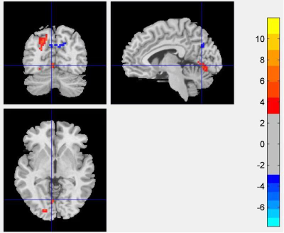 |
Resting State Functional Magnetic Resonance Imaging Study of Auricular Electro-acupuncture Therapy on Treatment-Resistant Depression
KE XU1, JILIANG FANG1, and XIAOJIAO LI1
1CHINA ACADEMY OF CHINESE MEDICAL SCIENCES, BEIJING, China
The aim of this study was to provide scientific basis for the therapeutic mechanism of auricular electro-acupuncture in treatment resistant depression patients. We evaluated the resting state functional connectivity before and after 8 week .Results showed that functional connectivity increased in the lingual gyrus and precuneus ,which is related to default mode network (DMN), might playing a potential part for the significant improvement of clinical symptoms in TRD patients.
|
|
3988.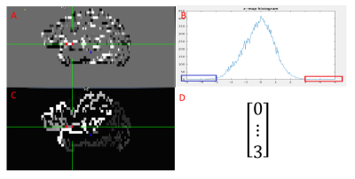 |
Seed-based resting state fMRI data analysis pipeline by using unsupervised machine learning
Mingyi Li1, Katherine Koenig1, Jian Lin1, and Mark Lowe1
1Diagnostic Radiology, Cleveland Clinic, Cleveland, OH, United States
We have developed an automatic pipeline to generate seed clusters and corresponding maps for rs-fMRI data analysis by using unsupervised machine learning method. It only needs manual participation in the end to review the candidate seed cluster locations and connectivity maps to make decision. In contrast to common anatomical seed scheme which usually consists of a small neighborhood surrounding a single voxel, seeds in our pipeline were determined functionally within large pre-defined ROI which can be derived by using automatic brain segmentation tools like FreeSurfer. The pipeline was tested successfully on rs-fMRI studies with accompanied task-based fMRI involving motor cortex.
|
3989.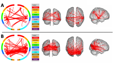 |
Behavioral performance prediction in aging with advanced resting-state imaging acquisitions
Scott James Peltier1,2, Michelle Karker2, Bruno Giordani3, Henry Paulson3, and Benjamin M Hampstead4,5
1Functional MRI Laboratory, University of Michigan, Ann Arbor, MI, United States, 2Biomedical Engineering, University of Michigan, Ann Arbor, MI, United States, 3Neurology, University of Michigan, Ann Arbor, MI, United States, 4Psychiatry, University of Michigan, Ann Arbor, MI, United States, 5Mental Health Service, VA Ann Arbor Healthcare System, Ann Arbor, MI, United States
Multi-band resting state data using two different protocols was collected in older subjects. Connectome-based predictive modeling yielded significant fits for measures of learning, memory, and language; with higher spatiotemporal sampling being more sensitive.
|
|
3990.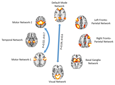 |
Functional connectivity between major resting state networks in Parkinson’s disease with Mild Cognitive Impairment
Karthik R Sreenivasan1, Xiaowei Zhuang1, Zhengshi Yang1, Zoltan Mari1, Jessica Caldwell1, Aaron Ritter1, Dietmar Cordes1, and Virendra Mishra1
1Cleveland Clinic Lou Ruvo Center for Brain Health, Las Vegas, NV, United States
The underlying cause of cognitive deficits in Parkinson’s disease patients with mild cognitive impairment (PD-MCI) is not well understood and there are no biomarkers to diagnose PD-MCI in early stages. The aim of the current study is to quantify the connectivity between the major resting state networks (RSNs) in PD–MCI using independent component analysis (ICA) and graph theory. Our results showed altered connectivity and reduced efficiency of the visual network which is implicated in several stages of PD and also showed that dysfunction exists in between large scale RSN connectivity which is correlated with behavioral scores.
|
|
3991.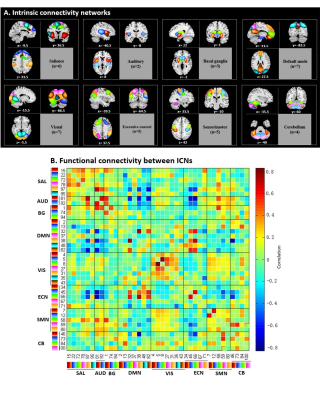 |
Different States of Dynamic Changes in Brain Functional Network Connectivity in Patients with Obsessive-compulsive Disorder
Hailong Li1, Xinyu Hu1, Xuan Bu1, Yingxue Gao1, Lianqing Zhang1, Lu Lu1, Shi Tang1, Yanlin Wang1, Yanchun Yang2, Qiyong Gong1, and Xiaoqi Huang1
1Huaxi MR Research Center (HMRRC), Functional and molecular imaging Key Laboratory of Sichuan Province, Department of Radiology, West China Hospital, Sichuan University, Chengdu, China, 2Department of Psychiatry, West China Hospital of Sichuan University, Chengdu, China
This study explored the temporal variability of functional connectivity among the brain networks in obsessive-compulsive disorder (OCD) patients. First, dynamic analysis suggested four distinct connectivity states. Relative to controls, OCD patients showed more frequent and larger-scale within and between-network connectivity changes at state II, whereas less frequent and smaller-scale between-network connectivity alterations appeared at state I and IV. Second, this study suggested a new network dysconnectivity model between SMN, DMN, cerebellum and visual network for OCD patients. These findings demonstrated the dynamic changes of brain network connectivity patterns in OCD, providing a new insight into OCD-related brain functional network alterations.
|
|
3992.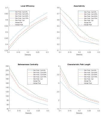 |
Simulation of postsurgical functional connectome from presurgical resting-state fMRI: optimization and validation
Henry Szu-Meng Chen1, Vinodh Kumar2, Sujit S Prabhu3, Kyle R Noll4, Sherise D Ferguson3, Ganesh Rao3, Ping Hou1, Jason M Johnson2, and Ho-Ling Anthony Liu1
1Imaging Physics, UT MD Anderson Cancer Center, Houston, TX, United States, 2Neuroradiology, UT MD Anderson Cancer Center, Houston, TX, United States, 3Neurosurgery, UT MD Anderson Cancer Center, Houston, TX, United States, 4Neuropsychology, UT MD Anderson Cancer Center, Houston, TX, United States
Post glioma-resection functional connectomic measures were simulated by modifying the presurgical resting-state fMRI data using the resection margin in 13 patients. The simulation uses atlas based approaches that removes the signal contribution in the resected area based on the portion of parcel removed. The simulated connectomic measures were found to approximate the actual postsurgical connectomic measures. BN246 atlas appears to be more stable in predicting the changes in connectomic measures than AAL90 atlas. The results show that derivation of postsurgical functional connectomic measures from presurgical data is feasible.
|
|
3993.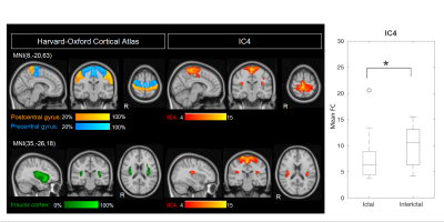 |
Functional connectivity during spontaneous migraine attacks compared to pain-free periods: a resting-state fMRI study
Raquel Pestana Araújo1, Patrícia Figueiredo1, Joana Pinto1, Pedro Vilela2, Isabel Pavão Martins3, and Raquel Gil-Gouveia2
1ISR-Lisboa/LARSyS and Department of Bioengineering, Instituto Superior Técnico, Universidade de Lisboa, Lisboa, Portugal, 2Hospital da Luz, Lisboa, Portugal, 3Department of Clinical Neurosciences, Faculdade de Medicina, Universidade de Lisboa, Lisboa, Portugal
Migraine is a severe neurological brain condition of cyclical nature, with intermittent attacks alternating with attack-free periods. In this work, we studied a group of patients with episodic migraine without aura during a spontaneous migraine attack and during a pain-free period, using BOLD resting-state fMRI. Results showed decreases in functional connectivity (FC) within a sensorimotor-insular cortex network and a left frontoparietal/executive control network. These FC changes were correlated with the attack duration and the pain intensity of the ongoing attack, respectively.
|
|
3994.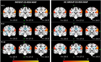 |
An ICA-based approach to study altered resting state functional networks in brain tumors
Manuela Moretto1,2, Erica Silvestri1,2, Marco Castellaro1,2, Mariagiulia Anglani3, Silvia Facchini1,4, Elena Monai1,4, Domenico D'Avella1,4, Alessandro Della Puppa5, Diego Cecchin1,6, Maurizio Corbetta1,4,7, and Alessandra Bertoldo1,2
1Padova Neuroscience Center, University of Padova, Padova, Italy, 2Department of Information Engineering, University of Padova, Padova, Italy, 3Neuroradiology Unit, University of Padova, Padova, Italy, 4Department of Neuroscience, University of Padova, Padova, Italy, 5Department of Neurosurgery, Neuroscience, Psychology, Pharmacology, and Child Health, University of Firenze, Firenze, Italy, 6Department of Medicine, Unit of Nuclear Medicine, University of Padova, Padova, Italy, 7Department of Neurology, Radiology, Neuroscience, Washington University School of Medicine, St Louis, MO, United States
Brain tumors can alter not only functions located in the perilesional area, but also the distal ones. Thus, the possibility to inform preoperatively surgeons about the state of preservation/alteration of a network could be a powerful aid for a better patient outcome. In this work we used independent component analysis (ICA) to map resting state networks (RSNs) at the single-subject level characterizing their alterations in terms of cosine similarity spatial patterns. Comparing the patient-specific spatial maps with those obtained for a group of healthy controls, we defined the presence of an alteration for each of the 44 analyzed RSNs.
|
|
3995.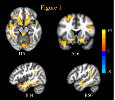 |
Investigating Mechanisms Underlying Autism Spectrum Disorders with Resting State fMRI Signal Complexity Metrics
Kaundinya Gopinath1, Elissar Andari1, and Larry Young1
1Emory University, Atlanta, GA, United States
Autism spectrum disorders (ASD) are characterized by impairments in social cognition, and oxytocin (OXT) system dysregulation. In this study, we employed resting state fMRI to examine brain function impairments in adult ASD, and the effects of OXT treatment. ASD patients exhibited increased fMRI signal complexity in social cognition and reward networks compared to healthy controls: indicating increased synaptic noise as a putative mechanism underlying ASD. Intranasal administration of OXT decreased synaptic noise in these regions, while increasing excitability of prefrontal cortex (PFC); thus indicating increased inhibitory control mediated through PFC as the mechanism underlying OXT induced brain rehabilitation in ASD.
|
|
3996.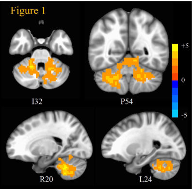 |
Resting state fMRI signal complexity metrics indicate cerebellar cholinergic system damage in Gulf War Illness
Kaundinya Gopinath1, Bruce Crosson1, Unal Sakoglu2, and Robert Haley3
1Emory University, Atlanta, GA, United States, 2University of Houston Clear-Lake, Houston, TX, United States, 3UT Southwestern Medical Center, Dallas, TX, United States
Around 200,000 veterans of the 1991 Gulf War (GW) suffer from GW illness (GWI), which is characterized by deficits in cognitive, emotion, perception and nociception domains. Previous studies have associated GWI with exposure to neurotoxic chemicals which impair the cholinergic system. Recently, an fMRI time-series signal complexity metric, multi-scale entropy (MSE) has been proposed as a potential biomarker of abnormal neural activity in brain disorders. In this study, we examined 23 GWI patients and 30 age-matched controls with resting state fMRI. GWI veterans exhibited abnormally increased MSE all across cerebellum, implicating cholinergic damage of cerebellum as a mechanism underlying GWI.
|
|
3997.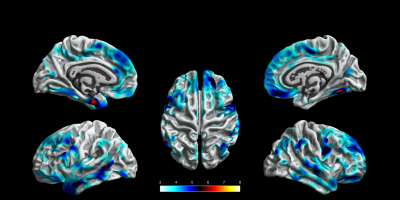 |
Surface-Based Morphometry of Brain in Benign Childhood Epilepsy with Centrotemporal Spikes
Zhengzhen Li1, Lisha Nie2, Xiaoxi Chen1, Heng Liu1, and Tijiang Zhang1
1Medical Imaging, the Affiliated Hospital of Zunyi Medical University, Zunyi, China, 2GE Healthcare,MR Research China, Beijing, China
This study investigated the cortical morphological changes of drug-naïve Benign childhood epilepsy with centrotemporal spikes (BECTS) using three-dimensional T1- weighted images(3D-T1WI) and explored the possible influence of cortical morphological changes on cognition.The results revealed aberrant morphology in thickness, gyrification and fractal dimension, in addition to Rolandic region. Cortical gyrification of right parahippocampal gyrus in BECTS may indirectly reflect the degree of language transmission damage or the result of contralateral compensation.
|
|
3998.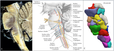 |
Functional Anatomy of Human Brainstem using resting state fMRI
Vinod Jangir Kumar1, Christian F. Beckmann2, Klaus Scheffler1, and Wolfgang Grodd3
1Department for High-field Magnetic Resonance, Max Planck Institute for Biological Cybernetics, Tuebingen, Germany, 2Donders Institute for Brain, Cognition, and Behavior, Centre for Cognitive Neuroimaging, Nijmegen, Netherlands, 3Max Planck Institute for Biological Cybernetics, Tuebingen, Germany Poster Permission Withheld
The brainstem engages in various brain functions. However, we still lack a detailed understanding of its underlying functional organization. Therefore, in this study, we analyzed 222300 fMRI scans of 62 subjects acquired at the HCP-7 Tesla project. We applied the instantaneous parcellation analysis (ICP) method and determined a reliable, reproducible, and stable functional anatomy of the brainstem. The results reveal that stable multi-scale functional anatomy exists in the brainstem. The spatial rich functional anatomy enables neuroscientists to better characterize brainstem organization and understand its function and role in various brain disorders.
|
|
3999.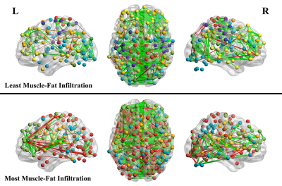 |
Resting-state Functional MRI Investigation of Whiplash Associated Disorder
James Higgins1, James Elliott2, and Todd Parrish1
1Northwestern University, Chicago, IL, United States, 2The University of Syndney, Sydney, Australia
Many individuals who suffer an initial whiplash injury develop a chronic condition known as whiplash associated disorder (WAD). In this study, 23 participants with WAD underwent resting-state fMRI along with a fat-water scan of the cervical region to assess muscle fat infiltration (MFI), which has been demonstrated as a marker of disorder severity. Brain network modularity was calculated from the RS-fMRI data and associations between modularity and clinical measures were investigated. An association was discovered between modularity and MFI which appeared to be independent of demographic variables as well as scan motion.
|
|
4000.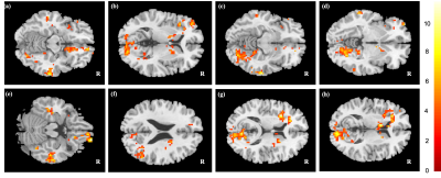 |
Mapping chemotherapy-induced brain functional network abnormality in breast cancer survivors
Kai-Yi Lin1, Vincent Chin-Hung Chen 2,3, Yuan-Hsiung Tsai 4, and Jun-Cheng Weng1,2,5
1Department of Medical Imaging and Radiological Sciences, Chang Gung University, Taoyuan, Taiwan, 2Department of Psychiatry, Chang Gung Memorial Hospital, Chiayi, Taiwan, 3School of Medicine, Chang Gung University, Taoyuan, Taiwan, 4Department of Diagnostic Radiology, Chang Gung Memorial Hospital, Chiayi, Taiwan, 5Medical Imaging Research Center, Institute for Radiological Research, Chang Gung University and Chang Gung Memorial Hospital at Linkou, Taoyuan, Taiwan
Chemo-brain is common among breast cancer survivors. However, some studies suggested cognitive function deficits may exist before chemotherapy initiation. Our study discovered the functional network alterations in women pre- (C-) and post-chemotherapy (C+) compared with health controls using resting-state functional MRI. The mfALFF showed changes in the prefrontal cortex, bilateral middle, right inferior temporal gyrus, right angular gyrus, left insula, and left caudate among three groups. Graph theoretical analysis demonstrated that the C+ group became inclined toward regular networks and the C- group became inclined toward random networks.
|
|
4001.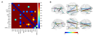 |
Altered brain language network in idiopathic peripheral facial paralysis patients with dysarthria
Wenwen Gao1, Xiaowei Han1, Haimei Li2, Yijiang Zhu3, Lei Du1, Li zhi Xie4, and Guolin Ma1
1China-Japan Friendship Hospital, Beijing, China, 2Fu Xing Hospital, Capital Medical University, Beijing, China, 3Anhui Provincial Hospital, Hefei, China, 4GE healthcare, China, Beijing, China
Dysarthria is a common symptom of facial paralysis (FP). The aim of this study was to investigate the functional alterations of the brain language network in FP patients with dysarthria using resting-state fMRI. We found that the functional connectivity between bilateral language regions was significantly decreased in these patients compared with healthy controls. The decrease of functional connectivity in the language network was positively correlated with the severity of oral paralysis in patients. To sum up, these data suggest that dysarthria caused by facial nerve paralysis may lead to a decrease of neural activity in the brain language network.
|
|
4002.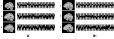 |
The Neural Basis for Olfactory Deficits in Alzheimer’s Disease
Peter James Castagna1, Rommy Elyan2, Paul Eslinger3, Qing Yang2, and Prasanna Karunanayaka2
1Psychiatry, Penn State University, Hershey, PA, United States, 2Radiology, Penn State University, Hershey, PA, United States, 3Neurology, Penn State University, Hershey, PA, United States
Deficits in odor-memory and odor-identification are among the first clinical symptoms of Alzheimer’s Disease (AD) and mild cognitive impairment (MCI); however, the neural basis of this observation remains unclear. In this study, we combined independent Component Analysis and olfactory functional magnetic resonance imaging to investigate the relationships between the olfactory network (ON) and the medial temporal lobe (MTL). Our study's results suggest that the functional connectivity between the MTL and ON may be aberrant in MCI and AD patients.
|
|
4003.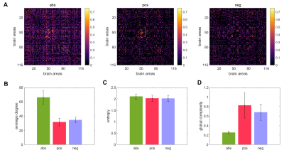 |
Estimating the complexity of fMRI-based brain networks
Roberto Sotero Sotero1, Lazaro Sanchez-Rodriguez1, and Narges Moradi1
1Department of Radiology, University of Calgary, Calgary, AB, Canada
Current measures of network complexity fail to capture the structural and functional diversity of brain networks. Here we use random walks processes to obtain a time series reflecting the complex structure of functional brain networks and use this time series to construct measures of local and global complexity. We found that complexity is significantly correlated to the strength of the connections in the network. For the positively correlated network this correlation is significantly weaker at the local scale compared to the global scale, whereas for the anticorrelation network the link is stronger at the local scale.
|
4004.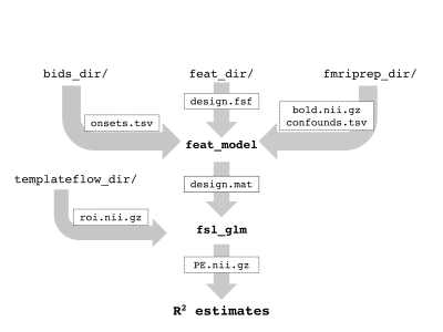 |
confounder: A BIDS/fMRIPrep app for efficiently assessing task-based GLM model fit with and without experimental confounds
Suzanne T Witt1, Kathryne Van Hedger1, Olivia Walton Stanley2, Ali Khan2, and Jörn Diedrichsen2
1BrainsCAN, University of Western Ontario, London, ON, Canada, 2University of Western Ontario, London, ON, Canada
Denoising task-based fMRI data is important for increasing SNR and has implications for data interpretation. There is not currently a method or tool available for directly comparing the relative impact of including experimental confounds in a first-level GLM. Here we present a new BIDS/fMRIPrep app, confounder, that allows users to efficiently assess model fit for first-level, task-based GLM models with and without experimental confounds. The app provides users with model-fit information such as regional R2, as well as additional information regarding data quality, such as the correlation between experimental confounds and predicted BOLD signal.
|
|
4005.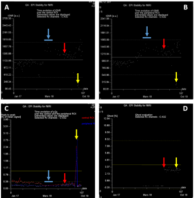 |
Fully automated fBIRN-like stability assessment: follow-up and case report at 7T
Franck Mauconduit1, Evelyn Eger2, Chantal Ginisty3, Valérie Berland3, Yann Lecomte3, Lionel Allirol3, Lucie Hertz-Pannier3, and Alexandre Vignaud1
1NeuroSpin (UNIRS) & Université Paris-Saclay, CEA, Gif-sur-Yvette, France, 2NeuroSpin (UNICOG) & Université Paris-Saclay, CEA, Gif-sur-Yvette, France, 3NeuroSpin (UNIACT) & Université Paris-Saclay, CEA, Gif-sur-Yvette, France
Functional Magnetic Resonance Imaging relies on weak blood oxygen level dependent signal variations. Therefore, it is extremely sensitive to MR scanner instabilities. A regular assessment of the device was shown to help preserving a good quality of fMRI data. It is also a powerful tool to compare systems. Here, we integrated a Quality Assessment based on fBIRN recommendations that can be automatically launched and reconstructed online to be able to immediately monitor system performances. We report cases where abnormal situations leading to suboptimal fMRI data were revealed which would have been missed if only relying on the manufacturer’s test service.
|
|
4006.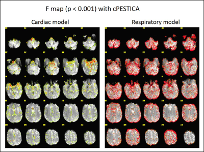 |
Physiologic noise correction for phase shifted cardiac fluctuation with the quality assurance of measured and estimated physiologic noise.
Wanyong Shin1 and Mark J. Lowe1
1Radiology, Cleveland Clinic, Cleveland, OH, United States
We have previously developed and publicly shared the physiologic noise estimation and removal software package, PESTICA (www.nitrc.org/projects/pestica). We present a new version of the PESTICA/RETROICOR software package, correcting cardiac phase shift and generating the goodness of fit with modeled physiologic noise and quality assurance of external physiologic signals based AFNI and Matlab. This study describes the updates, validation and demonstrate its usage.
|
|
4007.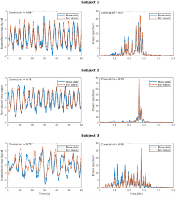 |
Correcting respiratory-related noise in brainstem fMRI using regressors derived from phase data
David Bancelin1, Pedro Lima Cardoso1, Beata Bachrata1,2, Andreas Ehrmann1, Siegfried Trattnig1,2, and Simon D. Robinson1,3,4
1High-Field MR Centre, Medical University of Vienna, Vienna, Austria, 2Christian Doppler Laboratory for Clinical Molecular MR Imaging, Vienna, Austria, 3Department of Neurology, Medical University of Graz, Graz, Austria, 4Centre for Advanced Imaging, University of Queensland, Queensland, Australia
Respiratory and cardiac data are generally used to properly account for the presence of physiological noise in fMRI data. Pneumatic belts can be unreliable, but we show that EPI phase data can be used to generate a reliable respiratory time series from which regressors are used in a GLM procedure to correct magnitude data. The efficacy of our method is compared with respect to (i) uncorrected magnitude data and (ii) magnitude data corrected using respiratory belt-derived regressors.
|
|
4008.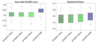 |
Optimum window-size in sliding-window dynamic functional connectivity analysis
Xiaowei Zhuang1, Zhengshi Yang1, Virendra Mishra1, Karthik Sreenivasan1, Bernick Charles1, and Dietmar Cordes1,2
1Lou Ruvo Center for Brain Health, Cleveland Clinic, Las Vegas, NV, United States, 2University of Colorado, Boulder, Boulder, CO, United States
In this abstract, we proposed an optimum window-size in a sliding-window approach for dynamic functional connectivity analysis. The proposed window-size was derived from the instantaneous period and energy of each intrinsic mode functions (IMF) obtained from empirical mode decomposition. IMFs track local periodic changes of non-stationary time series and therefore can capture subtle temporal variations. Using dynamic functional connectivity matrix computed with the proposed window-size as features, a higher accuracy was obtained in classifying cognitively impaired fighters from cognitively normal ones; and a larger behavioral variance was found in HCP data.
|
|
4009.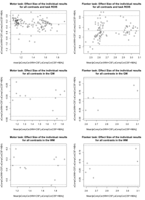 |
Optimizing aCompCor denoising in task fMRI: using WM as noise source area
Peter Van Schuerbeek1
1Radiology, UZ Brussel (VUB), Brussels, Belgium
Since neural BOLD responses were found in the white matter (WM), we hypothesized that using CSF and non-brain areas (NBA) as noise source areas, rather than WM and CSF, to denoise fMRI data with aCompCor, will positively affect the results of task fMRI studies. This hypothesis was tested by comparing the results of processing 2 task fMRI datasets once denoised with WM and CSF and once with CSF and NBA as noise source areas. Our results showed that denoising with aCompCor using CSF and NBA leads to higher effect sizes and a better overlap between individual activation maps.
|
|
4010.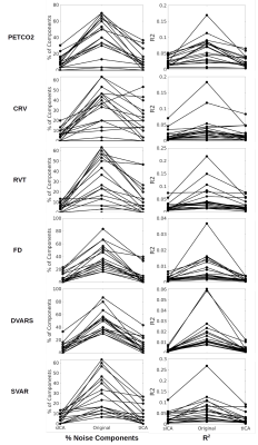 |
Performance of Temporal and Spatial ICA in Identifying and Removing Physiological Artifacts in resting-state fMRI
Ali Golestani1 and J Jean Chen2,3
1University of Toronto, Toronto, ON, Canada, 2Rotman Research Institute at Baycrest, Toronto, ON, Canada, 3Department of Biophysics, University of Toronto, Toronto, ON, Canada
Independent component analysis (ICA) has been used to identify and remove confounding factors in fMRI data. While spatial ICA (sICA) is more typical for fMRI, temporal ICA (tICA) can better distinguish between temporally independent but spatially overlapping components compared to sICA. We investigate if tICA and sICA perform differently in identifying different physiological components of the resting-state fMRI (rs-fMRI) signal. Our results show that noise sources with widespread effects across brain (e.g. PETCO2, FD, DVARS) are better identified by tICA. However, sICA performed significantly better than tICA in removing the effects of affine head motion parameters.
|
|
4011.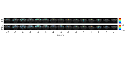 |
Comparison of Phase Synchrony Analysis and Pearson’s Correlation Coefficient in Resting State fMRI of Rat Brain
Yi-Cheng Wang1, Chia-Yu Huang1, Wen-Yen Chai2, Mey-Yu Yeh1, Hao-Li Liu2, and Fu-Nien Wang1
1National Tsing Hua University, Hsinchu, Taiwan, 2Chang Gung University, Taoyuan, Taiwan
Phase synchrony has been considered as an alternative method to measure connectivity in resting state functional MRI. Our results showed that the data with the correlation coefficient are monotonically increasing, but only when the correlation coefficient is larger than 0.4, phase synchrony and correlation coefficient showed linear dependence. The mappings of default mode network based on two analysis methods are similar in rat brains. However, care should be taken when interpreting data with lower phase synchrony (<0.4), since the phase synchrony of noise is also distributed in the range.
|
|
4012.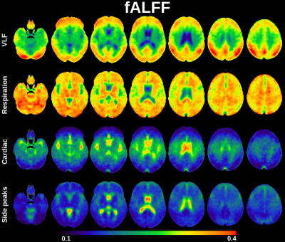 |
Amplitude modulation of cardiovascular brain pulsations
Lauri Raitamaa1, Vesa Korhonen1, Niko Huotari1, and Vesa Kiviniemi1
1Research Unit of Medical Imaging Physics and Technology (MIPT), University of Oulu, Oulu, Finland Ultra-fast 10 Hz whole brain fMRI with magnetic resonance encephalography (MREG) enables separation of cardiovascular waves from brain pulsation without temporal aliasing. This enables analysis of spectral characteristics of respiration and cardiovascular pulsations by extending ALFF and fALFF methods to cardiovascular and respiration frequencies . Modulation of the cardiovascular pulsation has been detected in ECG and SpO2 and in this study show the modulation of the cardiovascular brain pulsation in 3D brain in healthy controls and how age and blood pressure affects the modulation. |
|
4013.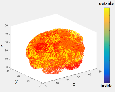 |
Classification of fMRI Study Participants Using Space-Filling-Curve Orderings of fMRI Brain Activation Maps
Unal Sakoglu1 and Lohit Ravi Teja Bhupati1
1Computer Engineering, University of Houston - Clear Lake, Houston, TX, United States
In this work, we develop a 3-D to 1-D ordering methodology for fMRI data, using a space filling curve, which is adaptive to brain's shape. We apply this ordering to fMRI activation maps from a schizophrenia study, compress the data, obtain features, and perform classification of schizophrenia vs normal controls. We compare the classification results with those of linear ordering, which has been the traditional method to convert the 3D fMRI data to 1D.
|
|
4014.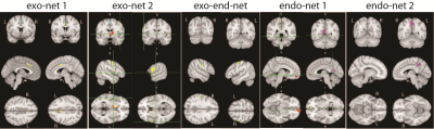 |
Brain-to-brain communication during eye contact: A data-driven approach based on the dfMRI
Ray Lee1, Nim Tottenham2, and Paul Sajda3
1Zuckerman Institute, Columbia University, New York, NY, United States, 2Psychology, Columbia University, New York, NY, United States, 3Biomedical engineering, Columbia University, New York, NY, United States
A comprehensive picture of brain-to-brain interaction during eye contact is presented at the brain network level. It consists of five dyadic brain networks (two exogenous, two endogenous, and one exogenous-to-endogenous network), which are derived from a novel data-driven approach based on the dyadic fMRI data. Our experimental results not only explicitly map out the intricate relations between imitation, empathy, mentalization, and face cognization network during eye contact, but also provide a quantitative measure on the information transmission between dyads. Thus, it effectively established the foundation for quantifying human social communication with dyadic fMRI.
|
|
4015.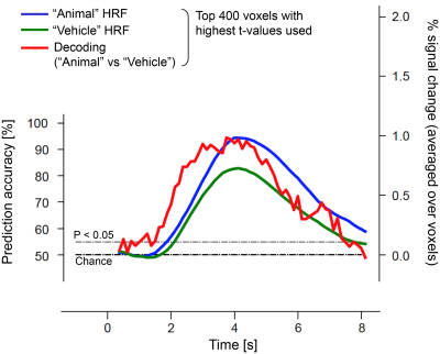 |
Event-related decoding of visual stimulus information using short-TR BOLD fMRI at 7T
Yoichi Miyawaki1,2,3, Daniel A Handwerker3, Javier Gonzalez-Castillo3, Laurentius Huber3,4, Arman Khojandi3, Yuhui Chai3, and Peter A Bandettini3
1The University of Electro-Communications, Tokyo, Japan, 2JST PRESTO, Tokyo, Japan, 3National Institutes of Mental Health, BETHESDA, MD, United States, 4University of Maastricht, Maastricht, Netherlands
Spatio-temporal dynamics of neural activity plays a pivotal role in forming our perception. Ultra-high field fMRI allows us to visualize fine spatial patterns of neural activity but little is known about its dynamics because it is typically believed to lack sufficient temporal resolution - due either to scanner or hemodynamic limitations. Here we demonstrate that event-related fMRI and multivariate pattern analyses allows us to decode visual information faster than the hemodynamic response, suggesting possibility to analyze neural information representation at high temporal resolution.
|
|
4016.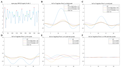 |
Voxel-wise Characterization of Hemodynamic Response Function in Brain White Matter Using Resting-State fMRI
Ting Wang1,2, D. Mitchell Wilkes3, Muwei Li2,4, Xi Wu1, John C. Gore2,4,5, and Zhaohua Ding2,3,5
1Department of Computer Science, Chengdu University of Information Technology, Chengdu, China, 2Institute of Imaging Science, Vanderbilt University, Nashville, TN, United States, 3Electrical Engineering & Computer Science, Vanderbilt University, Nashville, TN, United States, 4Department of Radiology and Radiological Sciences, Vanderbilt University Medical Center, Nashville, TN, United States, 5Department of Biomedical Engineering, Vanderbilt University, Nashville, TN, United States
This study investigated the hemodynamic response function (HRF) of BOLD effects in white matter (WM) voxels obtained from fMRI in a resting-state. It was found that the WM HRF can be derived by reference to GM avalanche activities. The derived resting-state WM HRFs have low magnitudes, delayed onsets, and prolonged initial dips compared with GM HRF. The time delay distribution patterns and correlation coefficient profiles for WM voxels depend on the selection of the reference GM region. These findings suggest that fMRI signals in WM are associated with those in GM and encode neural activities.
|
|
4017. |
BOLD signal simulation and fMRI quality control base on an active phantom: a preliminary study
Tiao Chen1, Jianfeng Qiu2, Yue Zhao3, Chuntao Jia3, and Zilong Yuan4
1Medical Engineering and Technology Research Center, Shandong First Medical University & Shandong Academy of Medical Sciences, Taian, China, 2Shandong First Medical University & Shandong Academy of Medical Sciences, Taian, China, 3Shandong University of Science and Technology, Qingdao, China, 4Hubei Cancer Hospital, Tongji Medical College, Huazhong University of Science and Technology, Wuhan, China
The purpose of this study was to design and manufacture an active BOLD simulation phantom, thus testing the stability and repeatability of the phantom. Three scanners were used to test the stability and repeatability of the BOLD signal detection of the phantom. It was found that the phantom had good stability and repeatability. The phantom can stably quantify the BOLD signal for fMRI quality control in different scanners. Consequently, the phantom can stably quantify the BOLD signal for fMRI quality control in different scanners.
|