
Digital Poster Session
Interventional: Interventional MR and Safety Issues
Interventional MR and Safety Issues
4111 -4125 Interventional MR and Safety Issues - Interventional: MR-Guided HIFU
4126 -4140 Interventional MR and Safety Issues - MR Thermometry Methods
4141 -4155 Interventional MR and Safety Issues - Interventional Devices & Methods
4156 -4170 Interventional MR and Safety Issues - MRI Safety & Intervention
4111.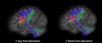 |
A method for post-surgical evaluation of targeting accuracy in transcranial focused ultrasound thalamic ablation
Benjamin T Newman1,2, Ana Untaroiu1, and T. Jason Druzgal1,2
1Department of Radiology & Medical Imaging, Division of Neuroradiology, University of Virginia Health System, University of Virginia, Charlottesville, VA, United States, 2Brain Institute, University of Virginia, Charlottesville, VA, United States
MR-guided focused ultrasound ablation targets small thalamic nuclei that are frequently difficult to distinguish from surrounding tissue. The ability to accurately evaluate the success in ablating the target nuclei is essential for predicting treatment outcome and evaluating surgical performance. We show microstructural changes in the thalamus that may prevent accurate post-surgical tractography. We propose a straightforward technique that reconstructs neuronal connections in the pre-surgical brain using the post-operative lesioned area as a seed-region for constrained spherical deconvolution based tractography. The proportion of tracts leading to regions known to be connected to the target nuclei is then evaluated.
|
|
 |
4112.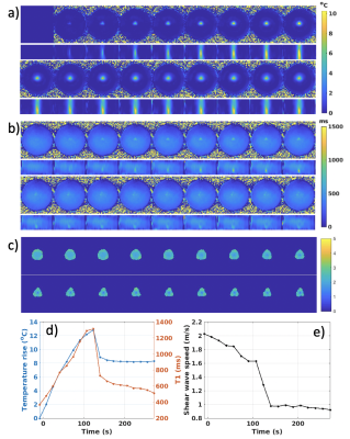 |
An efficient approach for simultaneous PRF-T1 MR thermometry and shear wave elastography to monitor MR-guided focused ultrasound therapies
Henrik Odéen1, Lorne Hofstetter1, and Dennis L Parker1
1Radiology and Imaging Sciences, University of Utah, Salt Lake City, UT, United States
Treatment endpoint assessment in MRgFUS is commonly performed by investigating non-perfused volume or accumulated thermal dose, but these measurements can be inaccurate and have low precision immediately after treatment and can be tissue type dependent. In this work we present a pulse sequence which can provide dynamic measurements of PRF thermometry (and hence also thermal dose), T1 and tissue mechanical properties through shear wave speed measurements. PRF and T1 can provide complete MR thermometry in all tissue types (aqueous and adipose), and together all parameters are useful in treatment endpoint assessment.
|
4113.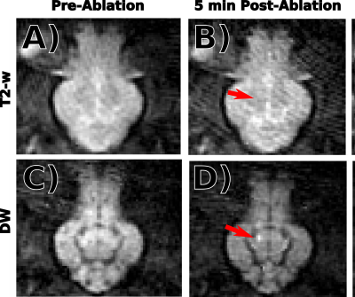 |
Intraoperative Diffusion Weighted MR Image Contrast during Focused Ultrasound Ablation in a Preclinical Swine Model
Steven P Allen1, Francesco Prada2, Jeremy Gatesman3, Xue Feng1, Helen Sporkin1, Yekaterina Gilbo1, Kim Butts Pauly4, and Craig H Meyer1,5
1Biomedical Engineering, University of Virginia, Charlottesville, VA, United States, 2Neurosurgery, University of Virginia, Charlottesville, VA, United States, 3Center for Comparative Medicine, University of Virginia, Charlottesville, VA, United States, 4Radiology, Stanford University, Stanford, CA, United States, 5Radiology, University of Virginia, Charlottesville, VA, United States This abstract presents a preclinical study of intraoperative diffusion weighted imaging (DWI) of focused ultrasound thermal lesions in the thalamus using a novel acquisition sequence. We found that DW imaging can visualize thermal lesions within 10 minutes of ablation. Our study indicates that MR-guided focused ultrasound lesions present a rapid and persistent decrease in apparent diffusion coefficient within minutes after ablation and that diffusion-based lesion classification methods can outperform T2-w based methods.
|
|
4114.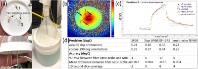 |
MR imaging Evaluation of an Interstitial Focused Ultrasound Probe used in Robotically Assisted Brain Ablation Procedures
MATTHEW TARASEK1, Eric Fiveland1, Chitresh Bhushan1, Goutam Ghoshal2, Tamas Heffter2, Katie Gandomi3, Paulo Alberto Carvalho3, Christopher Nycz3, Teresa Maiette4, Zahabiya Campwala4, Erin Jeannotte4, Michael Staudt4, E Clif Burdette2,
Gregory Fischer3, Julie Pilitsis4, and Desmond Yeo1
1GE Global Research, Niskayuna, NY, United States, 2Acoustic MedSystems Inc., Savoy, IL, United States, 3WPI, Worcester, MA, United States, 4Neurosurgery, Albany Medical College, Albany, NY, United States
In this work we evaluate a newly developed catheter-based therapeutic ultrasound (US) probe used for interstitial MR-guided high intensity focused US (iMRgFUS) procedures. Specifically, we optimize and down-select MR thermal imaging protocols in order to achieve maximum temperature measurement precision and volume coverage in the presence of image artifacts arising from the iMRgFUS probe design and operation.
|
|
4115.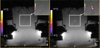 |
Enhancement of the MRgHIFU therapy using endovascular sono-sensitizers demonstrated in an experimental model of perfused tissue
Orane Lorton1, Ryan Holman1, Pauline Guillemin1, Stéphane Desgranges2, Laura Gui1, Lindsey A. Crowe3, François Lazeyras3, Antonio Nastasi4, Christiane Contino-Pépin2, and Rares Salomir1,3
1Image Guided Interventions Laboratory,University of Geneva, Faculty of Medicine, Geneva, Switzerland, 2University of Avignon, CBSA-IBMM (UMR5247), Avignon, France, 3Radiology Department, University Hospitals of Geneva,, Geneva, Switzerland, 4Research and Development Laboratory, Visceral and Transplantation Service, University Hospital Geneva, Geneva, Switzerland
Magnetic Resonance guided High Intensity Focused Ultrasound (MRgHIFU) is accepted for the non-invasive ablation of localized tumors. Thermal contrast between the target tissue and pre/post focal tissues can be improved for highly perfused tumors by injection of liquid core micro-droplets, used as endovascular sono-sensitizers. We are aiming to provide a substitution model for living organs or perfused ex vivo organs to facilitate the investigation of new sono-sensitizers and sonication paradigms. We are reporting an experimental model that is suitable for MR-guided HIFU studies, offering in situ tunable perfusion rate, multi-compartment structure, and tunable concentration of sono-sensitizers and dissolved gases.
|
|
4116.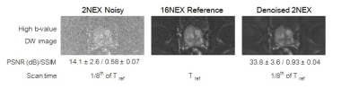 |
Feasibility of rapid diffusion-weighted imaging for monitoring of MR-guided prostate high intensity focused ultrasound ablation
Elena Kaye1 and Oguz Akin2
1Medical Physics, Memorial Sloan Kettering Cancer Center, New York, NY, United States, 2Radiology, Memorial Sloan Kettering Cancer Center, New York, NY, United States Poster Permission Withheld
MR-guided high-intensity focused ultrasound (HIFU) treatment of prostate cancer is a promising non-invasive approach. The outcomes of these treatments can be improved with more accurate visualization of ablative necrosis surrounding a tumor. Diffusion-weighted imaging (DWI) could provide valuable information about tissue viability without injection of contrast. However, currently DWI requires high number of excitations (NEX), averages, takes several minutes and is not practical for intraprocedural monitoring. To make DWI a practical tool for monitoring of prostate HIFU, we propose to replace high NEX DWI acquisitions, with NEX=2 acquisitions and subsequent deep-learning denoising of these image.
|
|
4117.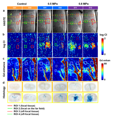 |
Monitoring of Focused Ultrasound Induced Stable Cavitation by Using Magnetic Resonance Imaging: In Vivo Experiments
Yu-Ting Jiang1, Cheng-Tao Ho1, Po-Hung Hsu2, Hao-Li Liu3, Chih-Kuang Yeh1, and Hsu-Hsia Peng1
1Department of Biomedical Engineering and Environmental Sciences, National Tsing Hua University, Hsinchu, Taiwan, 2Center for Advanced Molecular Imaging and Translation, Chang Gung Memorial Hospital, Taoyuan, Taiwan, 3Department of Electrical Engineering, Chang-gung University, Taoyuan, Taiwan
We aimed to monitor the focused ultrasound (FUS)-induced stable cavitation (SC) by using single-shot turbo spin-echo based sequence in in vivo experiments. The FUS were transmitted to the cortex ~2‐mm beneath the superior sagittal sinus of rats with acoustic pressure=0.5 or 0.8 MPa. A cavitation index map was computed to highlight the occurrence of SC at focus. In conclusion, the simultaneous acquisitions of spin-echo based images during FUS transmission and the proposed CI map can immediately localize the position occurring SC. The relevant information can be feedbacked to FUS facility for improving the procedure of BBB opening.
|
|
4118.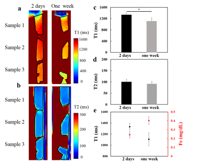 |
Evaluate thrombolysis effect of focused ultrasound in different thrombus ages by MRI
Yu-Ting Jiang1, Po-Hung Hsu2, Hao-Li Liu3, Chih-Kuang Yeh1, and Hsu-Hsia Peng1
1Department of Biomedical Engineering and Environmental Sciences, National Tsing Hua University, Hsinchu, Taiwan, 2Center for Advanced Molecular Imaging and Translation, Chang Gung Memorial Hospital, Taoyuan, Taiwan, 3Department of Electrical Engineering, Chang-gung University, Taoyuan, Taiwan
We aimed to evaluate the thrombolysis effect with variant focused ultrasound (FUS) and microbubbles (MB) conditions in different thrombus ages by MRI. Compared to one-week thrombi, 2-days thrombi presented longer T1 and similar T2, while the total iron content increased with thrombus ages. However, with identical thrombolysis conditions, there is no significant difference of weight loss between two different thrombi ages groups. In conclusion, to characterize the T1 and T2 relaxation times of thrombi with different ages can be helpful for evaluation of the thrombolysis effect with variant UK and FUS conditions in different thrombi ages.
|
|
4119.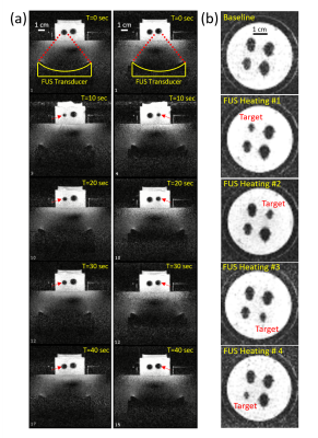 |
Selective fast focal switching of high-contrast-ratio magnetocaloric MRI labels with focused ultrasound
G. Wilson Miller1,2, Yekaterina Gilbo2, Stephen Dodd3, Alan P Koretsky3, Hatem ElBidweihy4, and Mladen Barbic5
1Radiology and Medical Imaging, University of Virginia, Charlottesville, VA, United States, 2Biomedical Engineering, University of Virginia, Charlottesville, VA, United States, 3National Institutes of Health, Bethesda, MD, United States, 4United States Naval Academy, Annapolis, MD, United States, 5HHMI - Janelia Research Campus, Ashburn, VA, United States
We describe the use of MRI-compatible focused ultrasound (FUS) for selective fast focal switching of high-contrast-ratio magnetocaloric MRI particle labels. Lanthanum-Iron-Silicon (La-Fe-Si) particles that have sharp first-order magnetic phase transitions at physiological temperatures and MRI field strengths were used as MRI labels. Non-invasive rapid thermal switching of these magnetocaloric MRI particle labels with focused ultrasound was clearly demonstrated at physiological temperatures within a clinical 1.5T MR scanner. We have thereby shown that high differential contrast ratio magnetocaloric MRI labels can be switched non-invasively, rapidly, and selectively under conditions relevant to clinical MRI.
|
|
4120.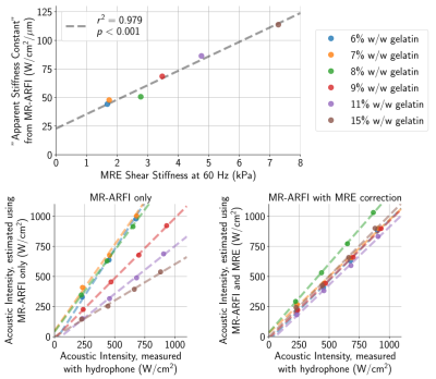 |
Improving in situ acoustic intensity estimation by augmenting MR acoustic radiation force imaging with MR elastography
Ningrui Li1, Pooja Gaur2, and Kim Butts Pauly2
1Electrical Engineering, Stanford University, Stanford, CA, United States, 2Radiology, Stanford University, Stanford, CA, United States
MR acoustic radiation force imaging (MR-ARFI) can be used to localize the focal spot for non-thermal transcranial ultrasound therapies. The acoustic radiation force is proportional to the applied acoustic intensity, meaning that tissue displacements measured with MR-ARFI can potentially be used to estimate the acoustic intensity at the target. However, variable brain stiffness is an obstacle to obtaining accurate acoustic intensity estimates. Using gelatin phantoms with varying stiffnesses, we demonstrate that stiffness information from MR elastography can be used in combination with MR-ARFI to improve in situ estimates of acoustic intensity. This could enable safer and more effective treatments.
|
|
4121.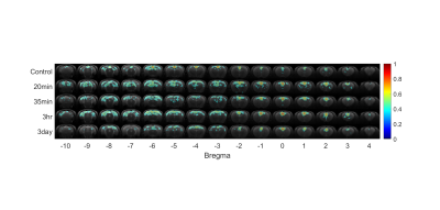 |
Focused Ultrasound Enhances Connectivity of Default Mode Network in Rat Brain
Chia-Yu Huang1, Yi-Cheng Wang1, Wen-Yen Chai2, Sheng-Min Huang1, Hao-Li Liu2, and Fu-Nien Wang1
1National Tsing Hua University, Hsinchu, Taiwan, 2Chang Gung University, Taoyuan, Taiwan
Focused ultrasound is a noninvasive, deep-penetrating, and spatially well-defined modality to modulate the neural activity. In this study, we used resting state functional MRI (rs-fMRI) to investigate the functionally connectivity after stimulation in the brain. Rats underwent focused ultrasound stimulation targeted unilaterally to the ventral posteromedial and ventral posterolateral thalamic nuclei (VPM/VPL) in the left thalamic region. The default mode network (DMN) showed hyper-correlation after sonication, and as time proceed, the correlation has a tendency to decline slowly in about 3 days.
|
|
4122.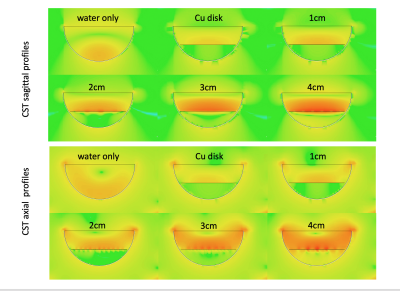 |
Designing RF Screens for MRI guided Fucused Ultrasound Applications
Rock Hadley1, Henrik Odeen2, Robb Merrill2, Sam Adams2, Viola Rieke2, Allison Payne2, and Dennis Parker2
1Radiology and Imaging Sciences, University of Utah, Salt Lake City, UT, United States, 2University of Utah, Salt Lake City, UT, United States
This work evaluates screens with different hole size in their ability to pass ultrasound and their ability to shield RF in a Transcranial MR guided Focused Ultrasound experiment. Hydrophone and radiation force balance studies were used to measure attenuation and peak pressure when passing ultrasound through the screens compared to water-only measurements. Electromagnetic simulations and MRI experiments were performed to measure RF shielding effectiveness of the screens by comparing their ability to change known artifacts in a transcranial transducer compared to a solid conductor.
|
|
4123.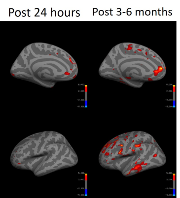 |
Brain Volumetric Changes following MR Guided Focused Ultrasound Treatment for Parkinson’s Disease
Justin Schumacher1, Li Jiang2, Jiachen Zhuo2, Howard Eisenberg3, Paul Fishman4, Dheeraj Gandhi2, and Rao Gullapalli2
1University of Maryland School of Medicine, Baltimore, MD, United States, 2Diagnostic Radiology and Nuclear Medicine, University of Maryland School of Medicine, Baltimore, MD, United States, 3Neurosurgery, University of Maryland School of Medicine, Baltimore, MD, United States, 4Neurology, University of Maryland School of Medicine, Baltimore, MD, United States
MR Guided Focused Ultrasound (MRgFUS) is a novel technique used to noninvasively lesion the brain for treatment of Parkinson’s Disease (PD). In this study 12 PD patients with asymmetric motor symptoms underwent MRgFUS pallidotomy and cortical thicknesses and subcortical volumes were measured pre and post treatment. Subcortical gray matter volume increases were observed 24 hours post treatment with bilateral decreases in basal ganglia gray matter volume 3-6 months post treatment. Cortical thicknesses decreases were observed in the frontal and temporal cortices of right-sided treated patients 3-6 months post treatment. These regions may play a role in the management of dyskinesias.
|
|
4124. |
MR guided blood-brain-barrier opening induced by focus ultrasound on Non-human primate
Hui Zhou1,2, Yang Liu3, Chao Zou1, Xiaojing Long3, Xin Liu1, and Hairong Zheng1
1Shenzhen Institutes of Advanced Technology,Chinese Academy of Sciences, Shenzhen, China, 2The Shenzhen College of Advanced Technology, University of Chinese Academy of Sciences, Shenzhen, China, 3Research center for medical AI, Shenzhen Institutes of Advanced Technology, Chinese Academy of Sciences, Shenzhen, China
Magnetic Resonance Imaging(MRI) is highly recommended for estimation of the feasibility and safety of BBB opening. We opened BBB in three different deep brain areas of an non-human primate with a single element ultrasound transducer working at 300kHz without detectable edema, hemorrhage and abnormal behavior. We found that the BBB in gray matter was easier to be disrupted than that in white matter, suggesting that for a local BBB opening in deep brain on non-human primate and human subjects, array transduces with relative large assemble surface are required to avoid the BBB disruption in cerebral cortex.
|
|
4125. |
Effects of mild hyperthermia and pulsed high intensity focused ultrasound in a mouse models of Pancreatic Ductal Adenocarcinoma
Ravneet Vohra1, Yak-Nam Wang2, Helena Son3, Stephanie Totten3, Joo Ha Hwang4, and Donghoon Lee1
1Radiology, University of Washington, Seattle, WA, United States, 2Applied Physics Laboratory, University of Washington, Seattle, WA, United States, 3Division of Gastroenterology, University of Washington, Seattle, WA, United States, 4Medicine, Stanford University, Stanford, CA, United States
Pancreatic cancer is expected to become the second leading cause of the cancer related deaths in the USA by 2020. The ineffectiveness of conventional chemotherapeutics in Pancreatic Ductal Adenocarcinoma (PDA) is thought to be largely due to the extensive stromal desmoplasia which inadvertently increases the interstitial fluid pressure. High intensity focused ultrasound (HIFU) sonication was performed with temperature monitored by MRI. The aim of this study was to develop a target treatment of pulsed HIFU treatment combined with mild hyperthermia on PDA and to monitor the response to the therapy using multi-parametric MRI as a non-invasive biomarker.
|
 Watch the Video
 View the Poster
4126. |
MR thermometry based on PRF using echo planar time-resolved imaging (EPTI)
Wending Tang1, Xing Wei2, Muheng Li1, Fuyixue Wang3,4, Zijing Dong3,5, Danna Wei6, Kawin Setsompop3,4,5, and Karen Ying1
1Department of Engineering Physics, Tsinghua University, Beijing, China, 2Orthopedic Department, Aerospace Center Hospital, Beijing, China, 3Athinoula A. Martinos Center for Biomedical Imaging, Massachusetts General Hospital, Charlestown, MA, United States, 4Harvard-MIT Health Sciences and Technology, Massachusetts Institute of Technology, Cambridge, MA, United States, 5Department of Electrical Engineering and Computer Science, Massachusetts Institute of Technology, Cambridge, MA, United States, 6Department of Biomedical Engineering, School of Medicine, Tsinghua University, Beijing, China
Proton resonance frequency (PRF) of water protons is widely utilized in MR thermometry. EPI sequence is often used to speed up the imaging rate, but suffers from geometric distortion and blurring. Here we proposed a rapid temperature mapping method through the EPTI sequence. EPTI sequence is a new multi-shot EPI technique capable of rapidly obtaining distortion-free images. Via this method, greater speed is achieved for monitoring temperature, which makes real-time temperature mapping possible.
|
|
4127. |
3D PRF Thermometry of the Liver
Waqas Majeed1, Sunil Patil1, Pan Su1, Rainer Schneider2, Henrik Odéen3, Dennis L. Parker3, John Roberts3, Jiachen Zhuo4, and Himanshu Bhat1
1Siemens Medical Solutions USA Inc., Malvern, PA, United States, 2Siemens Healthcare GmbH, Erlangen, Germany, 3Department of Radiology and Imaging Sciences, University of Utah, Salt Lake City, UT, United States, 4Department of Diagnostic Radiology and Nuclear Medicine, University of Maryland School of Medicine, Baltimore, MD, United States
Anatomical misregistration and background phase variation due to physiological motion are two major sources of inaccuracy in PRF thermometry of the liver. We propose a pipeline consisting of 3D segmented EPI acquisition, 3D deformable motion correction and PCA-based background phase correction for liver thermometry. 3D acquisition results in an increase in SNR and makes 3D registration possible. PCA-based background phase correction improves ΔT quality over a more commonly used dictionary-based method. The proposed pipeline was used to achieve 75th percentile ΔT bias and standard deviation values of 1.70°C and 1.65°C respectively over whole-liver in one healthy volunteer.
|
|
4128.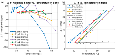 |
Development of T1-based Skull Thermometry using a 3D Spiral Ultra-Short Echo Time Sequence
Yekaterina K Gilbo1, Helen Sporkin2, Samuel W Fielden3, John P Mugler4, Grady W Miller4, Steven P Allen2, and Craig H Meyer2
1University of Virginia, Charlottesville, VA, United States, 2Biomedical Engineering, University of Virginia, Charlottesville, VA, United States, 3Department of Imaging Sciences & Innovation, Geisenger, Danville, PA, United States, 4Radiology and Medical Imaging, University of Virginia, Charlottesville, VA, United States
Skull temperature monitoring is important for MR-guided focused ultrasound (MRgFUS), as the skull is highly absorptive to acoustic energy. T1 thermometry uses T1 mapping to observe a linear increase in T1 with temperature but requires long acquisitions. Here we compare T1-weighted thermometry with T1 thermometry using a 3D spiral ultra-short-echo time sequence and show that T1 thermometry is feasible, but requires further acceleration, whereas T1-weighted thermometry results are nonlinear and inconsistent.
|
|
4129. |
On-the-fly 3D images generation from a single k-space readout using a pre-learned spatial subspace: Towards MR-guided therapy and intervention
Pei Han1,2, Fei Han3, Debiao Li1,2,4, Anthony Christodoulou2, and Zhaoyang Fan1,2,4
1Department of Bioengineering, UCLA, Los Angeles, CA, United States, 2Biomedical Imaging Research Institute, Cedars-Sinai Medical Center, Los Angeles, CA, United States, 3Siemens Healthineers, Los Angeles, CA, United States, 4Department of Medicine, UCLA, Los Angeles, CA, United States
A new real-time imaging scheme is proposed based on low-rank spatiotemporal decomposition. Briefly, a high-quality spatial subspace and a direct linear mapping from k-space navigator data to subspace coordinates are first learned from a “demo” scan. In the subsequent “live” scan, successive real-time images can be generated by a fast matrix multiplication procedure on a single instance of the k-space navigator readout (e.g. a single k-space line), which can be acquired at a high temporal rate. The method was demonstrated in vivo using a T1-T2 abdominal multitasking sequence.
|
|
4130.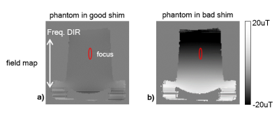 |
A k-space reconstruction method to increase the accuracy of PRF thermometry measurements
Shenyan Zong1,2, Chang-Sheng Mei3, Guofeng Shen1, and Bruno Madore2
1Biomedical Instrument Institute, School of Biomedical Engineering, Shanghai Jiao Tong University, Shanghai, China, 2Department of Radiology, Brigham and Women's Hospital, Harvard Medical School, Boston, MA, United States, 3Department of Physics, Soochow University, Taipei, Taiwan
It has been shown that spatial gradients in the background phase of an image can cause artifacts/errors in MRI temperature measurement. In the present work, a reconstruction method called 'k-space energy spectrum analysis' (KESA), originally developed in the context of partial-Fourier imaging, is adapted and tested here for its ability to correct for this particular problem. Compared to the previously-published image-based method already available for this purpose, the present k-space based method proved considerably more robust in the presence of high levels of noise, as tested with phantom data.
|
|
4131.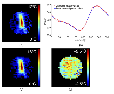 |
Reference-free PRFS thermometry using 3D spherical harmonic functions
Laura Gui1, Orane Lorton1, Max Scheffler2, and Rares Salomir1,2
1Image Guided Interventions Laboratory, Faculty of Medicine, University of Geneva, Geneva, Switzerland, 2Department of Radiology, University Hospitals of Geneva, Geneva, Switzerland
Magnetic resonance thermometry via the classic time-referenced PRFS method is perturbed by tissue motion and external magnetic field perturbations. We extend an existing 2-D reference-free PRFS thermometry method to 3-D data, by performing near-harmonic reconstruction of the background phase inside a sphere. Ex-vivo HIFU sonication experiments and in-vivo “zero-measurement” of a human brain showed good method accuracy and precision, fast computation times and similar results with the time-referenced PRFS method for motion-free scenarios. This study demonstrates the method’s feasibility for real time thermometry of moving tissues.
|
|
4132.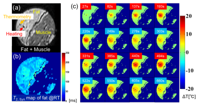 |
Thermometry of Fat-Water-mixed Tissues based on Synthesized Temperature Dependence of T2's of Methylene and Methyl in conjunction with PRF of water
Kagayaki Kuroda1, Satoshi Yatsushiro1, Yuta Hiranobu1, Takuya Watanabe1, Toru Niwa2, and Yutaka Imai2
1Department of Human and Information Sciences, School of Information Science and Technology, Tokai University, Hiratsuka, Japan, 2Department of Radiology, School of Medicine, Tokai University, Isehara, Japan
An practical MR thermometry technique for fat-water mixed tissues was proposed. Fatty tissue temperature was quantified by T2 synthesized from T2’s of methylene chain and terminal methyl obtained by chemical shift suppression of water and surrounding fatty acid components. While the aqueous tissue temperature was estimated by PRF shift of water proton. Those maps were then integrated into one based on the fat and water contents in each voxel. The resultant in vitro images demonstrated the usefulness of the technique.
|
|
4133.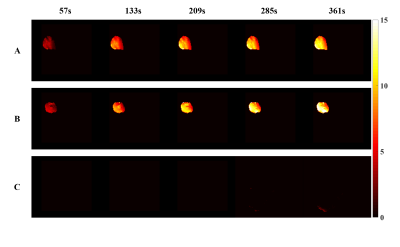 |
Real-Time Temperature Monitoring during MR Imaging Guided Photothermal Therapy
Boyu Zhang1, Xianfu Meng2, Yuwen Zhang1, Wenbo Bu2, and He Wang1,3
1Institute of Science and Technology for Brain-Inspired Intelligence, Fudan University, Shanghai, China, 2School of Chemistry and Molecular Engineering, East China Normal University, Shanghai, China, 3Human Phenome Institute, Fudan University, Shanghai, China
It is a challenge that accurate monitoring the spatial distribution of photothermal transducing agents (PTAs), meanwhile, real-time mapping temperature in photothermal therapy. Here, we propose an effective strategy that integrates T1 and MR temperature imaging for tracking nanoPTAs and real-time temperature monitoring in vivo. The synthesized nanoPTAs equip with favorable T1 performance and marginal susceptibility guaranteeing the accuracy of temperature mapping. The time resolution is 20 seconds, and the detection accuracy of temperature change is as low as 0.1K, which contributes to promoting the clinical application of photothermal therapy.
|
|
4134.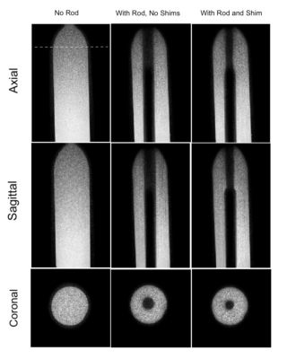 |
Active Shimming of Metallic Probes for Interventional MRI
Saikat Sengupta1 and Xinqiang Yan1
1Department of Radiology, Vanderbilt University Institute of Imaging Science, Nashville, TN, United States
Artifacts caused by large magnetic susceptibility differences between metallic probes and the surrounding tissue are a persistent problem in interventional MRI. In 2019, we presented the concept, design and modeling of active shims for metallic probes inspired from naval degaussing systems. Field disturbance induced by a metallic probe at 3 Tesla was modeled and an active shim insert design was presented to shim the field variation around the probe. In this abstract we present the first experimental results showing effective recovery of metallic probe induced signal loss at 3 Tesla.
|
|
4135.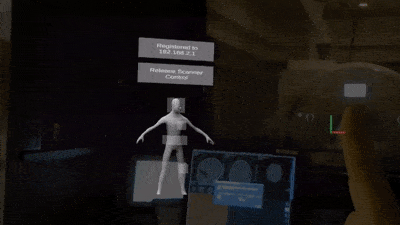 |
Scanner Control and Realtime Visualization via Wireless Augmented Reality
Andrew Dupuis1, Dan Ma2, and Mark A Griswold1,2
1Biomedical Engineering, Case Western Reserve University, Cleveland, OH, United States, 2Department of Radiology, School of Medicine, Case Western Reserve University, Cleveland, OH, United States
We implemented an augmented reality (AR) scanner interface in order to streamline the scanner console while allowing users to work natively in three dimensions. The Microsoft Hololens was leveraged to allow users to visualize and control the scanner wirelessly. A gesture based interface was created, with emphasis placed on simplification of the scan process to help untrained non-imaging personnel to interact with the scanner. The resulting system is simple to use and allows the operator to maintain direct sight of the patient throughout the scan.
|
|
4136. |
A feasibility study of Magnetic Resonance Fingerprinting for multi-contrast temperature mapping in both aqueous and adipose tissues
Megan E Poorman1,2, Rasim Boyacioglu3, William A Grissom4, Mark A Griswold3, and Kathryn E Keenan1
1Physical Measurement Laboratory, National Institute of Standards and Technology, Boulder, CO, United States, 2Department of Physics, University of Colorado Boulder, Boulder, CO, United States, 3Department of Radiology, Case Western Reserve Univeristy, Cleveland, OH, United States, 4Vanderbilt University Institute of Imaging Science, Nashville, TN, United States
Temperature monitoring in both adipose and aqueous tissues is important for guidance of thermal therapies in vivo. However, the proton resonant frequency shift for thermometry is only reliable in aqueous tissues. Temperature mapping in adipose has been explored using relaxation, but is limited by the accuracy and speed of the method used. MR Fingerprinting provides a framework for mapping differences due to multiple tissue properties simultaneously. This work explores adapting the MRF framework to allow temperature changes to be mapped directly in all tissue types, and simulates a dictionary update method that could offer improved temporal resolution over standard MRF.
|
|
4137. |
Temperature-induced chemical shift in single-shot echo-planar imaging
Chang-Sheng Mei1, Shenyan Zong2,3, Sheng Chen4, Guofeng Shen3, and Bruno Madore2
1Department of Physics, Soochow University, Taipei, Taiwan, 2Department of Radiology, Brigham and Women's Hospital, Harvard Medical School, Boston, MA, United States, 3Biomedical Instrument Institute, School of Biomedical Engineering, Shanghai Jiao Tong University, Shanghai, China, 4Shanghai Shende Medical Technology Co., Ltd, Shanghai, China
The proton resonance frequency (PRF) shift, whereby the Larmor frequency varies with temperature, is the effect that enables most currently-employed MR thermometry methods. The present work aimed to demonstrate that such temperature-related frequency changes can also cause non-negligible spatial distortions in single-shot EPI. In addition to demonstrating the effect, a solution is also proposed here. Phantom experiments are presented that demonstrate the problem and its solution.
|
|
4138. |
Multi-Radiofrequency Thermal Magnetic Resonance Targeting Glioblastoma Multiforme
Eva Oberacker1, Andre Kuehne2, Thomas Wilhelm Eigentler1, Cecilia Diesch1, Jacek Nadobny3, Pirus Ghadjar3, Peter Wust3, and Thoralf Niendorf1,2,4
1Berlin Ultrahigh Field Facility (B.U.F.F.), Max Delbrück Center for Molecular Medicine in the Helmholtz Association, Berlin, Germany, 2MRI.TOOLS GmbH, Berlin, Germany, 3Clinic for Radiation Oncology, Charité Universitätsmedizin, Berlin, Germany, 4Experimental and Clinical Research Center (ECRC), joint cooperation between the Charité Medical Faculty and the Max-Delbrück-Center for Molecular Medicine in the Helmholtz Association, Berlin, Germany
Ultrahigh field (UHF) MR employs higher radio frequencies (RF) than conventional MR and has unique potential to provide focal temperature manipulation and high resolution imaging (ThermalMR), as our simulations and experimental work demonstrated for f=300MHz. Here we use EMF simulations to demonstrate the added value for multi-frequency thermal intervention (f=300MHz-500MHz) to boost the efficacy of the heating paradigm for clinical target volumes (TVs) of two brain tumor patients. An optimization algorithm was developed to study RF power deposition in the TV for a single, double or triple frequency set permitting highest RF power deposition in the TV.
|
|
4139. |
MRI-Guided Cryoablation and Image-Guided Immunotherapy in Naturally Occurring Canine Osteosarcoma
Dara Kraitchman1, Cory Brayton1, Rebecca Krimins1, Byung-Hak Kang1, Zachary Millman1, Edward McCarthy1, and Brian Ladle1
Video Permission Withheld
1Johns Hopkins University School of Medicine, Baltimore, MD, United States
Improvements in outcomes with current chemotherapy and surgery treatment approaches for patients with osteosarcoma have plateaued over the past three decades necessitating new treatment paradigms. A novel therapy for osteosarcoma of MR-guided cryotherapy in conjunction with intratumoral injection of an immune adjuvant in the clinically relevant animal model of spontaneously occurring osteosarcoma in client-owned dogs with the intent of priming an adaptive immune response capable of delaying or preventing the occurrence of metastatic disease is demonstrated.
|
|
4140.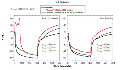 |
Investigation of the impact of RF heating on the treatment temperature during MRI-guided cryoablation at 1.5T
Aiming Lu1, David A Woodrum1, Christopher P Favazza1, Joel P Felmlee1, Brian T Welch1, Jacinta Browne1, and Krzysztof R Gorny1
1Radiology, Mayo Clinic, Rochester, MN, United States Poster Permission Withheld
MRI-guided cryoablation is a promising minimal invasive approach to treat localized tumors. Since the cryoneedles used in this system are metallic and the gas lines have metallic components, RF heating could occur during MR imaging. Our phantom study demonstrated temperature increase of more than 100 °C due to a sequence with moderate SAR of 2.1W/kg.In this work, we demonstrate that RF heating caused by the real-time monitoring MRI sequence could potentially affect the temperature course near the cryoneedles and therefore affect the treatment efficacy, especially when angiocath was used to aid the cryoneedle placement.
|
4141.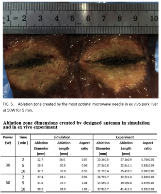 |
A Noval MR-Compatible and Clinically Available Microwave Needle for Spherical Ablation: Design and Ex Vivo Validation at a 0.7T
Xiaoyan Huang1, Yufu Zhou1, and Bensheng Qiu1
1University of Science and Technology of China, Hefei, China
A novel MR-compatible microwave needle is proposed to achieve spherical ablation and monitor the temperature of needle shaft. It has an asymmetrical dipole choke with titanium alloy tube and a conical tip made of solid zirconia to facilitate needle insertion into tissues. FDTD simulations are used to minimize reflection coefficient and maximize aspect ratio. This most optimal design was validated in ex vivo pork liver in a 0.7T open MR system and concluded an acceptable artifacts, reflection coefficient of -27.86 dB and near-spherical ablation zone with a length of 39.5 mm, a diameter of 34.5 mm at 50W, 5 min.
|
|
4142.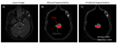 |
Tool for Image-guided Intracerebral Hemorrhage Evacuation: Automatic Segmentation of Clot, Edema, and Normal Brain Tissue
Thomas Lilieholm1, Alan McMillan1,2, Fang Liu1, Robert Moskwa1, Azam Ahmed3, and Walter F Block1,2,4
1Medical Physics, University of Wisconsin - Madison, Madison, WI, United States, 2Radiology, University of Wisconsin - Madison, Madison, WI, United States, 3Neurological Surgery, University of Wisconsin - Madison, Madison, WI, United States, 4Biomedical Engineering, University of Wisconsin - Madison, Madison, WI, United States
Improved image guidance is needed for neurosurgeons to reduce the residual remaining clot levels during minimally invasive evacuation of intracerebral hemorrhage (ICH) while not causing rebleeds. Neurosurgeons would benefit from a means to periodically render the clot volume against surrounding normal tissue during mechanical evacuation or pharmaceutical-based clot-busting. Using convolutional neural networks (CNN), we created machine learning models to automatically segment the constituent clot and edema induced by ICH cases using T2-weighted MR images. The CCN’s output results were found to be in agreement with manual segmentations of the same ICH cases.
|
|
4143.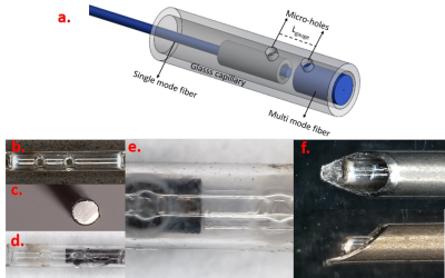 |
ENHANCED FPI BASED FORCE SENSOR DESIGNED FOR MRI COMPATIBLE BIOPSY NEEDLE
Dogangun Uzun1 and Ozgur Kocaturk1
1Institute of Biomedical Engineering, Bogazici University, Istanbul, Turkey
In this work, a novel design fiber optic force sensor with increased resolution thru Titanium coating is fabricated using Fabry-Pérot Interferometry (FPI) method and integrated into a biopsy needle to be used in interventional biopsy operations under MRI. The FPI based force sensor with improved resolution is tested via benchtop experiments after integrating into a MRI compatible biopsy needle. Sensor’s penetration detection, tissue characterization and differentiation capabilities are tested using different types of tissues. Overall system performance is tested via in vitro experiments under MRI using a commercial prostate phantom with leisons.
|
|
4144.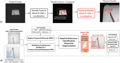 |
Physical Needle Localization using Mask R-CNN for MRI-Guided Percutaneous Interventions
Xinzhou Li1,2, Yu-Hsiu Lee3, Tsu-Chin Tsao3, and Holden H. Wu1,2
1Radiological Sciences, University of California, Los Angeles, Los Angeles, CA, United States, 2Bioengineering, University of California, Los Angeles, Los Angeles, CA, United States, 3Mechanical and Aerospace Engineering, University of California, Los Angeles, Los Angeles, CA, United States
Discrepancy between the physical needle location and the MRI passive needle feature could lead to needle localization errors in MRI-guided percutaneous interventions. By leveraging physics-based simulations with different needle orientations and MR imaging parameters, we designed and trained a Mask Regional Convolutional Neural Network (R-CNN) to automatically localize the physical needle tip and axis orientation based on the MRI passive needle feature. The Mask R-CNN framework was tested on a separate set of actual phantom MR images and achieved physical needle localization with median tip error of 0.74 mm and median axis error of 0.95°.
|
|
4145.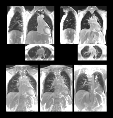 |
4D-MRI of Thorax and Abdomen with Variable Contrast at 1.5T
Orso Andrea Pusterla1,2,3, Francesco Santini2,3, Grzgorz Bauman2,3, Rahel Heule4, Alina Giger3, Philippe Claude Cattin3, Sairos Safai5, Sebastian Kozerke1, and Oliver Bieri2,3
1Institute for Biomedical Engineering, University and ETH Zurich, Zurich, Switzerland, 2Department of Radiology, Division of Radiological Physics, University Hospital Basel, Basel, Switzerland, 3Department of Biomedical Engineering, University of Basel, Allschwil, Switzerland, 4High Field Magnetic Resonance, Max Planck Institute for Biological Cybernetics, Tübingen, Germany, 5Center for Proton Therapy, Paul Scherrer Institute, Villigen-PSI, Switzerland
Time-resolved volumetric imaging (4D-MRI) of moving organs is essential for several clinical applications, e.g., in interventional treatments, radiation therapies, and high-intensity focused ultrasound ablations. In this work, an interleaved time-resolved data-navigator acquisition strategy for 4D-MRI with flexible contrast is developed offering versatile settings for thoracic and abdominal imaging: for both, image and navigator, not only the flip angle but also the basic acquisition type (pulse sequence kernel) can be chosen independently from either balanced SSFP (i.e., T2/T1-weighted) or spoiled gradient echo (i.e., T1-weighted).
|
|
4146. |
Geometric accuracy of real-time tumor tracking and dynamic beam adaptation using the Australian MRI-Linear accelerator
Paul Zhi Yuan Liu1, Urszula Jelen2, Bin Dong2, Gary Liney2, and Paul Keall1
1Image X Institute, The University of Sydney, The University of Sydney, Australia, 2Department of Medical Physics, Ingham Institute for Applied Medical Research, Liverpool, Australia
A real-time tumor tracking system has been developed for MRI-guided radiation therapy. This system allows for tumor motion to be monitored during radiotherapy and for the radiation beam to be modified to compensate for this motion. The tumor location was found during treatment through the use of a template matching algorithm and 2D cine-MR images. The tumor location information was sent to a tracking algorithm that modifies the shape of the beam in real-time to ensure the tumor target is irradiated. The tumor tracking system has been tested and found to be of submilimeter accuracy.
|
|
4147.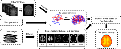 |
Development of a first principles-based model of brain thermoregulation and initial validation with MR chemical shift thermometry
Dongsuk Sung1, Peter A. Kottke2, Jason W. Allen3,4, Fadi Nahab4, Andrei G. Fedorov2,5, and Candace C. Fleischer1,3,5
1Department of Biomedical Engineering, Georgia Institute of Technology and Emory University, Atlanta, GA, United States, 2Woodruff School of Mechanical Engineering, Georgia Institute of Technology, Atlanta, GA, United States, 3Department of Radiology and Imaging Sciences, Emory University School of Medicine, Atlanta, GA, United States, 4Department of Neurology, Emory University School of Medicine, Atlanta, GA, United States, 5Petit Institute for Bioengineering and Bioscience, Georgia Institute of Technology, Atlanta, GA, United States
Brain temperature regulation is a key parameter after injury and ischemia. Experimental magnetic resonance (MR)-based thermometry is promising but not always clinically feasible. To complement MR thermometry measures, an improved thermal model of the brain based on first principles has been developed that rigorously ensures energy conservation in thermally interacting brain domains. As initial validation, a temperature map was generated using our model with MR angiography (MRA) and structural MR imaging (MRI) data, and compared with experimental chemical shift thermometry (CST) in the same subject. Biophysical models provide further insight into brain thermoregulation by complementing experimental thermometry measurements.
|
|
4148.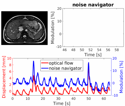 |
Noise navigator: An independent method to detect physiological motion during MRI-guided radiotherapy
Robin Navest1,2, Stefano Mandija1,2, Bjorn Stemkens1, Stefan Zijlema1,2, Anna Andreychenko1,3, Jan Lagendijk1, and Cornelis van den Berg1,2
1Radiotherapy, UMC Utrecht, Utrecht, Netherlands, 2Computational Imaging Group for MRI Diagnostics & Therapy, Center for Image Sciences, UMC Utrecht, Utrecht, Netherlands, 3Research and Practical Clinical Center of Diagnostics and Telemedicine Technologies of the Moscow Department of Healthcare, Moscow, Russian Federation
The noise navigator provides physiological motion detection completely independent of the imaging volume or contrast of the MR images and can be acquired on a much faster (millisecond) time scale than MRI. We believe that the noise navigator can be used as an independent verification method to detect the occurrence of motion in synergy with real-time MRI-guided radiotherapy and standalone during e.g. recontouring when typically no MRI is performed. For abdominothoracic radiotherapy, we investigated bulk motion and variable breathing as these have a relatively large amplitude (millimeters). Additionally, swallowing detection was investigated given its complications for head-and-neck radiotherapy.
|
|
4149. |
MRI-based motion characterisation of atrial fibrillation cardiac radiosurgery targets.
Suzanne Lydiard1,2, Beau Pontre3, Boris Lowe2, and Paul Keall1
1ACRF ImageX Institute, University of Sydney, Sydney, Australia, 2Auckland City Hospital, Auckland, New Zealand, 3University of Auckland, Auckland, New Zealand
Cardiac radiosurgery for atrial fibrillation (AF) is challenging; the target moves with both cardiac contraction and respiration and is in close proximity to critical structures. This study utilized non-contrast MRI to characterize AF cardiac radiosurgery target motion as well as relative target displacement to surrounding structures. The absolute and relative target motion seen in this study in combination with target proximity to the aorta and esophagus highlights the importance of carefully selecting cardiac radiosurgery technology and techniques. This MRI-based methodology could be useful in the AF cardiac radiosurgery clinical workflow to optimize treatment for the individual.
|
|
4150. |
Mixed reality neuronavigation for transcranial magnetic stimulation treatment
Christoph Leuze1, Supriya Sathyanarayana1, Mahendra T Bhati2, Noriah D Johnson2, Christopher C Cline2,3, Trishia El Chemaly1, Bruce Daniel1, Aapo Nummenmaa4,5, Amit Etkin2, and Jennifer A McNab1
1Radiology, Stanford University, Stanford, CA, United States, 2Psychiatry, Stanford University, Stanford, CA, United States, 3MIRECC, VA Palo Alto Health Care System, Palo Alto, CA, United States, 4Neuroscience, Massachusetts General Hospital, Charlestown, MA, United States, 5Radiology, Harvard Medical School, Charlestown, MA, United States
Transcranial magnetic stimulation (TMS) treatment is an important non-invasive treatment option for depressive patients who do not respond to medication. Effective TMS treatment is dependent on accurate stimulation intensity calibration and coil positioning, which is best achieved through image-based neuronavigation. Unfortunately, most TMS treatments use generalized scalp measurements, which are faster and cheaper but less accurate and less effective than image-based neuronavigation. We present a mixed reality neuronavigation system that allows the clinician to view brain MRI and targeting information superimposed on the patient’s head for an accelerated and simplified procedure
|
|
4151. |
Towards Quantitative Myocardial Perfusion: Catheter-Based Arterial Input Function Measurements with Flow-Normalization
Simon Stephan1, Simon Reiss1, Thomas Lottner1, Ali Caglar Özen1, and Michael Bock1
1Dept. of Radiology, Medical Physics, Medical Center University of Freiburg, Faculty of Medicine, University of Freiburg, Freiburg, Germany, Freiburg, Germany
For quantitative myocardial perfusion measurements with intra-arterial contrast agent (CA) injection precise knowledge of the arterial input function (AIF) is needed. Not necessarily absolute values of the AIF are needed if blood flow measurements are used to normalize the perfusion maps. In this work it is shown for a constant-flow phantom that this method works and probable issues for transferring the method to in-vivo measurements are discussed.
|
|
4152. |
Development of an optimal head localizer for stereotactic neurosurgery at 7.0T with minimal image geometric distortions
Aaron E Rusheen1,2, Elise M Berning1, Dane T Bothun1, Ben T Gifford1, Stephan J Goerss1, Kirk M Welker3, John Huston3, Kevin E Bennet1,4, Yoonbae Oh1,5, Charles D Blaha1, Kendall H Lee1,5, and Fagan J Andrew3
1Department of Neurosurgery, Mayo Clinic, Rochester, MN, United States, 2Medical Scientist Training Program, Mayo Clinic, Rochester, MN, United States, 3Department of Radiology, Mayo Clinic, Rochester, MN, United States, 4Department of Engineering, Mayo Clinic, Rochester, MN, United States, 5Department of Physiology and Biomedical Engineering, Mayo Clinic, Rochester, MN, United States
7.0T MRI provides precise visualization and targeting of brain structures for image-guided stereotactic neurosurgery. However, image localizers used by stereotactic systems do not exist for 7.0T scanners. Challenges in their development include: the small bore creates geometric constraints that disallow use of conventional localizers, and the increased B0 increases geometric distortion, affecting registration accuracy. Here, a skull-contoured localizer utilizing point fiducials was designed to attach to a novel stereotactic frame. Extracranial distortion was thoroughly mapped using several optimized imaging sequences. This data was used to optimally place fiducials on the localizer, improving registration accuracy.
|
|
4153.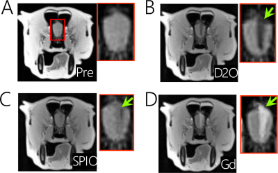 |
Deuterium oxide as a black water contrast medium for real-time MRI-guided endovascular neurointervention
Lin Chen1,2, Jing Liu1,3, Chengyan Chu1,4, Nirhbay Yadav1,2, Jiadi Xu1,2, Monica Pearl1, Piotr Walczak1,4, Peter van Zijl1,2, Miroslaw Janowski4, and Guanshu Liu1,2
1Department of Radiology and Radiological Science, The Johns Hopkins University School of Medicine, Baltimore, MD, United States, 2F.M. Kirby Research Center for Functional Brain Imaging, Kennedy Krieger Research Institute, Baltimore, MD, United States, 3The First Affiliated Hospital of Jinan University, Guangzhou, China, 4Department of Diagnostic Radiology and Nuclear Medicine, University of Maryland, Baltimore, MD, United States
The use of contrast media, such as Gadolinium-based contrast agents (GBCA), superparamagnetic iron oxide particles (SPIO) and carbon dioxide, can improve the conspicuity of MRI, thereby helping to improve the accuracy of transcathether infusions during endovascular neurointerventions. In this study, we exploited deuterium oxide (D2O) as a new MRI contrast medium for guiding intra-arterial hyperosmotic blood brain barrier (BBB) opening in experimental animal models. Compared to the SPIO-based approach, D2O MRI guidance was found to provide comparable results that can predict the territory of BBB opening (assessed by Gd-MRI).
|
|
4154. |
Intelligent Protocolling for Autonomous MRI
Keerthi Sravan Ravi1,2, Sairam Geethanath2, Patrick Quarterman3, Maggie Fung3, and John Thomas Vaughan Jr.2
1Biomedical Engineering, Columbia University, New York, NY, United States, 2Columbia University Magnetic Resonance Research Center, Columbia University, New York, NY, United States, 3GE Healthcare Applied Sciences Laboratory East, New York, NY, United States
MR Value can be interpreted as the ratio of image contrast to acquisition time. This can be maximised via intelligent protocolling that chooses an optimal combination of pulse sequence parameters. In this work, a Look Up Table (LUT) is consulted to obtain parameters that satisfy an acquisition time constraint. The LUT is populated with data collected from a vendor’s simulator, and a search algorithm is designed to prioritise preserving image contrast and achieving high through-plane resolution. Data is collected across four experiments from a healthy volunteer. LUT-derived and measured relative SNR values are compared and analysed.
|
|
4155.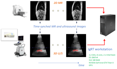 |
A test-retest analysis of motion compensation in image-guided radiation therapy using hands-free simultaneous MR-U/S system.
Jhimli Mitra1, Sydney Jupitz2, Bryan Bednarz2, James H. Holmes3, David Mills1, Lowell Scott Smith1, Heather Chan1, Aqsa Patel1, Eric Fiveland1, Warren Lee1, Alan McMillan3, Wes Culberson2, Michael Bassetti4, Andrew Shepard2,
Shourya Sarcar1, and Thomas K. Foo1
1General Electric Research, Niskayuna, NY, United States, 2Medical Physics, University of Wisconsin - Madison, Madison, WI, United States, 3Radiology, University of Wisconsin - Madison, Madison, WI, United States, 4Human Oncology, University of Wisconsin - Madison, Madison, WI, United States
An MR-compatible ultrasound probe was developed for hands-free, simultaneous MR-ultrasound image acquisition for image-guided radiation therapy in liver. Simultaneously acquired pre-treatment MR and U/S associates a pair of MR image and U/S volume to each respiratory state over multiple respiratory cycles without requiring physical MR image acquisition during the radiation treatment LINAC phase. Automatic fiducial tracking and respiratory state clustering were performed on the U/S volumes to determine expiration states for dose delivery. In a test-retest analysis, consistent and reproducible expiration states were obtained that can be used for targeted-dose delivery under LINAC.
|
4156. |
On the Carbon Footprint of ISMRM's Annual Meeting: Analysis and Implications
Christof Böhm1, Dominik Weidlich1, Sophia Kronthaler1, and Dimitrios C. Karampinos1
1TUM, Munich, Germany
Climate change is one of the defining issues of our time. Weather patterns are shifting and sea levels are rising. The most abundant greenhouse gas, accounting for about two-thirds of total greenhouse gas emissions, is carbon dioxide (CO2). CO2 is largely the product of burning fossil fuel and therefore the traveling towards the conference venue is expected to be a main contributor to the carbon footprint of the annual ISMRM meeting. This work estimates the C02 emissions of the past ISMRM meetings and tries to give data driven recommendation for future selection of ISMRM conference locations.
|
|
4157. |
Free-breathing 3D T2-weighted TSE using Cartesian acquisition with rewinded spiral profile ordering (rCASPR) for abdominal radiotherapy
Tom Bruijnen1,2, Osman Akdag1,2,3, Charlotte VM Bruel1,2,3, Bjorn Stemkens1,2, Tim Schakel1, Jan JW Lagendijk1, Cornelis AT van den Berg1,2, and Rob HN Tijssen1
1Department of Radiotherapy, University Medical Center Utrecht, Utrecht, Netherlands, 2Computational Imaging Group for MRI diagnostics and therapy, Centre for Image Sciences, University Medical Center Utrecht, Utrecht, Netherlands, 3Department of Biomedical Engineering, Eindhoven University of Technology, Eindhoven, Netherlands
T2-weighted turbo spin-echo (TSE) sequences are used for abdominal tumour contouring in MR-guided radiotherapy, but these sequences are generally not compatible with most respiratory-correlated 4D imaging methods. To overcome this limitation we propose a free-breathing T2-weighted TSE sequence based on a Cartesian acquisition with rewinded spiral profile ordering in combination with motion compensated image reconstruction. We showed that high quality free-breathing T2-weighted TSE scans are feasible with short scan times (<5 min), which have similar contrast to the conventional linear sampled scans while simultaneously quantifying the respiratory motion.
|
|
4158.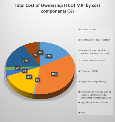 |
Total cost of ownership is a useful tool for life cycle management of MRI scanners and managing costs in an MRI department.
Elizabeth Jones1
1Radiology, NIH Clinical Center, Bethesda, MD, United States A total cost of ownership (TCO) model was developed for MRI scanners over the ownership life from fund allocation through decommissioning in an organization with outpatient, inpatient and research scanners. The TCO model included acquisition and installation costs; site modifications to comply with safety requirements; human resources including ancillary personnel; small equipment, supplies, contrast; service contracts; IT; overhead; and upgrades. The largest expense was human resources accounting for 35% of TCO. A cost reduction strategy is centralized remote scanning, reducing the total number of highly skilled technologists. TCO is tool for identifying hidden costs and comparing scenarios for managing costs. |
|
4159. |
Whole Body PET/MRI Biodistribution of F-18 fluorobenzylamide doxorubicin in an Intra-arterial Procedure
Parth Kumar1, Caroline D. Jordan1, Joseph E. Blecha1, Thomas R. Hayes1, Carol Stillson1, Aaron D. Losey1, Eric Mastria1, Colin Yee1, Bridget F. Kilbride1, Teri Moore1, Mark W. Wilson1, Henry F. VanBrocklin1, and Steven W. Hetts1
1Radiology and Biomedical Imaging, University of California, San Francisco, San Francisco, CA, United States
Recent studies have shown effective use of intravascular filtration devices to remove chemotherapy drugs; however, accurate quantification of the drug’s distribution on the device, within the targeted organ, and systemically, is necessary. We synthesized 18F analog of doxorubicin, fluorobenzylamide doxorubicin ([18F]FB-Dox), which was administered via an intra-arterial catheter, without a filter, and imaged under PET/MRI in vivo. [18F]FB-Dox showed a measurable decrease in the liver, and an increase in the bladder, kidney, and gall bladder over a 90-minute period after injection. This demonstrates a viable way to assess and track the baseline biodistribution of an intra-arterial chemotherapy procedure.
|
|
4160. |
Declutter the protocol tree: managing and comparing sequence parameters of multiple clinical systems using Python tools
Pim Pullens1,2, Pieter Devolder1, Tony Thienpont1, and Eric Achten1,2
1Radiology, University Hospital Gent, Gent, Belgium, 2Ghent Institute for functional and Metabolic Imaging, Ghent University, Gent, Belgium
Monitoring protocol and sequence changes is of key importance for efficiently running a clinical imaging department. Since an MRI protocol contains hundreds of parameters, it is almost impossible to manage protocols from the scanner console, especially in a clinical situation where console time for the MR physicist is very limited. We present a python-based platform that performs all these duties and show 4 potential use cases: 1) compare parameters from sequences that share the same name 2) same purpose, different name 3) differences in naming of Regions/Exams/Programs 4) real-life example of decluttering a pediatric neuro protocol.
|
|
4161. |
Electrodynamics and RF Applicator Concept for Sub-nanosecond Pulsed Thermal Magnetic Resonance
Egor Kretov1, Andre Kuehne2, Eva Oberacker1, Thomas Wilhelm Eigentler1, and Thoralf Niendorf1,2
1B.U.F.F., Berlin Ultrahigh Field Facility, Max-Delbrück Center for Molecular Medicine in the Helmholtz Association, Berlin, Germany, 2MRI.TOOLS GmbH, Berlin, Germany
This work presents electrodynamic considerations and an RF applicator concept that permit MR imaging and RF energy focusing inside of the human brain using sub-nanosecond broadband electromagnetic pulses. Such pulses can deposit RF energy in a small, focal area for thermal intervention. To meet this goal proof-of-principle is provided through EMF simulations using a dual-mode RF applicator that combines antipodal Vivaldi antennae with loop elements.
|
|
4162.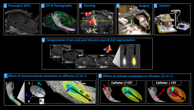 |
In vivo MRI measurement of microstructural constraints for direct drug delivery within the brain
Antonella Castellano1, Alice Segato2, Valentina Pieri1, Marco Vidotto2, Nicolò Pecco1, Marco Riva3,4, Riccardo Secoli5, Davide Danilo Zani6, Stefano Brizzola6, Marco Trovatelli7, Giuliano Ravasio6, Marcello Cadioli8, Lorenzo Bello4,9,
Ferdinando Rodriguez Y Baena5, Elena De Momi2, and Andrea Falini1
1Neuroradiology Unit and CERMAC, Vita-Salute San Raffaele University, IRCCS San Raffaele Scientific Institute, Milan, Italy, 2Department of Electronics, Information and Bioengineering, Politecnico di Milano, Milan, Italy, 3Department of Medical Biotechnology and Translational Medicine, Università degli Studi di Milano, Milan, Italy, 4Neurosurgical Oncology Unit, IRCCS Humanitas Clinical and Research Center, Rozzano (MI), Italy, 5Department of Mechanical Engineering, Imperial College London, London, United Kingdom, 6Department of Veterinary Medicine, Università degli Studi di Milano, Milan, Italy, 7Department of Health, Animal Science and Food Safety, Faculty of Veterinary Medicine, Università degli Studi di Milano, Milan, Italy, 8Philips Healthcare, Milan, Italy, 9Department of Oncology and Hemato-Oncology, Università degli Studi di Milano, Milan, Italy
Brain tissue microstructure may influence the efficient delivery of therapeutics within the brain. Diffusion Tensor Imaging (DTI) enables the depiction of tissue properties in vivo, and thus is potentially relevant for planning convection-enhanced delivery (CED) within the brain. We report on the quantitative assessment of the distribution of a Gadolinium solution infused by CED within the brain of a live ovine model. Infusate distributions were measured at multiple timepoints and compared to microstructural properties as depicted by DTI, thus demonstrating the impact of tissue features and catheter positioning on drug distribution in vivo.
|
|
4163.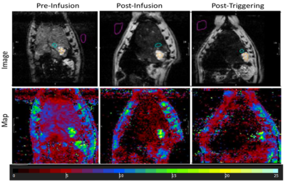 |
Magnetic field stimuli improve hepatic delivery of oxaliplatin to colorectal liver metastatic tumor bearing rats
Venkateswara Gogineni1, Dilip Maddirela1, Woo Ram Park2, Jaidip Jagtap1, Abdul Parchur1, Gayatri Sharma1, El-Sayed H Ibrahim1, Amit Joshi1, Andrew Larson2, Dong-Hyun Kim2, and Sarah B White1
1Medical College of Wisconsin, Milwaukee, WI, United States, 2Northwestern University, Chicago, IL, United States
Systemic chemotherapy is the standard of care for the treatment of unresectable metastatic colorectal cancer. However, new drug delivery platforms are needed to mitigate its associated toxicities. In this study, we develop and evaluate a liposome formulation that deliver oxaliplatin under magnetic field stimulus site selectively release in high concentration to alleviate the off-target effects in a rat model of colorectal liver metastases (CRLM). Quantitative MRI enabled assessment of changes in tumor characteristics over time for confirmation of procedural success and proper catheter-selective tumor targeting, which allows for high concentrations of oxaliplatin and improves survival outcomes in CRLM tumor-bearing rats.
|
|
4164.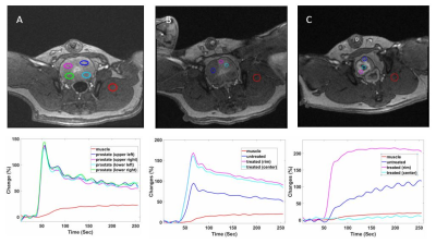 |
Diffusion and Dynamic Contrast-enhanced MRI for Evaluation of Prostate Response to 90Yttrium Radioembolization in A Canine Hyperplasia Model
Weiguo Li1,2, Kathleen Harris1, Ali Khan1, Simone Raiter1, Monica Matsumoto3, Andrew C Larson1, and Samdeep Mouli1
1Radiology, Northwestern University, Chicago, IL, United States, 2Research Resource Centers, University of Illinois at Chicago, Chicago, IL, United States, 3Radiology, University of Chicago, Chicago, IL, United States
Management options for localized prostate cancer have largely remained the same for the past 30 years. 90Yttrium (90Y) radioembolization is a novel radiotherapy approach that offers the potential to deliver high-dose radiation therapy with minimal non-target radiation. However, currently no accurate way exists to evaluate radiation effects noninvasively following embolization. In this study, we sought to apply diffusion and dynamic contrast-enhanced perfusion MRI to investigate assess the effects of treatment following 90Y prostate artery radioembolization in a dog prostate model.
|
|
4165.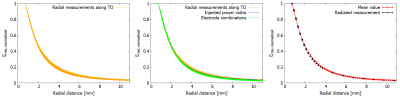 |
Approach to reduce the measurement volume to determine spatial distribution of power deposition around a straight lead according to ISO/TS 10974
Finya Ketelsen1,2, Jakob Kreutner3, Gregor Schaefers1,3, and Kevin Kröninger2
1MR:comp GmbH, Testing Services for MR Safety & Compatibility, Gelsenkirchen, Germany, 2TU Dortmund University, Dortmund, Germany, 3MRI-STaR - Magnetic Resonance Institute for Safety, Technology and Research GmbH, Gelsenkirchen, Germany
To determine the RF-induced heating of an active implant, it is necessary to measure the 3D distribution of the power deposition in the hotspot volume. By measuring the Erms distribution around a test object, different influences on the decay were investigated. It was shows that radial decay can be described with a single model along test object. This model is normalized to a defined distance. The measurement values along the TO at this distance, can be used to scale the model and the power deposition inside the hotspot volume can be calculated. This leads to an advantage in time saving.
|
|
4166.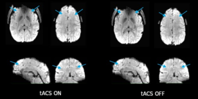 |
Concurrent alternating transcranial current stimulation and fMRI: subject safety and the data quality evaluation
Beni Mulyana1,2, Qingfei Luo1, Aki Tsuchiyagaito1, Ashkan Rashedi1, Jared Smith1, Masaya Misaki1, Duke Shereen3, Samuel Cheng2, Martin Paulus1, Hamed Ekhtiari1, and Jerzy Bodurka1,4
1Laureate Institute for Brain Research, Tulsa, OK, United States, 2University of Oklahoma, Tulsa, OK, United States, 3The Graduate Center, City University of New York, New York, NY, United States, 4Stephenson School of Biomedical Engineering, University of Oklahoma, Norman, OK, United States
We evaluated subject safety and the fMRI image quality in the presence of transcranial alternating current stimulation (tACS). Scalp temperatures under the tACS electrodes with and without stimulations were measured. Result indicated: i) the tACS stimulation itself had no harmful impact on scalp temperature; ii) tACS’s artifacts had negligible effects on fMRI images. Although the fMRI scan produced heating effects due to the RF energy deposition in tACS electrodes, the scalp temperatures remained below 37°C during a 12-min scan regardless of the presence of stimulations. Thus, concurrent tACS with fMRI has no impact on patient safety and image quality.
|
|
4167.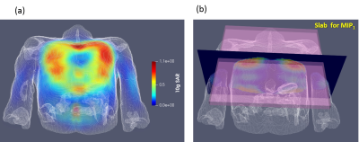 |
SAR Safety Analysis for a 8Tx/16Rx Coil Array Prototype for Human Cardiac MRI at 7 T
Maxim Terekhov1, Ibrahim A. Elabyad1, Carsten Kögler2, Theresa Reiter1,3, and Laura M. Schreiber1
1Chair of Cellular and Molecular Imaging, Comprehensive Heart Failure Center (CHFC), University Hospital Würzburg, Wuerzburg, Germany, 2Rapid Biomedical, Rimpar, Germany, 3Department of Internal Medicine l, University Hospital Wuerzburg, Wuerzburg, Germany
The purpose of this study was to simulate the variation of SAR caused by shifting the position of a cardiac array prototype when different B1-shimming phase vectors are employed. The variation of 10g average SAR distribution due to displacing the anterior array position on the thorax (central, shifted 5 cm to the right, left, head, foot and diagonal directions) was computed and analyzed using two human voxel models. The final goal is to determine maximal driving voltage of the array with adequate safety margins considering new hot-spots originated from possible array misplacement
|
|
4168.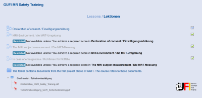 |
On the successful implementation of a first homogenized multicenter online safety training for ultrahigh field MRI
Astrid Wollrab1, Oliver Kraff2, Oliver Speck1,3,4, Harald H. Quick2,5, and Mark E. Ladd2,6
1Biomedical Magnetic Resonance, Otto-von-Guericke University, Magdeburg, Germany, 2Erwin L. Hahn Institute for Magnetic Resonance Imaging, University Duisburg-Essen, Essen, Germany, 3German Center of Neurodegenerative Diseases - DZNE, Magdeburg, Germany, 4Leibniz Institute for Neurobiology, Magdeburg, Germany, 5High-Field and Hybrid MR Imaging, University Hospital Essen, Essen, Germany, 6Medical Physics in Radiology, German Cancer Research Center - DKFZ, Heidelberg, Germany
One of the main tasks of the German Ultrahigh Field Imaging (GUFI) network was the establishment of a uniform online MRI safety training, created in cooperation with all German ultrahigh field (UHF) sites. Implemented with Moodle, the course provides basic MRI safety training and includes three lessons: "MRI Environment", "Subject Measurements" and "In Case of Emergencies". Participants have to pass tests, consisting of appropriate questions, at the end of each lesson. In total, 255 participants have successfully completed the course (since April 2018).
|
|
4169.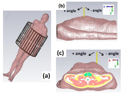 |
A simulation study of the SAR10g dependence of a microwave applicator for MR-guided ablation of hepatic tumors at 1.5T
Tom Dijkhuis1,2, Pim Hendriks2, Mark Burgmans2, Andrew Webb2, and Irena Zivkovic2
1Technical University of Delft, Delft, Netherlands, 2Radiology Department, Leiden University Medical Center, Leiden, Netherlands Magnetic resonance (MR) guidance of microwave ablation of hepatic tumors could help to overcome poor visualisation of the ablation zone by ultrasound, may allow better intraprocedural confirmation of technical success. The ablation applicator is a metallic rod and can potentially act as an ‘antenna’ during MR imaging which can produce unwanted localized heating in nearby tissue. In this abstract, we investigated via electromagnetic simulations the maximum SAR10g changes caused by applicators of different radius and length and for various insertion angles. |
|
4170.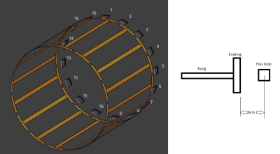 |
Application of pulse energy method (NEMA standard MS-8) for SAR measurement: location of current sensing flux loops and measurement quality
Xin Chen1, Shinji Mitsui2, Yoshinori Hamamura1, and Michael Steckner1
1Canon Medical Research USA, Inc., Mayfield Village, OH, United States, 2Canon Medical Systems Corporation, Otawara-shi, Japan
Pulse energy SAR measurement method (NEMA standard MS-8) uses current sensing flux loops to measure coil power loss, which varies in different imaging conditions. Modeling results suggest that using multiple flux loops distributed around Tx coil, as presently defined by the standard, may not give accurate results. For whole body birdcage coil loaded with a human subject, single flux loop spaced from endring and positioned at 3 or 9 o’clock may give more accurate results.
|