
Digital Poster Session
Engineering: RF Coils
Engineering
4018 -4033 RF coils - RF Coils 1
4034 -4047 RF coils - RF Coils 2
4048 -4063 RF coils - RF Coils 3
4064 -4078 RF coils - RF Coils 4
4079 -4094 RF coils - Dielectric Materials & Metamaterials for Coils
4095 -4110 RF coils - Multinuclear RF Coils
4018.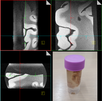 |
A tight-fit flexible high-impedance coil array for high-resolution imaging of small ex-vivo specimen using a human 7T scanner
Koji Fujimoto1, Bei Zhang2, Shin-ichi Urayama1, Atsushi Shima3, Nobukatsu Sawamoto4, Ryosuke Takahashi3, Tomohisa Okada1, and Martijn A. Cloos2
1Human Brain Research Center, Graduate School of Medicine, Kyoto University, Kyoto, Japan, 2Center for Advanced Imaging Innovation and Research (CAI2R) and Bernard and Irene Schwartz Center for Biomedical Imaging, Department of Radiology, New York University School of Medicine, New York, NY, United States, 3Department of Neurology, Kyoto University Graduate School of Medicine, Kyoto University, Kyoto, Japan, 4Department of Human Health Sciences, Kyoto University Graduate School of Medicine, Kyoto University, Kyoto, Japan
By borrowing concepts from the high-impedance glove coil, we propose an “elastic-tube coil” for imaging ex-vivo specimen at 7 Tesla. The elasticity of the sock makes it easy to setup and ensures a minimal distance between the coils and the specimen. Two high-impedance elements were stitched to an elastic tube suspended in a custom 3D printed housing. Volumetric gradient echo images of an ex-vivo human brain specimen were acquired with 60um-isotropic resolution.
|
|
4019. |
A Novel Clinical Friendly 7T T/R 32-Channel Head Coil Using Skipped-Rung Birdcage as Transmitter
Tsinghua Zheng1, Xiaoyu Yang1, Blaise Whitesell1, Martin Domondon1, Paul Taylor1, Samuel Musilli1, and Labros Petropoulos1
1Quality Electrodynamics, LLC, Mayfield Village, OH, United States
We propose a novel skipped-rung birdcage coil as the local transmitter for head imaging at 7 Tesla. This concept allows sizable openings on the coil anterior. The openings improve both patient comfort and workflow. A 7T head coil using the skipped-rung birdcage transmitter and a 32-channel receiver array was constructed and evaluated. The preliminary test results show a good transmitting efficiency and a good image quality compared with a commercial 7T head coil.
|
|
4020. |
Human colon imaging: simulation of novel SwiM RE-coils
Hamza Raki1,2, Kevin Tse Ve Koon1, Henri Souchay2, Simon A Lambert1, Fraser Robb3, and Olivier Beuf1
1Univ Lyon, INSA-Lyon, Université Claude Bernard Lyon 1, UJM-Saint Etienne, CNRS, Inserm, CREATIS UMR 5220, U1206, F-69100, Lyon, France, 2GE Healthcare, Buc, France, 3GE Healthcare, Aurora, OH, United States Coil-loop geometries were defined and simulated on electromagnetic (EM) software FEKO. Image intensity distribution was evaluated for different orientations with respect to B0. Overall, single loop and double loop with opposite current show complementary results. The switch between these two loops could be realized using MEMS (Micro Electro-Mechanical System) switches. This can reduce the dependency of the coil-sensitivity to its orientation with respect to B0 and can lead to the development of SwiM RE-coils (SWItches MEMS for Reconfigurable Endoluminal coils). |
|
4021. |
A Mono-surface 8Tx/16Rx Antisymmetric Dipole Antenna Array for Parallel Transmit Cardiac MRI in Pigs at 7T
Ibrahim A. Elabyad1, M. Terekhov1, and Laura M. Schreiber1
1Chair of Cellular and Molecular Imaging, Comprehensive Heart Failure Center (CHFC), University Hospital Wuerzburg, Wuerzburg, Germany
A mono-surface 8Tx/16Rx antisymmetric dipole antenna array was designed and tested for parallel transmit (pTx) cardiac magnetic resonance imaging (cMRI) in pigs at 7T. The antisymmetric array comprised of a mono-surface 16-dipoles arranged so that two central L-shaped dipoles were anti-symmetrically flanked by 7-dipoles on either side. Combined FA, SNR, and g-factor maps were acquired in phantom using the dipole antenna array and compared with an 8Tx/16Rx loop array. After $$$B_1^+$$$-shimming using the dipole array, the SNR was enhanced by ~20% and FA by ~42%. Dipole antenna array has demonstrated ~20-times improvement in RSD of the FA after $$$B_1^+$$$-shimming.
|
|
4022. |
Development and RF Shimming of an 8Tx/16Rx Antisymmetric Transceiver Coil Array for Parallel Transmit Cardiac MRI in Humans at 7T
Ibrahim A. Elabyad1, M. Terekhov1, and Laura M. Schreiber1
1Chair of Cellular and Molecular Imaging, Comprehensive Heart Failure Center (CHFC), University Hospital Wuerzburg, Wuerzburg, Germany
To develop and optimize an 8Tx/16Rx antisymmetric transceiver coil array with improved characteristic for static phase $$$B_1^+$$$-shimming and parallel receive for human cardiac MRI at 7T. The array design was based on antisymmetric loop configurations in 2-different directions (L/R and A/P). EM-simulations were carried out in phantom and 4-human models (Duke, Ella, Gustav, and Laura). $$$B_1^+$$$-shimming has been carried out for 16-elements with three different cost functions to improve $$$B_1^+$$$-field homogeneity, $$$Tx_{eff}$$$ and weighted combination of both within Duke and Ella models. The hardware and imaging performance of the antisymmetric array was validated through EM-simulations and phantom MR-measurements at 7T.
|
|
4023.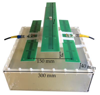 |
Dipole Decoupling with Parasites
Gregor F. Neumann1,2, Mark E. Ladd1,2, and Arthur W. Magill1
1Medical Physics in Radiology, German Cancer Research Center (DKFZ), Heidelberg, Germany, 2Physics & Astronomy, Heidelberg University, Heidelberg, Germany
We investigate the decoupling of a pair of fractionated dipoles using an additional passive parasitic dipole. Decoupling is optimised either geometrically, by adjusting the parasite length and standoff distance from the active dipoles, or by adding resistance and reactance to the parasite centre. Adjusting two parameters allows control over the amplitude and phase of the decoupling signal induced via the parasite, producing very strong decoupling at the target frequency (better than -40 dB). The effect is demonstrated using EM simulation and verified with bench measurements.
|
|
4024.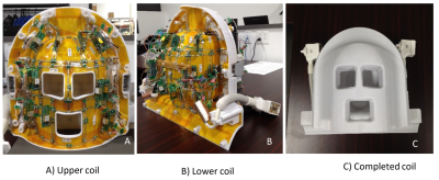 |
A 30-Channel Single Layer Transceiver Head Coil at 3.0T
Haoqin Zhu1, Xiaoyu Yang1, Michael Wyban1, Yiping Guan2, and Yoshinori Hamamura2
1Quality Electrodynamics, LLC (QED), Mayfield Village, OH, United States, 2Canon Medical Research USA, Inc, Mayfield Village, OH, United States
A novel open design 30ch transceiver head coil without using a local RF power source is proposed. Each coil element has no detuning circuit, couples to the system whole body coil inductively, and generates a uniform B1+ field in transmit mode. The same coil element also acts as a typical phased array element in receive mode. The transceiver feature of the coil may benefit the SAR demanding simultaneous multi-slice (SMS)/multi-band (MB) and high-resolution imaging and still keep the clinical friendly openings. In addition, the same coil element approach simplifies the coil circuit and improves the signal-to-noise ratio (SNR).
|
|
4025. |
A Novel Ultra-Flexible High-Resolution AIR (Adaptive imaging receive) 64-Channel Bilateral Phased Array for 3T Brachial Plexus MRI
Yun-Jeong Stickle1, Clyve Follante1, Mark Giancola1, David Anderson1, Fraser Robb1, Victor Taracila1, Robert Stormont2, Holly Blahnik3, Simone Winkler4, and Darryl Sneag5
1MR Engineering, GE Healthcare Coils, Aurora, OH, United States, 2MR Engineering, GE Healthcare, Waukesha, WI, United States, 3MR Apps&WF, GE Healthcare, Waukesh, WI, United States, 4MRI Engineering, Weill Cornell Medicine, New York, NY, United States, 5Radiology and Imaging, Hospital for Special Surgery, New York, NY, United States
An ultra-flexible AIR 64-Channel bilateral phased array coil is described for acquiring high-sensitivity images of the brachial plexus by wrapping coil elements snugly around the cervical, shoulder, axillary, and arm regions. This coil comprises a foam posterior and flexible flaps with a low loss malleable conductor optimized for zero reactance. An exceptionally low noise preamplifier is tolerant of a wide range of loading. This coil facilitates imaging large fields of view bilateral brachial plexi, as well as higher resolution unilateral imaging for detailed evaluation of nerve fascicular architecture and other pathology, thereby optimizing setup time and patient comfort.
|
|
4026. |
Developing High Channel Count Receive Arrays for Human Brain Imaging at 10.5T
Nader Tavaf1,2, Russell L. Lagore1, Steve Jungst1, Shajan Gunamony3, Jerahmie Radder1, Andrea Grant1, Edward Auerbach1, Steen Moeller1, Kamil Ugurbil1, Gregor Adriany1, and Pierre-Francois Van de Moortele1
1Center for Magnetic Resonance Research (CMRR), University of Minnesota Twin Cities, Minneapolis, MN, United States, 2Department of Biomedical Engineering, University of Minnesota Twin Cities, Minneapolis, MN, United States, 3Centre for Cognitive Neuroimaging, University of Glasgow, Glasgow, Scotland
Highly decorrelated, high channel count receive arrays are a prerequisite to capturing the signal-to-noise ratio and acceleration performance potential of ultra-high field MRI. A self-decoupled 32-channel receive array was built for human brain imaging at 10.5T. Noise correlation and signal-to-noise ratio (SNR) of the RF coil were measured in phantom experiments at 10.5T. SNR was compared to a commercial 32-channel receiver array at 7T. Noise correlation matrices demonstrated effective decoupling of receive elements. Experimental SNR measurements demonstrated on average 60% higher overall SNR at 10.5T compared to 7T.
|
|
4027. |
A 4-Channel Conformal Hybrid Coil Array for Rat Limb Imaging at 7T
Jan Paska1,2, Amparo Ruiz1,2, and Jose Raya1,2
1Center for Advanced Imaging Innovation and Research (CAI2R), NYU School of Medicine, New York, NY, United States, 2Center for Biomedical Imaging, Department of Radiology, NYU School of Medicine, New York, NY, United States
We aim to image the rat limb in longitudinal follow up studies using contrast agents at 7T. Consistent positioning and a high and uniform SNR is required over the ROI. Commercial surface coil arrays offer a high SNR close to the surface but are difficult to position and have poor penetration. Therefore we decided to design a dedicated coil array taking into account the animal's anatomy. We optimized the design in simulations, compared it to a commercial surface coil array in a phantom, and show first in-vivo images.
|
|
4028. |
Multilayer Radiofrequency Coils: A Novel Surface Coil Design for Improved B1+ Efficiency and Signal-to-Noise
Tony Zhou1,2, Justin YC Lau1,2, Jack JJJ Miller1,2,3, Navjeet Chhina4, Christopher Randell4, and Damian J Tyler1,2
1Department of Physiology, Anatomy and Genetics, University of Oxford, Oxford, United Kingdom, 2Oxford Centre for Clinical Magnetic Resonance Research, University of Oxford, Oxford, United Kingdom, 3Department of Physics, University of Oxford, Oxford, United Kingdom, 4PulseTeq LTD, Chobham, United Kingdom
Surface transmitters are often B1+ limited for X-nuclear applications due to hardware limitations of broadband power amplifiers. For both X-nuclear MRI and proton MRI, improved signal detection is always desired. Commonly, signal is measured using surface radiofrequency loop coils or coil arrays. We propose a novel surface coil design using stacked copper layers from one continuous track for increased magnetic flux and sensitivity over a central region of the coil. ‘Multilayer’ designs promise increased B1+ efficiency and Signal-to-Noise, seen using electromagnetic simulations and verified experimentally when compared to conventional loop coils over a range of diameters.
|
|
4029. |
An Implantable MRI Micro-coil of Blood vessel for High Sensitivity and High SNR Imaging at high field MRI.
Sana Ullah1 and Hyoungsuk Yoo1
1Department of Biomedical Engineering, Hanyang University, Seoul, Republic of Korea
The importance of implantable micro-coil has been rapidly increased due to its advantages in diagnosing intravascular and brain diseases. In this study, the receive-only conformal-shaped micro-coil was designed and presented. Our research demonstrated that homogeneous B1- magnetic field distributions and receptive sensitivity around the blood-vessel walls were achieved by using the proposed micro-coil.
|
|
4030. |
An Inductive Coupling Neck Coil for Expanding Effective Coverage and Increasing SNR at 3.0T
Guangqi Li1, Xinyuan Wang2, Zhi Zhao2, Wanshan Li2, Yishi Wang3, and Hua Guo1
1Center for Biomedical Imaging Research, Department of Biomedical Engineering, School of Medicine, Tsinghua University, Beijing, China, 2Tsimaging Healthcare Ltd, Beijing, China, 3Philips Healthcare, Beijing, China
The image uniformity and signal to noise (SNR) is not high enough for extra-cranial imaging with the existing Head-Neck coil due to the limited channel number of the neck section. Traditionally, minimizing the coupling between nearest-neighbor coils is necessary for eliminating signal crosstalk. However, in this work, we propose an inductive coupling coil design to increase effective coverage without any signal interference, thereby breaking through the limitation of the channel number. Extra-cranial imaging has better image uniformity and 16.5% higher SNR using the new neck coil at 3.0T.
|
|
4031. |
Development of a biplanar volume coil for an open 0.1 T MRI system
Maksym Yushchenko1, Najat Salameh1, and Mathieu Sarracanie1,2
1Laboratory for Adaptable MRI Technology (AMT lab), Dpt of Biomedical Engineering, University of Basel, Allschwil, Switzerland, 2Medical Image Analysis Center (MIAC AG), Basel, Switzerland Poster Permission Withheld
An open, biplanar volume RF coil was built and tested as a transceiver with a homogeneous B1 at 4.2 MHz. We demonstrate its feasibility and performance in a low-field, open biplanar magnet. While giving good sensitivity, this geometry provides easy access to the object from multiple sides. In addition to simpler positioning of patients, this potentially opens the way for improved MRI applications where access is primarily required such as interventional MRI.
|
|
4032. |
Flexible torso array coil for Fast Field-Cycling MRI at 8.5 MHz
Robert S Stormont1,2, Broche M Lionel1, P. James Ross1, Gareth R. Davies1, and David J. Lurie1
1School of Medical Sciences, Aberdeen Biomedical Imaging Centre, University of Aberdeen, Aberdeen, United Kingdom, 2Magnetic Resonance Imaging, GE Healthcare, Waukesha, WI, United States An array coil for 0.2 T reception has been developed for colon and heart studies in a FFC-MRI system. The array consists of 3 element posterior/anterior sections, with 16cm loops arranged in a “venn” pattern. Minimizing conductor losses and noise figure over varying loading is particularly important in a low field system. A two-turn litz arrangement was chosen offering good flexibility and Qu/Ql ratios. Interfacing electronics component topologies were simplified losses keping losses to a minimum. Matching, blocking, decoupling, and preamplification reside at element. Packaging is flexible textiles and substrates. The resulting array conforms and retains performance with varying anatomy. |
|
4033. |
RF endoluminal coils with NMR electro-optical conversion and transmission for colon wall imaging: Initial finding
Paul Nobre1, Alice Ferrando1, Gwenaël Gaborit2,3, Raphaël Sablong1, and Olivier Beuf1
11Univ. Lyon, CREATIS ; CNRS UMR 5220 ; INSERM U1206 ; INSA-Lyon ; UJM-Saint Etienne ; Université Lyon1 ; 69616 Villeurbanne, France, Lyon, France, 2University of Savoie, IMEP-LAHC, UMR 5130, 73376 Le Bourget-du-Lac, France, Le Bourget-du-Lac, France, 3KAPTEOS, 73376 Sainte-Hélène-du-Lac, France, Sainte-Hélène-du-Lac, France
Receive surface coils in MRI have proven to enable higher spatial or temporal resolution. However, the proximity of the galvanic connexions with the body when the coil is used as receiver coil together with a volume coil can lead to safety issues, in particular for inner coil. This configuration can induce currents in coaxial cable and local SAR increase can be induced. Replacing the galvanic connexions with optical ones could prevent those risks and enable safe colon wall imaging with endoluminal coils. First experimental demonstration of an electro-optic conversion based on Pockel’s effect is demonstrated at 4.7T.
|
 View the Poster
 Watch the Video
4034. |
A tight-fit 8-channel transceiver array for simultaneous EEG-fMRI at 7-Tesla
Gavin Paterson1, Paul McElhinney1, Marios G Philiastides1, and Shajan Gunamony1
1University of Glasgow, Glasgow, United Kingdom
Simultaneous EEG-fMRI exploits complementary information from the two modalities and offer insights into the dynamics of the brain function. In this work, we developed a tight-fit 8-channel transceiver head coil which facilitates the use of a 64-electrode EEG setup for simultaneous EEG-fMRI at 7T. Influence of the EEG cap on transmit and receive performance of the coil is presented in this work.
|
|
4035. |
Signal-to-noise measurement of a 4-channel parallel radiofrequency transmission system for MRI at 3 T
Benson Yang1,2, Chih-hung Chen2, and Simon J Graham1,3
1Physical Sciences, Sunnybrook Research Institute, Toronto, ON, Canada, 2Electrical and Computer Engineering, McMaster University, Hamilton, ON, Canada, 3Medical Biophysics, University of Toronto, Toronto, ON, Canada
Parallel radiofrequency transmission (pTx) is a highly researched topic. As a result, researchers have implemented different in-house system designs to investigate its clinical benefits. The performance specifications of these custom systems are often unknown. Signal-to-noise (SNR) ratio analysis is an important consideration for optimal imaging. This work uses a simple multi-purpose method to conduct SNR analysis and noise characterization of a 4-channel pTx system integrated on an existing 3 T MRI system.
|
|
4036. |
Open-Sourced, Stereotax-Integrated, High-Fidelity and Adjustable 4-Channel Receive Array for Primate Brain Imaging at 3T
Marc Fischer1, Karthik Gopalan1, Ana Arias1, and Michael Lustig1
1EECS, UC Berkeley, Berkeley, CA, United States
High quality MRI scans of nonhuman primates allow researchers to gain a deeper understanding of the brain. Most commercially available coils are designed for human imaging and are not compatible with common animal head restraint systems. We describe the design and testing of a 4-channel coil array that interfaces with a commercially available sterotax. Each channel is integrated with a modified Loc-line that enables the positioning of each coil independently in order to get the coils as close to the subject as possible. SNR maps reveal exceptional performance and good coil decoupling compared to three commercially available human coils.
|
|
4037. |
32-Channel Lightweight Dipole RF Array for Cardiac MRI at 7.0 Tesla
Thomas Wilhelm Eigentler1, Andre Kuehne2, Laura Boehmert1, Eva Oberacker1, Soeren Lippert1, and Thoralf Niendorf1,2,3
1Berlin Ultrahigh Field Facility, Max-Delbrück-Center for Molecular Medicine in the Helmholtz Association, Berlin, Germany, 2MRI.TOOLS GmbH, Berlin, Germany, 3Experimental and Clinical Research Center (ECRC), a joint cooperation between the Charité Medical Faculty and the Max-Delbrück Center for Molecular Medicine, Berlin, Germany
A lightweight, high-density transceiver RF array of 32 compact self-grounded bow-tie antennas was developed, manufactured, evaluated, and applied for cardiac MRI at 7.0 T. Our work contributes to the technological basis for future clinical assessment of parallel transmit techniques of the heart for optimization of RF antenna placement and RF antenna count with the goal to approach ultimate SNR in ultrahigh field MRI of the heart.
|
|
4038. |
Validation and Safety Approval of a Dual-Mode Head Coil for pTx Applications In Vivo at 7 Tesla
Sydney N. Williams1, Sarah Allwood-Spiers2, Paul McElhinney1, Yuehui Tao3, John E. Foster2, David A. Porter1, and Shajan Gunamony1,4
1Imaging Centre of Excellence, University of Glasgow, Glasgow, United Kingdom, 2MRI Physics, NHS Greater Glasgow and Clyde, Glasgow, United Kingdom, 3Imaging Centre of Excellence, Siemens Healthcare Ltd. United Kingdom, Glasgow, United Kingdom, 4MR CoilTech, Glasgow, United Kingdom
Following regulatory approval of single-transmit MRI at 7 tesla, there is a rapidly growing interest in clinical MRI at this field strength. However, the wider use of diagnostic MRI at 7T will require imaging in parallel-transmit (pTx) mode to reduce B1+ inhomogeneity. Previous work introduced a dual-mode head coil that operates in both transmit modes and this study investigates the use of this coil for the pTx case. It also describes the safety procedure that was followed to ensure safe operation for human scanning using the real-time SAR supervision on a commercial scanner.
|
|
4039.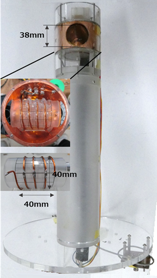 |
Development of variable-temperature MR microimaging system with a large-diameter solenoid coil for a vertical wide bore superconducting magnet
Naoya Takagawa1, Masaya Ishikawa2, and Yasuhiko Terada1
1Institute of Applied Physics, University of Tsukuba, Tsukuba, Japan, 2Department of Forest Science, Graduate School of Agricultural and Life Sciences, The University of Tokyo, Tokyo, Japan
We have developed a large-diameter, solenoid probe for a wide bore NMR superconducting magnet that can image samples at different temperatures. The controllable temperature range was -20 to 20 °C. To demonstrate the performance, plant buds were imaged at different temperatures, and the frozen process was visualized.
|
|
4040.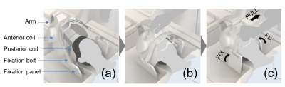 |
Evaluation of signal-to-noise ratio of a size-adaptable 16-channel head array
Kohjiro Iwasawa1, Yosuke Otake2, Kazuyuki Kato2, Hideta Habara2, Masayoshi Dohata2, Hisaaki Ochi1, and Toru Shirai1
1Research & Development Group, Hitachi, Ltd., Tokyo, Japan, 2Healthcare Business Unit, Hitachi, Ltd., Tokyo, Japan Poster Permission Withheld
In order to increase both coil setting efficiency and coil sensitivity, a size-adaptable tight-fit 16ch head array which enables one-action-fixation was investigated at 3 T. The anterior part of the head array is a flexible coil, and can fit to the patient when the fixation belt is tightened. SNR increased by 1.2-1.4 times across 1-99 percentile adult head sized phantoms against a conventional 15ch rigid head coil. SNR improvement could also be seen in vivo with a healthy volunteer.
|
|
4041.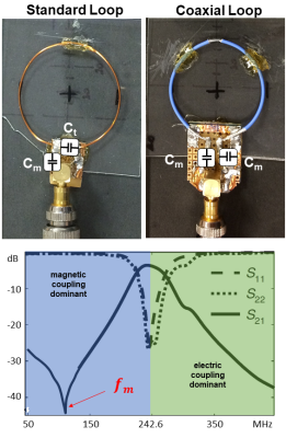 |
Comparison of magnetic and electric coupling coefficients for coaxial coils and standard loop coils
Bernhard Gruber1,2 and Elmar Laistler1
1Division MR Physics - Center for Medical Physics and Biomedical Engineering, Medical University Vienna, Vienna, Austria, 2Massachusetts General Hospital, Harvard Medical School, A. A. Martinos Center for Biomedical Imaging, Charlestown, MA, United States
Coaxial coils as highly flexible elements of RF coil arrays have been proposed, and superior inter-element decoupling over standard loops has been reported, but so far without convincing explanation of the underlying mechanism. In an attempt for an explanation measurements of the magnetic and electric coupling coefficients for coaxial and standard loop coils were performed, and showed differences in the S21 findings and the coupling parameters, which are topic of further investigations.
|
|
4042.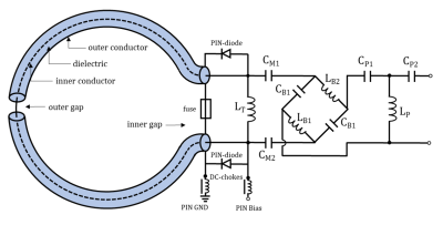 |
Optimization and miniaturization of Rx-only coaxial coil interfacing.
Michael Obermann1,2, Lena Nohava1,2, Raphaela Czerny1, Roberta Frass-Kriegl1, Sigrun Roat1, Michael Pichler1, Jacques felblinger3, Jean-Christophe Ginefri2, and Elmar Laistler1
1Division MR Physics, Medical University of Vienna, Vienna, Austria, 2IR4M, Université Paris-Saclay, Orsay, France, 3Université de Lorraine, Nancy, France
Coaxial coils have been introduced recently, the way of interfacing these coils impacts the achievable SNR. In this work, the interfacing circuitry is studied to optimize sensitivity and ensure safety. Performance tests on the bench and in the MR scanner were done for 8-cm 123 MHz coaxial coils, operated at their self-resonance. It was found that a high inductance across the port and symmetric matching capacitors increase SNR. A balun and a phase shifter for preamp decoupling are described, the correct positioning of a fuse is demonstrated. Miniature preamplifiers and toroid inductors minimize size and weight to obtain maximum flexibility.
|
|
4043. |
An automated 32 channel VNA RF switch for high density coil prototyping
Dimitri Welting1, Carel C. van Leeuwen1, Alexander J.E. Raaijmakers1, and Dennis W.J. Klomp1
1Department of Radiology, University Medical Center Utrecht, Utrecht, Netherlands
An automated 32 channel vector network analyzer switch was developed in-house, to be used for high density coil arrays. Using this device full S11 and S21 measurements of 32 channels can be acquired on the bench in less than 2.5 minutes. Performance of the device was evaluated using a 32 channel 7T headcoil and a 8 channel 7T dipole body array.
|
|
4044. |
Strategies to reduce coupling for continuous acquisition of a Bloch-Siegert spatial encoding in low-field MRI device
BAOSONG WU1, Charles Rogers III1, Sajad Hosseinnezhadian1, Yonghyun Ha1, Kartiga Selvaganesan1, Gigi Galiana1, and R. Todd Constable1
1Department of Radiology and Biomedical Imaging, Yale University, NEW HAVEN, CT, United States
This work describes methods and systems to implement Bloch-Siegert(BS) spatial encoding using simultaneous transmit and receive in low-field MRI. The key challenge is to reduce coupling power in receiver chains so that the on-resonance signal can be acquired during the off-resonance irradiation. The developments include combining filters and coil decoupling methods. Together these methods suppress irradiation leakage more than 60dB which, for an anticipated offset B1 field of 60 dBm, allows for spatial encoding in low-field MRI device.
|
|
4045. |
Optimized Quadrature Head Coil Improves SNR in the Brain at 6.5mT
Neha Koonjoo1,2,3, Sheng Shen1,4, Charlotte R Sappo5, and Matthew S Rosen1,2,3
1Department of Radiology, A.A Martinos Center for Biomedical Imaging/MGH, Charlestown, MA, United States, 2Harvard Medical School, Boston, MA, United States, 3Department of Physics, Harvard University, Cambridge, MA, United States, 4State Key laboratory for Power Transmission Equipment and System Security and New Technology, Chongqing University, Chongqing, China, 5Institute of Imaging Science, Vanderbilt University, Nashville, TN, United States
High performance RF coil design is challenging at ultra-low field as rules of thumb learned from high-field may not apply. Previously, we built a 2-channel quadrature head coil with orthogonal B1 fields using an air-core transformer to null the mutual coupling between the coil, however this was unsuccessful in boosting the SNR beyond that of our single-channel spiral volume coil for brain imaging at 276 kHz. Here, a new optimized quadrature coil with a capacitive decoupling element and a new outer layer coil was designed. Images acquired show an expected √2-factor or more enhancement in combined maximum SNR.
|
|
4046. |
The feasibility study about the open RF loop & the simple RF circuit topology
Seunghoon Ha1 and Adam Morris1
1Philips Healthcare, Pewaukee, WI, United States Poster Permission Withheld
It is introduced that the open loop coil with the simple and efficient RF circuit topology with the identical or better coil performance nevertheless less RF components usage. It is analyzed and measured to prove this proposal. It is compared to the conventional loop coil by thermal test and Q-measurements.
|
|
4047.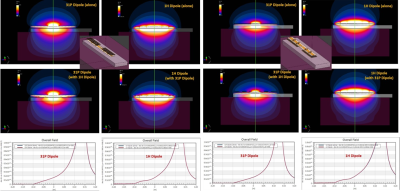 |
Stacked dipole arrays for 31P and 1H MRI in the body at 7T
Ria Forner1, Ettore Flavio Meliadò2,3, Martijn Lunenburg3, Catalina Arteaga de Castro3, Ladislav Valkovič4, Christopher T. Rodgers4,5, Alexander Raaijmakers2,6, and Dennis Klomp2
1Radiology, UMC Utrecht, Utrecht, Netherlands, 2UMC Utrecht, Utrecht, Netherlands, 3TeslaDC, Zaltbommel, Netherlands, 4Oxford Centre for Magnetic Resonance Research, University of Oxford, Oxford, United Kingdom, 5Dept of Clinical Neurosciences, University of Cambridge, Cambridge, United Kingdom, 6Biomedical Engineering, Eindhoven University of Technology, Utrecht, Netherlands
While an RF birdcage integrated behind the bore-liner of a 7T MRI can provide relatively uniform 31P excitation, it requires substantial system integration activities. Here we present an alternative approach using an array of stacked dipole antennas where one is tuned to 1H and the other to 31P. The setup is significantly easier to configure and while 31P B1+ uniformity is somewhat compromised, B1+ is more efficient when compared to an integrated birdcage.
|
4048. |
Sensitivity Enhancement at 7T Brain MR imaging Using Wireless Coupled-Split-Ring-Resonators Array
Akbar Alipour1, Gaurav Verma1, Michael Bush2, Judy Alper1, and Priti Balchandani1
1Biomedical Engineering and Imaging Institute, Icahn School of Medicine at Mount Sinai, New York, NY, United States, 2Siemens Medical Solutions USA, Inc, New York, NY, United States
Image quality in the brain periphery at ultra high field MRI is severely limited by low sensitivity. In this work we illustrate an efficient method for enhancing imaging sensitivity at 7T in a cadaver brain. The method is based on the application of a wireless coupled-split-ring-resonator array that amplifies the MR signal during acquisition, which can alleviate the low sensitivity problem in the brain periphery when low flip angle and low SAR sequences are needed. Initial ex-vivo 7T MR brain imaging results showed that 2-fold to 6-fold SNR gain was obtained using this array.
|
|
4049. |
RF field extension and improvement by using floating strip conductors for parallel microstrip coil for PET/MRI insert
Md Shahadat Hossain Akram1, Takayuki Obata2, and Taiga Yamaya1
1Advance Nuclear Medicine Sciences, National Institute of Radiological Sciences (NIRS-QST), Chiba, Japan, 2Molecular Imaging and Theranostics, National Institute of Radiological Sciences (NIRS-QST), Chiba, Japan
For parallel transmit coils, maximum field-intensity is seen for the ROI close to each coil and, peripheral imaging region in between the coils usually show minimum field-intensity. For this reason, a compact orientation of multiple coils is usually used to surround the ROI. An approach of field extension to the regions in between coils is presented here for a 4-channel microstrip coil. Multiple narrow floating copper strips (width 4 mm) were used in between coils. For three such strips, image signal intensity between the coils almost doubled, that resulted in 20% increase in image homogeneity and 25% increase in SNR.
|
|
4050. |
The Very RF Hungry Caterpillar Trap (Highly Flexible, Distributed System of Toroid Cable Traps)
Ekin Karasan1, Victor Taracila2, Fraser Robb2, and Michael Lustig1
1Department of Electrical Engineering, University of California, Berkeley, CA, United States, 2GE Healthcare, Coils, Aurora, OH, United States “In the light of the moon a little egg lays on a leaf”[7] Management of cabling connecting coil arrays to system remains the nightmare of RF-engineers. Coupling within arrays to cabling reduces performance, and coupling to transmit field causes high shield currents. Consequently, traps are placed on conductors. To provide high blocking, traps are rigid, large and often heavy, hindering flexibility. Instead of a few high blocking traps, we propose a distributed system of small, elastic traps. We leverage self-shielded resonant toroids forming a caterpillar-like structure. We show that this design can attenuate shield currents while being robust to flexing. |
|
4051.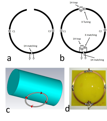 |
Design and evaluation of a dual-tuned circular dipole antenna up to 14T MRI
Suk-Min Hong1, Chang-Hoon Choi1, Jörg Felder1, and N. Jon Shah1,2,3,4
1Institute of Neuroscience and Medicine 4, INM-4, Forschungszentrum Jülich, Jülich, Germany, 2Institute of Neuroscience and Medicine 11, INM-11, JARA, Forschungszentrum Jülich, Jülich, Germany, 3JARA - BRAIN - Translational Medicine, Aachen, Germany, 4Department of Neurology, RWTH Aachen University, Aachen, Germany
When the field strength of MRI is increased to 14T, the capacitor values required to tune a conventional loop coil become extremely small. For 7T MRI, the circular dipole antenna has been introduced and evaluated as the array structure and new decoupling method. In this study, we modified a circular dipole antenna to a dual-tuned circuit by using an LC trap. The coil operates as a circular dipole antenna for the proton signal as the trap blocks 1H current and operates as a conventional loop at the X-nuclei frequency. The dual-tuned circular dipole antenna was evaluated up to 14T frequency.
|
|
4052. |
A Patient-Friendly Coil For Bilateral Carotid Artery Imaging
Bili Wang1, Justin Ho 1, Jerzy Walczyk1, Thanh D Nguyen2, Bei Zhang1, and Ryan Brown1
1Radiology, New York University Langone Health, New York, NY, United States, 2Radiology, Weill Cornell Medical College, New York, NY, United States
Carotid MRI imaging can be vital in preventing heart disease and stroke but is difficult due to variable neck anatomy. Traditional rigid coils have a hard time achieving a close fit on all patients which can result in SNR loss that is essential for delineating the thin vessel wall. We designed and implemented an 8-channel flexible coil by incorporating the resonators into tailored cloth sleeves with integrated semi-rigid skeletons and a head cradle that provided the means for straightforward patient positioning and comfort, and a contoured fit for high SNR.
|
|
4053. |
A radiolucent 32-channel high impedance coil receive array to accelerate 3D imaging on hybrid 1.5 T MR-linac systems
Stefan Emiel Zijlema1,2, Luca van Dijk1, Jan J.W. Lagendijk1, Dennis W.J. Klomp3, Catalina S. Arteaga de Castro4, Rob H.N. Tijssen1, and Cornelis A.T. van den Berg1,2
1Department of Radiotherapy, UMC Utrecht, Utrecht, Netherlands, 2Computational Imaging Group for MRI diagnostics and therapy, Centre for Image Sciences, UMC Utrecht, Utrecht, Netherlands, 3Department of Radiology, UMC Utrecht, Utrecht, Netherlands, 4Tesla Dynamic Coils, Zaltbommel, Netherlands High impedance coils (HICs) offer major advantages for use during MRI-guided radiotherapy (MRIgRT), as they lack lumped elements that can attenuate radiation. Furthermore, their flexibility and low channel coupling simplify high-density array development and enable on-body placement. Here, we present a fully functional 32-channel HIC array, confirmed its radiation transparency (radiolucency) and compared the imaging performance with the current clinical (LIC-based) array. Dosimetrically, no clinically significant attenuation was caused by the array. Imaging-wise, the prototype showed higher SNR values and lower g-factors, thus allowing for faster imaging. In conclusion, our 32-channel array can accelerate all imaging for MRIgRT applications. |
|
4054.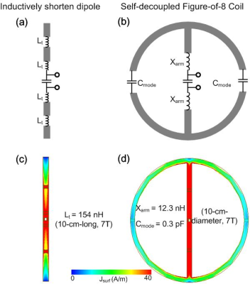 |
Self-decoupled Folded Dipole Antenna
Ming Lu1,2,3, John C. Gore1,2, and Xinqiang Yan1,2
1Vanderbilt University Institute of Imaging Science, Nashville, TN, United States, 2Department of Radiology, Vanderbilt University Medical Center, Nashville, TN, United States, 3College of nuclear equipment and nuclear engineering, Yantai University, Yantai, China
In ultra-high-field MRI, both curl-free (dipole-like) and divergence-free current patterns (loop-like) are desired for better parallel transmission performance as well as to approach the ultimate receive performance for deep tissues. But decoupling dipole antennas is still a challenging topic. However, as shown in previous work, the strongest coupling in a dipole array is actually end-to-end between elements. Another challenge when using dipoles in dense arrays comes from their length, 45 cm at 300 MHz. We propose a novel, folded figure-of-eight (Fo8) configuration to shorten the dipole and simultaneously make it self-decoupled from others.
|
|
4055.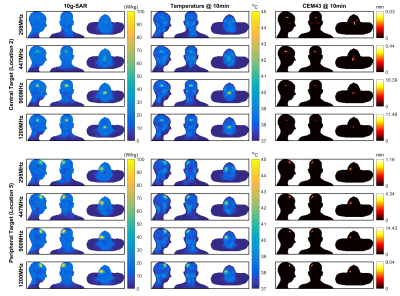 |
Simulation of Focused RF Heating using a High-Channel-Count RF Array and a Maximum SAR Algorithm
Koray Ertan1,2, Joshua De Bever1, Mihir Pendse1, Paolo Decuzzi2, and Brian Rutt1
1Department of Radiology, Stanford University, Stanford, CA, United States, 2Italian Institute of Technology, Genoa, Italy
A 70-channel parallel transmit RF coil array intended for focused RF applications was designed and simulated at four frequencies (298 MHz, 447 MHz, 900 MHz and 1200 MHz) using a Ella body model. A previously proposed maxSAR algorithm was used to focus SAR in target regions while limiting the SAR in background tissues. Spatial maps of optimized SAR distribution are shown for several target locations and all frequencies. SAR distributions together with temperature and CEM43 maps suggest that clinical levels of focused hyperthermia can be achieved using high channel count parallel transmit RF coils and the maxSAR algorithm.
|
|
4056. |
Development and Evaluation of a pTx Transceiver Cardiac Array Based on Y-Shape Central 3-Loop Arrangement for Cardiac MRI in Pigs at 7T
Ibrahim A. Elabyad1, M. Terekhov1, D. Lohr1, and Laura M. Schreiber1
1Chair of Cellular and Molecular Imaging, Comprehensive Heart Failure Center (CHFC), University Hospital Wuerzburg, Wuerzburg, Germany
An 8-element anterior array based on central 3-elements Y-shape arrangement was combined with a rectilinear 8-elements posterior array to form a dedicated 8Tx/16Rx pTx transceiver cardiac array for pigs was developed. The hardware and imaging performance of the pig array was validated through EM-simulations, phantom and ex-vivo MR-measurements at 7T. Combined SNR, FA, g-factor maps, and high-resolution ex-vivo cardiac images were acquired with an in-plane resolution of 0.3mm×0.3mm. A SNR of 36±23 was achieved within the heart of the pig. Parallel imaging with acceleration factor (R=4) was possible while keeping the mean g-factor within the heart region at 1.14.
|
|
4057. |
Endoluminal imaging with MEMS in series with loop coil for active decoupling
Hamza Raki1,2, Kevin Tse Ve Koon1, Isabelle Saniour1, Henri Souchay2, Simon A Lambert1, Fraser Robb3, and Olivier Beuf1
1Univ Lyon, INSA-Lyon, Université Claude Bernard Lyon 1, UJM-Saint Etienne, CNRS, Inserm, CREATIS UMR 5220, U1206, F-69100, Lyon, France, 2GE Healthcare, Buc, France, 3GE Healthcare, Aurora, OH, United States
A Receiver-Endoluminal-Coil (REC) integrating an active-decoupling circuit based on MEMS switch in series with the loop (sMEMS) was built and characterized on bench and at 1.5T. The results were compared to a conventional PIN-diode REC. Although the quality factor of sMEMS was significantly lower (34%) than the one of PIN-diode, efficient (|S11|<-0.1dB) and fast (delays<8µs) active-decoupling were obtained for the sMEMS. Obtained MR images display no hyper intensity or artifacts due to active-decoupling failure. SNR-values and SNR-isocontours of sMEMS were similar to those of PIN-diode for GRE and lower for FSE sequences. sMEMS can bring new coil-design possibilities for endoluminal-imaging.
|
|
4058. |
Feasibility study of volume RF coils constructed using coupled H-shaped dipole antennas for MR imaging at ultrahigh fields
Shasha Yue1, Zhe Wang1, Cheng Fang1, Nan Li2, Yan Hou1, Kun Zhang1, Rong Xue3,4,5, Ye Li2,6, and Xiaoliang Zhang7
1Institute of Biophysics, Chinese Academy of Sciences, Beijing, China, 2Lauterbur Imaging Research Center, Shenzhen Institutes of Advanced Technology, Chinese Academy of Sciences, Shenzhen, China, 3State Key Laboratory of Brain and Cognitive Science, Beijing MRI Center for Brain Research, Institute of Biophysics, Chinese Academy of Sciences, Beijing, China, 4University of Chinese Academy of Sciences, Beijing, China, 5Beijing Institute for Brain Disorders, Beijing, China, 6Shenzhen Key Laboratory for MRI, Shenzhen, China, 7Department of Biomedical Engineering, State University of New York, Buffalo, NY, United States, Buffalo, NY, United States
The feasibility of constructing the non-array volume RF coil using the coupled dipole antennas has been explored. The electromagnetic coupling among the dipoles is relatively weak and not readily to form a volume coil unless massive and dense dipoles are employed. An H-shaped dipole is proposed as the basic resonant elements to construct the ultrahigh field volume RF coil. The simulation and imaging experiment results show that the proposed H-shaped dipoles can achieve sufficient electromagnetic coupling with a much-reduced number of dipoles and form a volume coil with a practical coil length and uniform B1 fields in an efficient way.
|
|
4059.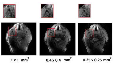 |
A flexible five-channel shielded-coaxial-cable transceive neck coil for high resolution carotid imaging at 7T
Irena Zivkovic1, Thomas Ruytenberg1, and Andrew Webb1
1Radiology Department, Leiden University Medical Center, Leiden, Netherlands
Imaging the carotid arteries at 7T requires a multichannel array which allows B1-shimming and conforms to different neck sizes. The major challenge is to minimise coupling between closely-spaced coils and to make the coupling relatively insensitive to loading conditions. We have designed a five channel transceive array composed of shielded-coaxial-cable coils placed on the anterior part of the neck and conforming to the anatomy. Coil flexibility has been demonstrated by imaging subjects with different neck circumferences. In vivo black-blood images were acquired with very high in-plane spatial resolution (0.25 x 0.25 mm2) with clear depiction of the vessel walls.
|
|
4060.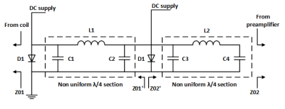 |
An automatic tuning free pin diode shunt switch with artificial non-uniform transmission line λ/4 sections for receive only surface array coils.
Miheer Pradeep Mayekar1, Rohit Apurva1, Bhaskara Naik1, and Rajesh Harsh1
1Technology Innovation Department, Society for Applied Microwave Electronics Engineering and Research, Mumbai, India
The excessive high-power during transmission saturates or in worst case damages the preamplifier. In shunt switch an artificial (lumped component) uniform λ/4 section is inserted between two shunt diodes to increase its isolation. To restore the preamplifier-decoupling performance degraded by λ/4 section, it is required to add lossy tuning components e.g. varactor diodes to change the λ/4 section’s phase. We are proposing a pin-diode shunt RF switch with two lumped component non-uniform λ/4 sections which will maintain the preamplifier decoupling performance during reception sans complex tuning components and provides additional isolation of around 10-db as compared to uniform λ/4 sections.
|
|
4061.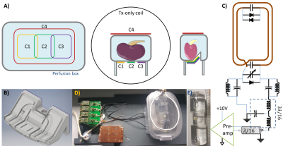 |
A Dedicated 17O Rx Array to Assess Renal Metabolism of Donor Kidneys
Ali Caglar Özen1,2, Johannes Fischer1, Hao Song1, Yanis Taege1, Christian Schuch3, Rianne Schutter4, Cyril Moers4, Ronald JH Borra5, and Michael Bock1
1Dept. of Radiology, Medical Physics, Medical Center - University of Freiburg, Faculty of Medicine, University of Freiburg, Freiburg, Germany, 2German Consortium for Translational Cancer Research Partner Site Freiburg, German Cancer Research Center (DKFZ), Heidelberg, Germany, 3NUKEM Isotopes GmbH, Alzenau, Germany, 4Department of Surgery – Organ Donation and Transplantation, University Medical Center Groningen, Groningen, Netherlands, 5Medical Imaging Center, University Medical Center Groningen, Groningen, Netherlands
Direct 17O-MRI can be used to measure renal metabolism in perfused kidneys in an organ transplantation setup. To optimize SNR, a dedicated 17O Rx array was designed that fits into a perfusion box used for functional metabolism tests of the donor kidneys. The increased filling ratio resulted in higher SNR compared to the volume and surface Tx/Rx coils. In combination with a 17O birdcage Tx coil for homogeneous excitation the 4-element Rx array could also be used for parallel imaging.
|
|
4062.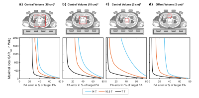 |
UHF MRI at 14 T: Initial transmit performance analysis of 8-channel local RF arrays using fractionated dipoles
Marco L. Wittrich1, Andreas K. Bitz1,2, Jonathan K. Stelter1, Mark E. Ladd1,3,4, and Thomas M. Fiedler1
1Medical Physics in Radiology, German Cancer Research Center (DKFZ), Heidelberg, Germany, 2Electromagnetic Theory and Applied Mathematics, Faculty of Electrical Engineering and Information Technology, FH Aachen – University of Applied Sciences, Aachen, Germany, 3Erwin L. Hahn Institute for MRI, University Duisburg-Essen, Essen, Germany, 4Faculty of Physics and Astronomy and Faculty of Medicine, University of Heidelberg, Heidelberg, Germany Numerical simulations were used to design and evaluate antenna arrays for body imaging at 14T. Previously presented fractionated dipole designs for 7T and 10.5T were adapted for 14T. Pulse optimization was performed in volumes with different sizes. In general, the RF shim performance decreases at higher RF frequency. The performance of 14T arrays in ROIs located in the body center is further limited by local SAR limits, as the SAR efficiency ($$$B_1^+$$$ normalized to $$$\sqrt{\operatorname{SAR}_{10g,max}}$$$) is lower in the body center. |
|
4063.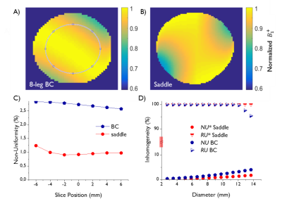 |
Saddle Coil Design for MRI/MRS at 14.1 Tesla: A Favorable Alternative to the Quadrature Birdcage Coil
Claudia Christina Zanella1, Jérémie Daniel Clément1,2, Daniel Wenz3, Bernard Lanz1, and Rolf Gruetter1
1Laboratory for Functional and Metabolic Imaging (LIFMET), EPFL, Lausanne, Switzerland, 2School of Biomedical Engineering and Imaging, King's College London, London, United Kingdom, 3Center for Biomedical Imaging – Animal and Imaging Technology (CIBM-AIT), EPFL, Lausanne, Switzerland
A single channel 1H saddle coil and a quadrature 1H 8-leg birdcage coil were compared in terms of $$$B_1^+$$$ homogeneity and $$$B_1^+$$$ efficiency for preclinical small-animal imaging at 14.1 Tesla. Electromagnetic field simulations showed that the saddle coil generated a more homogeneous $$$B_1^+$$$ field for all regions of interest (up to 13.7 mm central disk diameter) than the birdcage coil. Saline phantom measurements showed a more constant flip angle excitation upon multi-slice imaging in axial direction and a 66% higher $$$B_1^+$$$ efficiency for the saddle coil.
|
4064.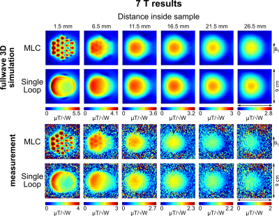 |
Multi-loop radio frequency coil elements
Roberta Frass-Kriegl1, Sajad Hosseinnezhadian2, Marie Poirier-Quinot2, Elmar Laistler1, and Jean-Christophe Ginefri2
1Division MR Physics, Center for Medical Physics and Biomedical Engineering, Medical University of Vienna, Vienna, Austria, 2IR4M (Imagerie par Résonance Magnétique et Multi-Modalités), UMR 8081, Université Paris-Sud/CNRS, Université Paris-Saclay, Orsay, France
FDTD simulations and experimental B1 mapping at 3T and 7T demonstrate that multi-loop coils (MLCs), i.e. coils made of small loops in series, provide a significant transmit efficiency boost over conventional loop coils at close distance in sample noise dominated settings. Further away, the performance of MLCs is comparable to single loop coils. The MLC principle brings additional degrees of freedom for coil design and optimization and appears advantageous for single coils as well as individual elements of arrays, especially for applications with large target area and shallow target depth, e.g. skin imaging or high resolution MRI of brain slices.
|
|
4065.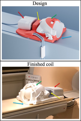 |
A custom-made design for integrating a magnetic field monitoring system into a 32ch MRI head coil
Yoojin Lee1, Michael Kennedy1, and Zoltan Nagy1
1Laboratory for Social and Neural Systems Research (SNS lab), University of Zurich, Zurich, Switzerland
While magnetic field probes can concurrently measure the spatio-temporal magnetic field dynamics during the acquisition of spiral or EPI readout, incorporating such field probes into a head coil requires careful planning and execution. The final set-up must conform to a wide array of boundary conditions for a safe and easy-to-use installation. The specific aims of this project were installing magnetic field probes into a Siemens 32ch coil and make that set-up compatible with a 3T Philips Achieva scanner. It was a high risk/gain project that was achievable only with a widely interdisciplinary approach and the kind input of colleagues.
|
|
4066. |
Dual Band Lattice Balun for Multinuclear MRI
Charlotte R Sappo1,2, William A Grissom1,2,3,4, John C Gore1,2,3,4, and Xinqiang Yan2,3
1Biomedical Engineering, Vanderbilt University, Nashville, TN, United States, 2Vanderbilt University Institute of Imaging Science, Vanderbilt University, Nashville, TN, United States, 3Radiology, Vanderbilt University, Nashville, TN, United States, 4Electrical and Computer Engineering, Vanderbilt University, Nashville, TN, United States
Baluns are transformers commonly used to connect RF coils to coaxial cables. For dual-tuned RF coils, baluns suppress the common-mode current at the Larmor frequencies for both proton and X-nuclear. This can be realized by utilizing separated single-frequency baluns in series or utilizing dual-band baluns. The LC lattice balun is widely used in RF coils since it can be built directly on the coils’ feeding board and uses minimal space. In this work, we introduce a dual-band Lattice balun design to fit the multinuclear MRI application. We analyzed, simulated and constructed the dual-band lattice balun for the 7T H/Na application.
|
|
4067.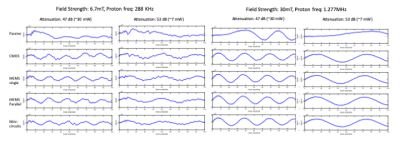 |
Active Transmit/Receive Switches for Low Field Magnetic Resonance (<100mT)
Charlotte R Sappo1,2,3, Michele N Martin3, Sheng Shen4,5, Neha Koonjoo4,6, Anthony B Kos3, William A Grissom1,2, Matthew S Rosen4,6, and Karl F Stupic3
1Biomedical Engineering, Vanderbilt University, Nashville, TN, United States, 2Vanderbilt University Institute of Imaging Science, Vanderbilt University, Nashville, TN, United States, 3National Institute of Standards and Technology, Boulder, CO, United States, 4Massachusetts General Hospital, A.A. Martinos Center for Biomedical Imaging, Boston, MA, United States, 5Electrical Theory and New Technology, Chongqing University, Chongqing, China, 6Physics, Harvard University, Cambridge, MA, United States
Low-field MRI is of increasing interest due to its low cost, improved safety, portability, and low power requirements. A transmit/receive switch is an essential piece of hardware used to dynamically connect a coil to either the transmitter or receiver. Current low field systems typically use passive crossed-diode TR switches. For RF excitation with small flip angles, there may not be sufficient forward bias voltage to turn on the diodes in passive switches, which limits fast imaging and MR fingerprinting. In this study we evaluate alternative active T/R switches for low power experiments and compare them, at 6.7 and 30 mT.
|
|
4068.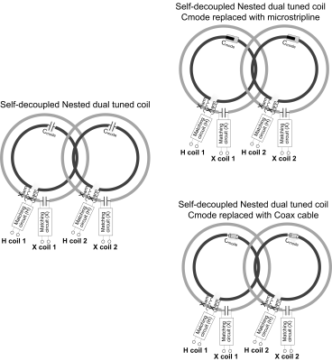 |
Flexible Self-decoupled Dual-tuned Array
Xinqiang Yan1,2 and John C. Gore1,2
1Department of Radiology, Vanderbilt University Medical Center, Nashville, TN, United States, 2Institute of Imaging Science, Vanderbilt University, Nashville, TN, United States
Highly flexible or wearable dual-tuned coil arrays appear to be good choices for increasing the SNR for both protons and X-nuclei and for improving patient comfort. However, complex couplings arise in a dual-tuned coil array. In this work, we have designed self-decoupled coils within a flexible, nested, dual-tuned array, in which impedances are redistributed to achieve high inter-element isolation.
|
|
4069.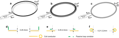 |
Differential Mode Self-Decoupled Coils
Ming Lu1,2,3, William A. Grissom1,2,4, John C. Gore1,2,4, and Xinqiang Yan1,2
1Vanderbilt University Institute of Imaging Science, Nashville, TN, United States, 2Department of Radiology, Vanderbilt University Medical Center, Nashville, TN, United States, 3College of nuclear equipment and nuclear engineering, Yantai University, Yantai, China, 4Department of Biomedical Engineering, Vanderbilt University, Nashville, TN, United States
Self-decoupled coils (SDCs) were recently described which can solve complex coupling issues in dense arrays. SDCs have a simple structure and represent a general approach. However, dipole mode in SDCs make it sensitive to the load. In this work, we introduced a differential mode to the design of SDCs to alter the necessary value of Cmode and a coils’ robustness to loading. This differential-mode SDC can be seen as a combination of differential-mode decoupling with self-decoupling. We found that suitable Cmode can choose to increase coil robustness as well as avoid added coil loss.
|
|
4070. |
Routing algorithm for the interconnection of closed wire loops for MR gradient and MR shim coils
Philipp Amrein1, Feng Jia1, Sebastian Littin1, and Maxim Zaitsev1
1Dept. of Radiology, Medical Physics, University Medical Center Freiburg, Freiburg, Germany
An automatized solution is presented for the generation of a single wire track from closed loops for complex coil and supporting structure topologies. The algorithm includes a mesh parameterization, a toplogical analysis, route optimization between groups of loops and finally opening and interconnection of the loops.
|
|
4071. |
Compact and Reproducible Microstrip Power Splitters for Array-Compressed Parallel Transmission at 7T
Gabriela Gallego1, Charlotte Sappo1,2, Xinqiang Yan2,3, and William A Grissom1,2,3,4
1Department of Biomedical Engineering, Vanderbilt University, Nashville, TN, United States, 2Vanderbilt University Institute of Imaging Science, Nashville, TN, United States, 3Department of Radiology, Vanderbilt University, Nashville, TN, United States, 4Department of Radiology, Vanderbilt University Institute of Imaging Science, Nashville, TN, United States
Parallel transmission (pTx) with an array of radiofrequency (RF) coils enables spatially uniform excitation with lower SAR in high-field MRI. Performance improves with the number of coils. Currently, 7T scanners have a limited number of transmit channels due to their high cost and complexity. Array-compressed pTx (acpTx) networks comprise unequal power splitters that sit between the transmit amplifiers and coils, enabling a small number of channels to optimally drive a large number of coils. This study presents the design of low-loss unequal power splitter building blocks with minimal size that can be combined in stages for a variety of applications.
|
|
4072. |
A 2kW RF power amplifier for 7T proton imaging with digital pre-distortion
Stephan Orzada1,2, Mark E. Ladd1,3,4, and Harald H. Quick1,2
1Erwin L. Hahn Institute for MRI, University of Duisburg-Essen, Essen, Germany, 2High-Field and Hybrid MR Imaging, University Clinic Essen, Essen, Germany, 3Medical Physics in Radiology, German Cancer Research Center (DKFZ), Heidelberg, Germany, 4Faculty of Physics and Astronomy and Faculty of Medicine, University of Heidelberg, Heidelberg, Germany
A 2 kW peak RF amplifier for 32-channel 7T proton imaging is presented. It is equipped with a digital pre-distortion providing a high linearity up to peak output power. The amplifier can be used for signal bandwidths of 1 MHz, making it suitable for almost any proton MR application.
|
|
4073.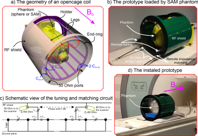 |
Experimental Realization of a Clinical Opencage Head Coil for Ultra-High Field MRI
Anton Nikulin1, Marc Dubois2,3, Tania S.Vergara Gomez2,3, Djamel Berrahou4, Frank Kober3, Alexandre Vignaud5, Redha Abdeddaim2, Julien de Rosny1, and Abdelwaheb Ourir1
Video Permission Withheld
1Institut Langevin, ESPCI Paris, CNRS, PSL University, Paris, France, 2CNRS, Centrale Marseille, Institut Fresnel, Aix Marseille Univ, Marseille, France, 3CNRS, CRMBM, Aix Marseille Univ, Marseille, France, 4Multiwave Innovation SAS, Marseille, France, 5CEA, DRF, JOLIOT, NeuroSpin, UNIRS, Universit´e Paris-Saclay, Gif-sur-Yvette, France
A conventional birdcage head coil at 7T is not well suited for certain applications such as motion control. We propose an original coil based on a birdcage with a wide lateral aperture that provides access to the object under examination. We show that a sufficiently homogeneous magnetic field distribution can be obtained by optimizing the current distribution of such an “Opencage”. That coil was optimized using a full wave simulation and tested experimentally. The performance of an Opencage coil was compared to a commercial birdcage coil. Eventually, the Opencage coil is shown as a tradeoff between field homogeneity and access.
|
|
4074. |
An 8-channel Loop Coil Array for Body Imaging Using Coupled Line Phase Shifters at 7T
Haiwei Chen1, Lei Guo1, Aurelien Destruel1, Mingyan Li1, Ewald Weber1, Feng Liu1, and Stuart Crozier1
1School of Information Technology and Electrical Engineering, The University of Queensland, Brisbane, Australia
A large-size loop coil suffers from the current phase inversion along the loop surface and therefore weak the current intensity at the phase inversion points. A new RF coil using coupled line phase shifters is presented for body imaging at 7T MRI. This design can retain an in-phase current along the loop when the perimeter is larger than 1.5$$$\lambda$$$ ($$$\lambda$$$ is the wavelength in free space). An 8-channel coil array using this new design is simulated and compared with a conventional loop array. The results indicate that improved $$$B^{+}_{1}$$$ shimming can be achieved in a large region of interest.
|
|
4075.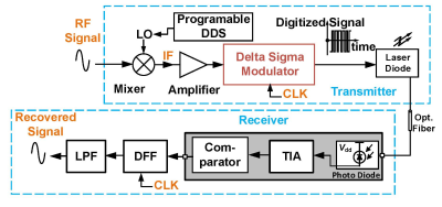 |
Optical Transmission of Digitized MRI Signals Using Delta-Sigma Modulation
Mingdong Fan1, Xi Gao2, David Ariando2, Shinya Handa3, Labros Petropoulos3, Xiaoyu Yang3, Hiroyuki Fujita3, Michael Martens1, Robert Brown1, and Soumyajit Mandal2
1Physics, Case Western Reserve University, Cleveland, OH, United States, 2EECS, Case Western Reserve University, Cleveland, OH, United States, 3Quality Electrodynamics (QED), Mayfield Village, OH, United States
The electromagnetic interference between conductive cables is becoming an issue as the number of MRI receive channels increases. Optical fibers are seen as one of the potential alternatives. Analog optical links have been investigated due to their relatively simple RF coils structure and minimum modification to the MRI system, but they are often limited by the electrical and optical nonlinearities and degradation of noise figure. In this study, we propose a digital optical transmission system based on delta-sigma modulation (DSM) that aims to provide high dynamic range (DR) for MRI signal transmission.
|
|
4076.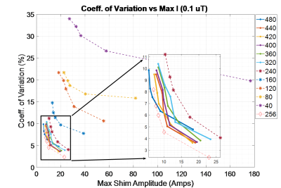 |
RF Shim Flexibility with Multi-Surface-Loop Arrays Over Varying Head Geometries
Benjamin M Hardy1,2, Yurui Gao2,3, and Adam W Anderson2,3
1Department of Physics and Astronomy, Vanderbilt University, Nashville, TN, United States, 2Vanderbilt University Institute of Imaging Science, Nashville, TN, United States, 3Department of Biomedical Engineering, Vanderbilt University, Nashville, TN, United States
Magnetic and electric field values are simulated with up to 480 Radio Frequency surface loops 1 cm in radius surrounding realistic head models. The work investigates the capability of B1+ shimming alone with increased degrees of freedom. Transmit efficiency, power limitations, and target homogeneity across the brain volume inform design choices for the array. Evidence of the effects of subject to subject variation can be seen in the optimized shim weights magnitude and phase variation for a choice 256-element geometry optimized over 20 unique head models oriented at 3 head angles.
|
|
4077. |
Feasibility of Forward and Reversed Preamplifier Decoupling (FPD and RPD) Techniques for a Low Field MRI System at 1 MHz
Sajad Hosseinnezhadian1, Yonghyun Ha1, Kartiga Selvaganesan1, Charles Rogers III1, Baosong Wu1, Gigi Galiana1, and R. Todd Constable1
1Department of Radiology and Biomedical Imaging, Yale University School of Medicine, New Haven, CT, United States
This study investigated the feasibility of applying forward and reversed preamplifier decoupling techniques for low and high impedance coils, respectively, at 1 MHz. At a low field MRI, e.g. 1 MHz, the choice of components needed for interface circuitry together with availability of preamplifiers with low input impedance is challenging. In this study we demonstrated that using a high impedance coil with a lower number of turns achieved better decoupling levels than that of a low impedance coil. In addition, the minimum requirement for the input resistance of a preamplifier at 1 MHz was investigated.
|
|
4078.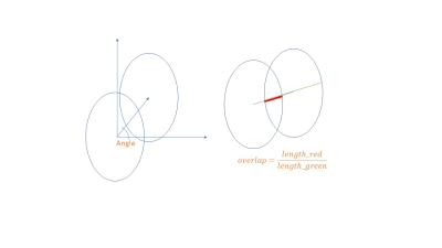 |
Critical Overlap for Elliptical Arrays
Victor Taracila1 and Fraser Robb1
1GE Healthcare, Aurora, OH, United States
Multi-channel receive RF coils for MRI are usually comprised of an array of loops. These loops come in various shapes – circles, rectangles, hexagons, octagons, etc. Coil designers try to decrease the coupling between the loops through overlapping – partial overlapping of the two closed contours allows reduction of magnetic coupling between the loops to minimum. This configuration is called a critical overlap. While the critical overlap for circular loops is mentioned in multiple papers, a similar recipe is missing for elliptical loops. In this work we calculate the overlap coefficients for isotropic uniform elliptical arrays.
|
4079. |
Efficient 3D-Characterization of Metamaterial Devices for Enhanced Magnetic Resonance Imaging
Robin Niklas Wilke1, Endri Stoja2, Kyra Bekaan1,3, Dennis Philip1, Jürgen Jenne1, Diego Betancourt2, Simon Konstandin1, Reinhold Umathum1, Thomas Bertuch2, and Matthias Günther1,4
1MR Physics, Fraunhofer MEVIS, Bremen, Germany, Bremen, Germany, 2Antenna Technology and Electromagnetic Modelling, Fraunhofer Institute for High Frequency Physics and Radar Techniques FHR, Wachtberg, Germany, 3University of Oldenburg, Oldenburg, Germany, 4MR-Imaging and Spectroscopy, Faculty 01 (Physics/Electrical Engineering), University of Bremen, Bremen, Germany
We present a dedicated RF-measurement setup consisting of custom-build Tx/Rx coils, an automated positioning system and a vector network analyzer that is suitable for an efficient characterization of MR-metamaterial devices which is crucial during the design stage. We measure complex scattering parameters over a large volume in air and obtain dispersion curves of a magneto-inductive lens and a wire-resonator metasurface that have been designed for 3T applications. Strong field enhancement effects are observed close to the metasurface as well as the positional dependency of extrema in dispersion curves.
|
|
4080.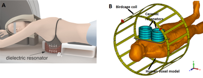 |
Novel ceramic-based resonators for bilateral breast imaging at 3T
Viacheslav Ivanov1, Alena Shchelokova1, Anna Andreychenko1,2, and Alexey Slobozhanyuk1
1Faculty of Physics and Engineering, ITMO University, Saint-Petersburg, Russian Federation, 2Research and Practical Clinical Center of Diagnostics and Telemedicine Technologies of the Moscow Heathcare Department, Moscow, Russian Federation
We propose a concept for targeted bilateral breast imaging. A practical demonstration of the concept features a pair of hollow dielectric cylindrical resonators based on a composite material with very high permittivity. The resonators are electromagnetically coupled to the body birdcage coil and each other. Electromagnetic simulations demonstrated that symmetric B1+ field mode, inherent in interacting resonators, can be efficiently excited at 123 MHz via the body birdcage coil. Simulations with realistic voxel models let us estimate transmit efficiency and radiofrequency safety of the proposed device and demonstrated that the concept promising for clinical applications.
|
|
4081. |
The manipulation of displacement current wave in ultra-High Dielectric Constant Helmet on B1 field distribution in human head at 3T
Navid Pourramzangandji1, Christopher Sica2, Sebastian Rupprecht3, Michael Lanagan4, and Qing Yang1
1Department of Neurosurgery, PennState University College of Medicine, Hershey, PA, United States, 2Department of Radiology, PennState University College of Medicine, Hershey, PA, United States, 3HyQ research Solution, State College, PA, United States, 4Department of Engineering Science and Mechanics, Pennsylvania State University, State College, PA, United States
A displacement current wave is formed in an ultra-high dielectric constant (uHDC) helmet, which perturbs transmission B1 field uniformity within the human head as a result of phase retardation. With computer modeling, we manipulated the displacement current by segmenting the helmet into multiple pieces to control the propagation of the induced displacement current wave to “shim” the B1 field inside a human head model. We demonstrated that with a 4-segmented helmet, we can obtain a uniform B1+ distribution within the human brain with enhanced transmit efficiency.
|
|
4082. |
The displacement current wave on a high dielectric constant (HDC) helmet and its effect on B1 Field at 10.5 T (447 MHz)
Navid Pourramzangandji1, Sebastian Rupprecht2, Myung-Kyun Woo3, Michael Lanagan4, Bei Zhang5, Riccardo Lattanzi5, Russell L. Lagore3, Jerahmie Radder3, Gregor Adriany3, Brian Rutt6, Kamail Ugurbil3, and Qing Yang1
1Department of Neurosurgery, PennState University College of Medicine, Hershey, PA, United States, 2HyQ research Solution, State College, PA, United States, 3Department of Radiology, Center for Magnetic Resonance, University of Minnesota Research, Minneapolis, MN, United States, 4Department of Engineering Science and Mechanics, Pennsylvania State University, State College, PA, United States, 5Department of Neurosurgery, The Bernard and Irene Schwartz Center for Biomedical Imaging, New York University, New York, NY, United States, 6Radiology, Stanford University, Stanford, CA, United States
We observed a propagating wave of displacement current on a continuous high dielectric constant (HDC) helmet at 10.5T with an eight channel dipole array in CP mode. As a secondary current source of B1, the distribution and phase retardation of the displacement wave on the helmet leads to an interesting problem for utilizing HDC material for B1 enhancement. We extracted the B1+ field originated by the HDC helmet by subtracting the primary field (within the human head model without helmet) from the total field (field in the presence of the helmet).
|
|
4083.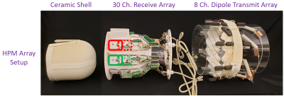 |
Improved Brain Imaging with an Integrated High-Permittivity Material Head Array
Karthik Lakshmanan1,2, Jerzy Walczyk1,2, Sebastian Rupprecht3, Michael T Lanagan3, Qing X Yang3, Ryan Brown1,2,4, and Christopher M Collins1,2,4
1Bernard and Irene Schwartz Center for Biomedical Imaging, Department of Radiology, NYU School of Medicine, New york, NY, United States, 2Center for Advanced Imaging Innovation and Research (CAI2R), Department of Radiology, NYU School of Medicine, New york, NY, United States, 3HyQRS LLC, State College, PA, United States, 4The Sackler Institute of Graduate Biomedical Science, New york, NY, United States
High electric permittivity materials (HPM) have been used to improve transmit efficiency (B1+) and receive sensitivity in targeted regions. In this work we demonstrate improvements in the entire brain by designing and implementing an integrated HPM shell for a head coil with 8-channel transmit and 30-channel receive arrays. The coil with HPM insert provides gains of approximately 55% in transmit efficiency and 20% in SNR compared to an identical coil without the insert.
|
|
4084. |
High-Permittivity Dielectric Probes for Ultra-High Field MR Microscopy at 17.2 T
Marine A.C. Moussu1,2, Luisa Ciobanu3, Elodie Georget2, Stanislav Glybovski4, Andrew G. Webb5, Stefan Enoch1, and Redha Abdeddaim1
1Institut Fresnel, Aix Marseille Université, Marseille, France, 2Multiwave Imaging, Marseille, France, 3DRF/Joliot/Neurospin/UNIRS, Gif-sur-Yvette, France, 4ITMO University, Saint-Petersbourg, Russian Federation, 5Leiden Univ. Medical Center, Leiden, Netherlands
We propose a theoretical model predicting the Signal-to-Noise Ratio (SNR) of dielectric antennas for Magnetic Resonance Imaging. The SNR is estimated with a semi-analytical approach describing the first transverse electric mode of a high-permittivity ring resonator. When the ring is properly excited with a feeding loop, it induces a strong magnetic field in the sample while dielectric losses are limited due to low electric field intensity in this region. The proposed model is validated with numerical simulations regarding microscopy applications and its prediction confirmed experimentally at 17.2 T.
|
|
4085.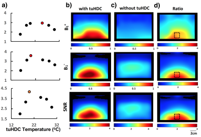 |
Integrating RF coil with low-loss tuHDC ceramics at room temperature for large improvements in B1 fields and SNR, denoising for UHF 23Na MRSI
Hannes M. Wiesner1, Byeong-Yeul Lee1, Xiao-Hong Zhu1, Maryam Sarkarat2, Michael T. Lanagan2, Qing X. Yang3, and Wei Chen1
1Department of Radiology, CMRR, University of Minnesota Medical School, Minneapolis, MN, United States, 2Department of Engineering Science and Mechanics, The Pennsylvania State College of Engineering, University Park, PA, United States, 3CNMRR, Department of Neurosurgery, The Pennsylvania State College of Medicine, Hershey, PA, United States Poster Permission Withheld
Integrating ultrahigh-dielectric constant (uHDC) materials with RF coils have improved MR imaging performance. In this work, we present a novel approach based on the permittivity tunable (tuHDC) ceramics, which has an extremely high tunability of permittivity from 2000-15000 by changing the ceramic temperature between few to 40 °C, for 23Na MRSI at UHF. We demonstrate that the optimal temperature and performance of the tuHDC ceramic for 23Na MRSI at 7T was at room temperature. We report spatially dependent increases in the RF transmission and reception fields and SNR with a large factor of ~4 accompanied by moderate global denoising effect.
|
|
4086.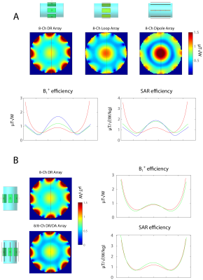 |
Dielectric Resonators and Dipole Antennas Combined: New Approach in Radio Frequency Coil Design for Ultrahigh Field MRI
Daniel Wenz1 and Rolf Gruetter1,2,3,4
1Center for Biomedical Imaging - Animal Imaging and Technology (CIBM-AIT), Ecole Polytechnique Fédérale de Lausanne (EPFL), Lausanne, Switzerland, 2Laboratory of Functional and Metabolic Imaging (LIFMET), Ecole Polytechnique Fédérale de Lausanne (EPFL), Lausanne, Switzerland, 3Department of Radiology, University of Lausanne, Lausanne, Switzerland, 4Department of Radiology, University of Geneva, Geneva, Switzerland
Dielectric resonators (DR) can be advantageous over conventional loop elements, especially in ultrahigh field MRI. We noticed, that it is possible to combine DR in transverse electric mode with dipole antennas (DA), by placing them exactly above each other. In this study we explore the benefits of such combination in a multi-channel array configuration. We present the results of electromagnetic field simulations in which we compared an 8/8-channel DR-DA array with: 8-channel loop array, 8-channel DA array and 8/8-channel loop-DA array.
|
|
4087.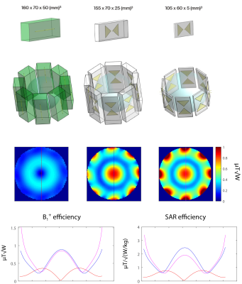 |
Dielectrically-Shortened Dipole Antennas for MRI at 7.0 T: Thick or Thin?
Daniel Wenz1 and Rolf Gruetter1,2,3,4
1Center for Biomedical Imaging - Animal Imaging and Technology (CIBM-AIT), Ecole Polytechnique Fédérale de Lausanne (EPFL), Lausanne, Switzerland, 2Laboratory of Functional and Metabolic Imaging (LIFMET), Ecole Polytechnique Fédérale de Lausanne (EPFL), Lausanne, Switzerland, 3Department of Radiology, University of Lausanne, Lausanne, Switzerland, 4Department of Radiology, University of Geneva, Geneva, Switzerland
Shortening dipole antennas using dielectric materials provides an opportunity to design very high-channel count dipole arrays which should boost signal-to-noise ratio and enable higher acceleration factors in parallel MRI at 7.0 Tesla. In this study, we compare different dipole antennas shortened using D2O. We demonstrate, that the height of the dielectric block becomes a critical design parameter when there is no direct contact between the block and the sample and what we know about dielectric block design should be revisited. Our results indicate, that miniaturizing dipole antennas, and employing them in an 8-channel configuration, is feasible without significant performance decrease.
|
|
4088.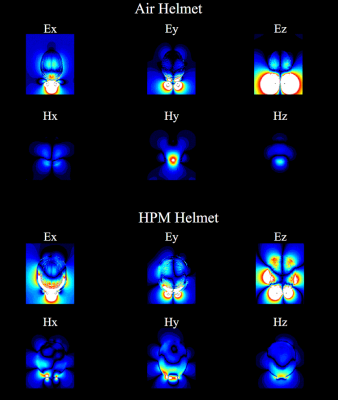 |
Distribution and Propagation of Fields and Displacement Currents in a high-permittivity helmet
Giuseppe Carluccio1,2, Qing X Yang3, and Christopher Michael Collins1,2
1Radiology, Center for Advanced Imaging Innovation and Research (CAI2R), New York, NY, United States, 2Radiology, Bernard and Irene Schwartz Center for Biomedical Imaging, New York, NY, United States, 3Radiology, Penn State University, Hershey, PA, United States
High-permittivity material (HPM) helmets have provided significant increase in transmit efficiency and local SNR. However, the physical mechanisms that explain such improvements are still not fully explored. In this work, through electromagnetic simulations, we analyze the fields propagation when a loop is placed near the occipital lobe with and without the presence of an HPM helmet. The analysis is performed observing the field propagation through time, the direction and the amplitude of the Poynting vector, and the current density distribution.
|
|
4089. |
SNR Comparison of a Receive Array Coil When Mounted on a High-Permittivity Helmet Former vs. Moved Closer to the Head
Giuseppe Carluccio1,2 and Christopher Michael Collins1,2
1Radiology, Center for Advanced Imaging Innovation and Research (CAI2R), New York, NY, United States, 2Radiology, Bernard and Irene Schwartz Center for Biomedical Imaging, New York, NY, United States
High-permittivity materials (HPM) have provided promising results in terms of higher local SNR, such as when an HPM helmet former is used in combination with a receive coil array. Since the vicinity of the coil affects the SNR and the presence of the HPM helmet prevents placement of the coil closer to the head, in this work we compare the SNR distribution when the receive array coil is mounted on top of an HPM helmet to when it is moved closer to the head. Results show that the array coil with the HPM helmet provides an overall higher SNR.
|
|
4090. |
Assessment of planar coil noise performance with discrete uHDC material
Christopher T Sica1, Navid Pourramzan Gandji2, and Qing X Yang2
1Radiology, PennState University College of Medicine, Hershey, PA, United States, 2Neurosurgery, PennState University College of Medicine, Hershey, PA, United States
A study of the noise performance of a planar coil array in the presence of ultra-high dielectric constant (uHDC) material was undertaken. We measured changes in the noise standard deviation and correlation matrix of a 4 channel flex array due to manipulation of uHDC block locations, with blocks either fully overlapping a single channel or straddling two channels. Overlapping a block with a coil element increased the noise of that element, while reducing next-neighbor noise correlations. Further research into this effect could lead to the design of more optimal dielectric configurations that achieve greater SNR.
|
|
4091. |
NEAR-FIELD ELECTROMAGNETIC CLOAKING OF METALLIC ELONGATED IMPLANTS TO REDUCE RF-INHOMOGENEITY ARTIFACTS AND SAR ELEVATION
Umberto Zanovello1 and Luca Zilberti1
1Istituto Nazionale di Ricerca Metrologica (INRIM), Torino, Italy
The eddy currents induced by the RF magnetic field B1 due to the presence of a conductive object, such as a metallic implant, may compromise both the RF coil transmit and receive sensitivity homogeneity; especially in the periprosthetic areas. A further effect may be an increase of the local SAR which can affect the overall safety of the MRI exam. In this study, the outcomes of an electromagnetic cloaking strategy based on the application of a suitable coating to elongated conductive implants are shown. The coating restores the coil sensitivities and SAR distribution to those obtained without the implant presence.
|
|
4092.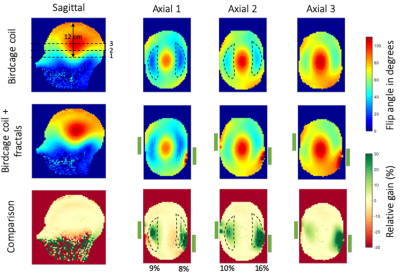 |
Passive RF shimming with fractal metasurfaces in a 7T head coil
Tania S. Vergara Gomez1,2, Marc Dubois1,2, Kaizad Rustomji3, Elodie Georget3, Tryfon Antonakakis3, Stanislas Rapacchi1, Frank Kober1, Stefan Enoch2, and Redha Abdeddaim2
1Aix Marseille Univ, CNRS, CRMBM, Marseille, France, 2Aix Marseille Univ, CNRS, Centrale Marseille, Institut Fresnel, Marseille, France, 3Multiwave Imaging, Marseille, France
B1+ inhomogeneities in the temporal lobes of the brain are common while using a birdcage coil at 7T. Different passive RF shimming approaches have already targeted this problem, at the cost of decreasing the B1+ field in other important areas or being uncomfortable to the patient. Here, we present a new approach based on two resonant fractal metasurfaces printed on ultra-thin flexible substrate. We show that these resonators improve the B1+ field amplitude simultaneously in each temporal lobe without affecting the B1+ field amplitude in other parts of the brain.
|
|
4093.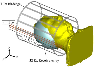 |
Passive RF shimming for 7T head coil with hybridized meta-atom: In vivo investigation
Marc Dubois1,2, Lisa Leroi3, Zo Raolison4, Redha Abdeddaim1, Tryfon Antonakakis4, Julien De Rosny5, Alexandre Vignaud3, Pierre Sabouroux1, Elodie Georget4, Benoit Larrat3, Gérard Tayeb1, Nicolas Bonod1, Alexis Amadon3, Franck
Mauconduit3, Cyril Poupon3, Denis Le Bihan3, and Stefan Enoch1
1Institut Fresnel, Aix Marseille Université, CNRS, Marseille, France, 2CRMBM, Aix Marseille Université, CNRS, Marseille, France, 3CEA, DRF, JOLIOT, NeuroSpin, UNIRS, Gif sur Yvette, France, 4Multiwave Imaging, Marseille, France, 5ESPCI Paris, PSL Research University, CNRS, Institut Langevin, Paris, France
We show that hybridized meta-atom can be used to improve transmit homogeneity in a 7T head birdcage coil equipped with a 32-channel receive array. Our results demonstrates the enhancement of transmit signal in vivo with the possibility to fill one of the gap usually observed in the brain temporal lobes. This metamaterial based passive shimming strategy provides a cost effective, long-lasting solution without any impact on the patient’s comfort during the examination.
|
|
4094.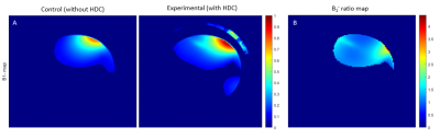 |
Improving B1 fields and SNR for Proton MRI and MRSI at 7T using 4-piece Helmet filled with High Dielectric Constant (HDC) Material
Soo Han Soon1, Byeong-Yeul Lee1, Hannes M. Wiesner1, Matt Waks1, Parker Jenkins1, Navid Pourramzan Gandji2, Sebastian Rupprecht2, Qing X. Yang2, Xiao-Hong Zhu1, and Wei Chen1
1CMRR, Department of Radiology, University of Minnesota, Minneapolis, MN, United States, 2CNMRR, Department of Neurosurgery, Penn State University, State College, PA, United States
Ultrahigh-field (UHF) MR systems allow non-invasive images with high sensitivity and spatiotemporal resolution. High dielectric constant (HDC) material utilized with RF coils has been shown to be promising for further improving RF transmitting power efficiency and increasing the signal-to-noise ratio (SNR) in previous studies. This work focuses on the application of a 4-piece HDC helmet-shaped former to different types of coils to demonstrate the consistency of improvement in B1 fields at 7T. The technology could enhance SNR and efficiency of RF transmission for brain 1H MR imaging application at UHF.
|
4095.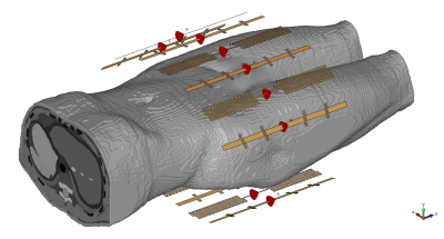 |
Multinuclear Virtual Observation Points
Arthur W. Magill1 and Mark E. Ladd1,2
1Medical Physics in Radiology, German Cancer Research Center (DKFZ), Heidelberg, Germany, 2Physics & Astronomy, Heidelberg University, Heidelberg, Germany
Parallel transmission (PTx) is a valuable tool for UHFMRI, but complicates SAR monitoring considerably. The Virtual Observation Point (VOP) algorithm has been crucial in enabling online SAR monitoring during PTx. To date, PTx has only been applied for proton, but as static field strengths continue to increase it becomes interesting to apply PTx methods for other nuclei. This abstract investigates how to apply the VOP algorithm to a transmit array operating at more than one frequency.
|
|
4096.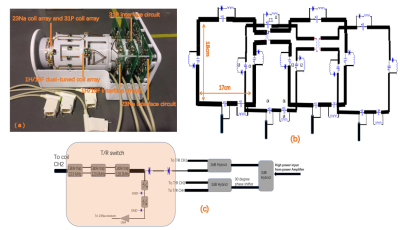 |
A four-frequency 1H/19F/23Na/31P RF coil system for simultaneous multinuclear MRI and MRS at 3.0T
xing yang1, Nan Li1, Feng Du1, Kai Wang2,3, Xin Liu1, Hairong Zheng1, and Ye Li1
1Shenzhen Institutes of Advanced Technology,Chinese Academy of Sciences, Shen'zhen, China, 2Molecular Imaging Research Center, Harbin Medical University, Harbin, China, 3TOF-PET/CT/MR Center, The Fourth Hospital of Harbin Medical University, Harbin, China
Most of current multinuclear MR experiments are performed by doing mono-nuclear acquisitions separately, followed by registering images obtained from each one. To reduce the total measurement time and experiment registration error, a novel four-frequency 1H/19F/23Na/31P RF coil system was developed and evaluated. Our performance evaluations showed that all coil arrays were sufficiently matched, decoupled and detuned when working simultaneously, and the unbalance of amplitude and phase increments generated by the interface circuits were well controlled, thus can be expected to generate a uniform B1+ field and high SNR images for simultaneous 1H/19F/23Na MRI and for 31P MRS.
|
|
4097.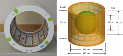 |
Evaluation of a Four-Ring Birdcage Head Coil for 1H Imaging and 13C Spectroscopy at 3 Tesla
Joseph Murphy-Boesch1, Kazu Yamamoto2, Ananda Kumar3, Peter L. Choyke4, and Murali Krishna Cherukuri2
1NINDS, National Institutes of Health, Bethesda, MD, United States, 2NCI, National Institutes of Health, Bethesda, MD, United States, 3Lambda-Z Technologies, Baltimore, MD, United States, 4NCI, NIH, Bethesda, MD, United States
Dissolution DNP with Hyperpolarized 13C requires receive coils optimized for sensitivity and a transmit coil capable of delivering strong B1 fields for the carbon frequency. Here, a dual-tuned, four-ring birdcage head coil is described that can produce a transmit field of >90 microTesla and serve as a receive coil for an MRI system with few receivers. Hyperpolarized 13C spectra localized using 2D-CSI were obtained in 6 seconds from a human pancreatic tumor xenograph grown in the flank of a nude mouse. SNR of 13C spectra obtained from a tumor volume of <1 ml was suitable to measure the lactate/pyruvate ratio.
|
|
4098.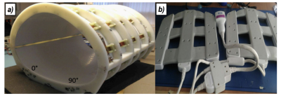 |
RF coil system for accelerated 129Xe hyperpolarized imaging at 1.5T
Claudio Puddu1, Madhwesha Rao1, Guilhem J. Collier1, Adam Maunder1, Ho-Fung Chan1, Oliver Rodgers1, Xiaojun Xu1, Nicola De Zanche2, Fraser Robb3, and Jim M. Wild1
1POLARIS,University of Sheffield, Sheffield, United Kingdom, 2Department of Medical Physics, Cross Cancer Institute, Edmonton, AB, Canada, 3GE Healthcare, Aurora, OH, United States
This abstract describes the development of a separate transmit/receive setup of coils for human chest 129Xe MRI at 1.5T. The transmit coil is an asymmetrical birdcage type designed to fit the scanner bore. The lumped components are calculated in consideration of the mutual coupling with the 1H body coil of the scanner, enabling the scanning of anatomical images and localization with the birdcage inside the bore. Furthermore, a parallel imaging strategy for 129Xe ventilation imaging has been tested to demonstrate accelerated acquisition times.
|
|
4099.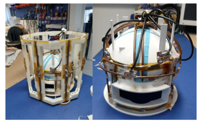 |
6-channel RF array for hyperpolarized 129Xe brain MRI at 3T
Claudio Puddu1, Madhwesha Rao1, Oliver Rodgers1, Adam Maunder1, and Jim Wild1
1POLARIS,IICD department, University of Sheffield, Sheffield, United Kingdom
Dissolved phase hyperpolarised 129Xe is an emerging technique for perfusion imaging in the brain using inhaled gas as a perfusion tracer. As the dissolved 129Xe signal is inherently weak when compared to the gas phase signal, RF coil sensitivity is critical for high SNR 129Xe brain MRI. In this work, the design, development and testing of a novel 6 channel receiver and birdcage transmit coil RF system for dissolved phase 129Xe MRI in the human brain at 3T is demonstrated.
|
|
4100.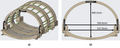 |
Asymmetrical Body Birdcage Coil for Xenon-129 Imaging at 3T
Wolfgang Loew1, Christopher Ireland1, Matthew Willmering2, Ronald Pratt1, Hui Wang3, Kaley Bridgewater1, Peter van der Meulen4, Charles Dumoulin1, and Jason Woods2
1Imaging Research Center, Cincinnati Children's Hospital Medical Center, Cincinnati, OH, United States, 2Center for Pulmonary Imaging Research, Cincinnati Children's Hospital Medical Center, Cincinnati, OH, United States, 3Philips Healthcare, Gainesville, FL, United States, 4Philips Healthcare, Best, Netherlands
Hyperpolarized 129Xe imaging is increasingly viewed as a viable tool for assessing lung structure and function. An asymmetrical whole-body xenon birdcage design for homogeneous transmit performance, previously evaluated through electromagnetic field simulations, was constructed. Coil homogeneity was evaluated with a high-pressure thermally polarized xenon Boltzmann cylinder phantom and hyperpolarized 129Xe gas. MR images of the phantom and the hyperpolarized gas confirmed homogeneous excitation.
|
|
4101.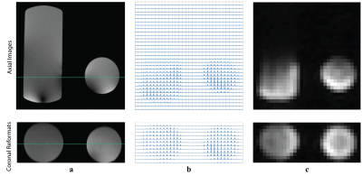 |
Multinuclear RF coil for 13C and 1H breast MR imaging at 3.0 T
Madhwesha Rama Rao1, Kristin Granlund2, Hedvig Hricak2, Jim M Wild1, and Kayvan R Keshari2
1University Of Sheffield, Sheffield, United Kingdom, 2Memorial Sloan Kettering Cancer Center, New York, NY, United States
Hyperpolarized 13C MRI enables the investigation of metabolism of breast lesions for diagnosis and to monitor disease progression. Acquiring 13C MR images along with 1H MR images using a multi-nuclear RF coil enables accurate co-registration of the images, thereby providing complementary structural and functional images. In this work, we propose a design for multinuclear 13C-1H RF coil array for breast MR imaging, and assess the performance using both RF and MRI measurements. Both 13C and 1H MR images were obtained of using phantoms.
|
|
4102.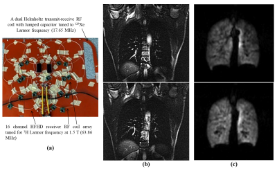 |
Highly-Flexible Highly-Decoupled 1H RF Coils for MR imaging of the lungs as utilized within a 129Xe Transmit-Receive RF Coil at 1.5 T
Madhwesha Rama Rao1, Fraser J Robb1,2, Victor Taracila2, Weixing Zhang2, and Jim M Wild1
1University Of Sheffield, Sheffield, United Kingdom, 2GE Healthcare, Aurora, OH, United States
Flexibility of a RF coil constructed with flexible printed circuit board is reduced when it is co-housed with another RF coil, as typically applied for multinuclear MR imaging. In this work, for multinuclear 129Xe-1H lung imaging application, we propose to use highly-flexible multiple linked resonators for 1H RF coil along with 129Xe RF coil. These highly-flexible 1H RF coils are also highly-decoupled at 129Xe Larmor frequency. For the demonstration of multi-nuclear lung imaging, sixteen receive only highly-flexible highly-decoupled 1H RF coils were fixed on to a transmit-receive 129Xe RF coil, and 129Xe and 1H
lung images were obtained.
|
|
4103.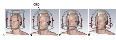 |
Numerical evaluation of a domed and folded two-ring birdcage coil for 1H/31P at 3T MRI
Suk-Min Hong1, Chang-Hoon Choi1, Jörg Felder1, and N. Jon Shah1,2,3,4
1Institute of Neuroscience and Medicine 4, INM-4, Forschungszentrum Jülich, Jülich, Germany, 2Institute of Neuroscience and Medicine 11, INM-11, JARA, Forschungszentrum Jülich, Jülich, Germany, 3JARA - BRAIN - Translational Medicine, Aachen, Germany, 4Department of Neurology, RWTH Aachen University, Aachen, Germany
For double-frequency coils, it has been shown that four-ring birdcage coils provide minimum losses at X-nuclei frequencies. However, to avoid coupling between two rings, the length of the outer parts needs to be extended, increasing the unusable area at the service-end. In this study, we redesigned one side of the conventional birdcage coil as a dome shape by using end-caps. The domed birdcage coil was evaluated in simulations in both the single- and double-tuned condition. The performance of the folded-ring, domed and double-tuned birdcage coil was also evaluated and compared to that of a conventional single-tuned birdcage coil.
|
|
4104.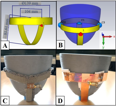 |
A Wearable and Flexible 23Na Transmit/Receive Breast Coil at 3T
Busra Kahraman Agir1, Basak Bayrambas2, Antoine Klauser3, Francois Lazeyras 3, Esin Ozturk Isik2, and Korkut Yegin4
1The Graduate School of Natural and Applied Sciences, Dokuz Eylul University, Izmir, Turkey, 2Institute of Biomedical Engineering, Bogazici University, Istanbul, Turkey, 3Department of Radiology and Medical Informatics, University of Geneva, Geneva, Switzerland, 4Department of Electrical and Electronic Engineering, Ege University, Izmir, Turkey
A novel wearable and flexible 23Na transmit/receive breast coil made from a highly conductive thread was introduced for 3 T. Comparison studies were conducted with a 23Na traditional breast coil having the same geometry as the flexible one. Sensitivity maps of 23Na traditional breast coil and 23Na flexible textile breast coil were similar. In addition, 23Na flexible textile breast coil provided better SNR performance when different NaCl concentration were to be imaged. In conclusion, applicability of textile coils on 23Na MRI was demonstrated by means of similar SNR results of 23Na flexible textile breast coil and 23Na traditional breast coil.
|
|
4105. |
A dual-frequency surface coil design comprised of a single loop for both proton and deuterium magnetic resonance imaging at 16.4T
Guangle Zhang1, Wei Zhu1, Xiao-Hong Zhu1, Hannes M. Wiesner1, Tao Wang1, and Wei Chen1
Video Permission Withheld
1Center for Magnetic Resonance Research, Department of Radiology, University of Minnesota, Minneapolis, MN, United States
X-nuclear MRS imaging (MRSI) plays critical roles in studying cellular energy metabolism in health and diseases. However, it requires one 1H coil for structural imaging and B0 shimming and another coil for X-nuclear MRS, leading to coil coupling and performance degradation. Here we developed a simple, low-cost technique to construct dual-frequency surface coil(s) operating at proton and X-nuclear frequencies. For the proof of concept, we conducted 1H MRI and 2H MRSI study at 16.4T; and more importantly, we made a two-channel dual-frequency transceiver coil array to demonstrate the potential for large volume imaging at ultrahigh field.
|
|
4106.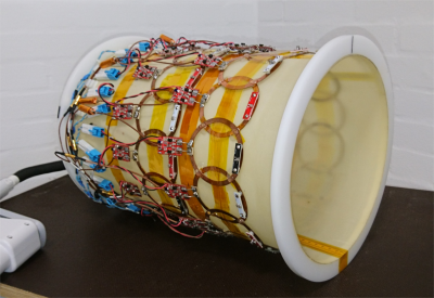 |
Fixed Geometry 28-Channel Array for Full Head Coverage of Hyperpolarized 13C MRS
Juan D. Sánchez-Heredia1, Aws Ali1,2, Wenjun Wang1, Rie B. Olin1, Vitaliy Zhurbenko1, and Jan H. Ardenkjær-Larsen1
1Department of Health Technology, Technical University of Denmark, Kgs. Lyngby, Denmark, 2Department of Electrical Engineering, Technical University of Denmark, Kgs. Lyngby, Denmark
We describe the design and performance of a 28-channel RF receive array optimized for human head 13C imaging at 3T. The array employs a fixed and symmetric cylindrical geometry to provide predictable prior information of the coil sensitivity profiles, which can be used to resolve practical challenges of hyperpolarized parallel imaging and to improve performance. The results show good SNR and spatial encoding of the different elements, with only small performance difference between the individual elements.
|
|
4107.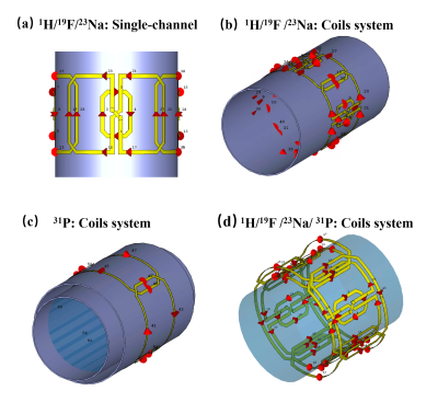 |
A 16-channel quadruple-nuclear RF coil system for 1H / 19F / 23Na/ 31P MR imaging and spectroscopy at 3T
Nan Li1, Xing Yang1, Feng Du1, Guoming Chen2, Xiaoliang Zhang3, Xin Liu1, Hairong Zheng1, and Ye Li1
1Shenzhen Institutes of Advanced Technology, Chinese Academy of Sciences, Shenzhen, China;, shenzhen, China, 2Department of Biomedical Engineering, Chongqing University of Technology, Chongqing 400054, China, Chongqing, China, 3Department of Biomedical Engineering, State University of New York at Buffalo, NY, United States., Buffalo, NY, United States
Acquisition of multiple nuclear species in MRI/MRS is significant for obtaining metabolic imaging in vivo and biological information. In this study, a16-channel RF quadruple-nuclear coils system capable of 1H / 19F / 23Na/ 31P imaging was designed. Numerical electromagnetic simulation was employed to validate the feasibility and performance of the proposed RF coil system by the scattering parameters and B1+field distributions.
|
|
4108.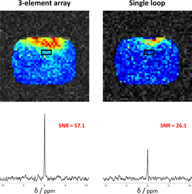 |
Comparison of a 3-channel 31P array to a single loop coil for cardiac MRS at 7 T
Sigrun Roat1, Martin Vít2,3, Stefan Wampl1, Albrecht Ingo Schmid1, and Elmar Laistler1
1Center for Medical Physics and Biomedical Engineering, Medical University of Vienna, Vienna, Austria, 2IKEM, Institute for Clinical and Experimental Medicine, Praha, Czech Republic, 3Technical University of Liberec, Liberec, Czech Republic
Cardiac 31P MRS is a powerful noninvasive tool in the assessment of myocardial energy metabolism, but it is inherently limited by low SNR, which can be mitigated by increasing B0. In order to fully exploit the SNR gain, elaborate RF coil development is necessary. In this work we compare the performance of a standard single loop coil with an optimized 3-element array in simulation and measurement. We validated the superior performance of the array experimentally, with a 2.2-fold SNR increase in localized spectroscopy, and in simulation yielding a 1.7 and 2.2 times higher PE and SE for the array, respectively.
|
|
4109.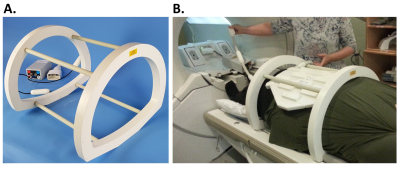 |
Large field of view Sodium-23 and Carbon-13 imaging at 3T using a dedicated multinuclear birdcage body coil
Joshua D Kaggie1, Titus Lanz2, Mary McLean1, Frank Riemer3, Rolf F Schulte4, Martin J Graves1, Fiona J Gilbert1, and Ferdia A Gallagher1
1Radiology, University of Cambridge, Cambridge, United Kingdom, 2Rapid Biomedical GmbH, Rimpar, Germany, 3MMIV, Radiology, Haukeland University Hospital, Bergen, Sweden, 4GE Healthcare, Munich, Germany
There has been a significant increase in carbon-13 and sodium-23 MRI research over the last decade, both in the areas of technical development and clinical applications. However, the lack of whole-body transmit systems for these nuclei limits large field of view imaging and uniform excitation. Here we demonstrate the imaging of natural abundance sodium and carbon signal from the abdomen using a 50 cm long 4-rung birdcage (40 cm inner bore) for abdominal 23Na/13C MRI. This whole body 23Na/13C coil has the potential to be used for a wide range of clinical applications.
|
|
4110.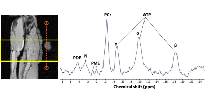 |
A 13C/31P surface coil to visualize metabolism and energetics in the brain
Manushka V. Vaidya1, Georgios Batsios1, Bei Zhang2, Ryan Brown2, Pavithra Viswanath1, Jan Paska2, Gerburg Wulf3, Aaron K. Grant4, Sabrina Ronen1, and Peder E.Z. Larson1
1Department of Radiology, University of California San Francisco, San Francisco, CA, United States, 2Center for Advanced Imaging Innovation and Research, and Center for Biomedical Imaging, New York University School of Medicine, New York, NY, United States, 3Department of Hematology-Oncology, Beth Israel Deaconess Medical Center, Boston, MA, United States, 4Department of Radiology, Beth Israel Deaconess Medical Center, Boston, MA, United States
In this work, we constructed a 13C/31P surface coil for studying cancer metabolism and bioenergetics in the brain. In a single scan session, hyperpolarized 13C-pyruvate MRS and 31P MRS were carried out for a healthy rat brain. A commercial volume proton coil was used for anatomical localization and B0 shimming. Results show good detection of 13C labeled lactate, alanine, and bicarbonate in addition to ATP, phosphocreatine, inorganic phosphate, phosphodiesters and phosphomonoesters from 31P MRS. The coil enables obtaining complementary information within a scan session, thus reducing the number of trials and minimizing biological variability for studies of metabolism and bioenergetics.
|