
Digital Poster Session
Preclinical/Animal Studies: Cancer Imaging: Preclinical & Miscellaneous
Preclinical/Animal Studies
4705 -4719 Cancer Imaging: Preclinical & Miscellaneous - Preclinical: Models & Methods
Session Topic: Cancer Imaging: Preclinical & Miscellaneous
Session Sub-Topic: Preclinical: Models & Methods
Digital Poster
Preclinical/Animal Studies
4705.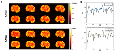 |
3D High Resolution T1 and T2 Mapping at Ultrahigh Fields Using Magnetic Resonance Fingerprinting
Yuning Gu1, Yun Wu2, Hongyi Yang2, Yong Chen3, Jesse I. Hamilton4, Nicole Seiberlich3,4, Charlie Androjna5, Kai Zhong2, and Xin Yu1,3,6
1Department of Biomedical Engineering, Case Western Reserve University, Cleveland, OH, United States, 2High Magnetic Field Laboratory, Chinese Academy of Sciences, Hefei, China, 3Department of Radiology, Case Western Reserve University, Cleveland, OH, United States, 4Department of Radiology, University of Michigan, Ann Arbor, MI, United States, 5Cleveland Clinic Pre-Clinical Magnetic Resonance Imaging Center, Cleveland Clinic Foundation, Cleveland, OH, United States, 6Department of Physiology and Biophysics, Case Western Reserve University, Cleveland, OH, United States
This study demonstrates the development of a 3D T2-prepared MRF sequence, in combination with a stack-of-spirals trajectory, for simultaneous T1 and T2 mapping of an ex vivo macaque monkey (Macaca mulatta) brain with a voxel size of 0.35x0.35x1 mm3. Retrospective evaluation of the undersampling accuracy showed that, for an undersampling rate of up to 16, the normalized root mean square error was within 6% and 15% in T1 and T2 mapping, respectively. These results suggest that 3D simultaneous T1 and T2 mapping can be in <30 min with unprecedented spatial resolution.
|
|
4706. |
A Rabbit Model of Intracranial Atherosclerotic Disease for Pathologic Validation of Vessel Wall MRI
J Scott McNally1, Adam de Havenon1, Seong-Eun Kim1, Chuanzhuo Wang2, Matthew Zabriskie1, Dennis Parker1, Hediyeh Baradaran1, and Matthew David Alexander3
1University of Utah, Salt Lake City, UT, United States, 2Shengjing Hospital of China Medical University, Shenyang, China, 3Radiology and Imaging Sciences, University of Utah, Salt Lake City, UT, United States
Two rabbit model types (Watanabe Heritable Hyperlipidemic and Apolipoprotien E knockout) were studied with vessel wall MRI (vwMRI) and confirmatory histopathological analysis to evaluate for presence of intracranial atherosclerosis (ICAD). Disease was reliably produced in both rabbit types. Sensitivity and specificity of vwMRI for detection of ICAD were 89.5% and 91.9%, respectively. When excluding mild cases to assess accuracy vwMRI for detecting moderate to severe ICAD lesions, sensitivity improved to 100% with unchanged specificity.
|
|
4707. |
Longitudinal voxel-based analysis in Alzheimer’s disease transgenic marmosets
Fumiko Seki1,2, Seiji Shiozawa2, Sho Yoshimatsu2, Yuji Komaki1, Marin Nishio3, Erika Sasaki1, and Hideyuki Okano2
1Central Institute for Experimental Animals, Kawasaki, Japan, 2Department of Physiology, Keio University School of Medicine, Tokyo, Japan, 3Department of Radiological Science, Tokyo Metropolitan University, Tokyo, Japan
This study investigated whether MRI could detect brain abnormalities in transgenic marmoset models of Alzheimer’s disease (AD). Magnetization transfer (MT) contrast-MRI have a potential to detect the presence of amyloid plaques, which could be present in the early stage of the diseases. Voxel-based analysis was conducted to explore whether any difference existed between AD models and healthy marmosets (young-to-middle-aged adults). Significant increase of MT-ratios imaging was observed in posterior cingulate cortex whereas decrease was observed in unilateral inferior temporal cortex. This result supported MT-imaging was sensitive to early abnormalities. Continuous evaluation until old age is worthwhile to be clinically relevant.
|
|
4708. |
Diffusion kurtosis imaging features of renal cell carcinoma: a preliminary study
Lijuan Wang1, Qingqiang Zhu1, Weiqiang Dou2, Jing Ye1, and Jingtao Wu1
1Northern Jiangsu People’s Hospital, Yangzhou, China, 2GE Healthcare, Beijing, China
We aimed to investigate if diffusion kurtosis imaging (DKI) can be applied to differentiate different types of renal cell carcinoma (RCC). For MD, a significant higher value was shown in CCRCC (3.12±0.26) than the rest RCCs (1.03±0.25 for PRCC, 1.78±0.31 for ChRCC and 1.68±0.30 for CDC, p<0.05). For MK,KA and RK, a significant higher value was shown in PRCC (1.52±0.27, 1.93±0.36, 1.21±0.37) than the rest RCCs (1.16±0.23 1.29±0.26, 0.83±0.31 for ChRCC; 0.88±0.21, 1.19±0.33, 0.62±0.29 for CDC and 0.41±0.09, 0.53±0.13, 0.36±0.10 for CCRCC, p<0.05).
|
|
4709.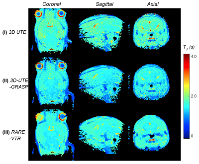 |
Fast 3D T1 mapping with isotropic high resolution using ultrashort TE MRI
Jin Zhang1, Karl Kiser1, and Sungheon Gene Kim1
1New York University School of Medicine, New York, NY, United States
It remains challenging to acquire 3D isotropic high resolution T1 mapping in in vivo MRI experiments. The increase of T1 mapping resolution and coverage are typically limited by the scan time. The purpose of this study is to investigate the feasibility of estimate 3D isotropic high resolution T1 mapping using the variable flip angle (VFA) method1 and 3D-UTE-GRASP sequence2 for small animal imaging at 7T within 3 minutes.
|
|
4710.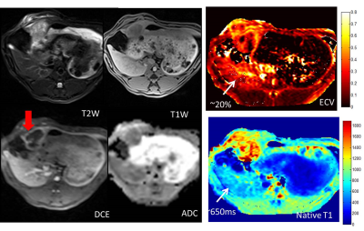 |
Extracellular Volume Fraction (ECV) of Liver Tumors: Mapping of VX2 Tumors in Rabbits before and after Therapy.
Dana C Peters1, Jonatan C. Ferm1, Lynn Savic1, Steffen Huber1, John Walsh1, Daniel Coman1, Fahmeed Hyder1, Mingde Lin1, James S Duncan1, Douglas Rothman1, Albert J Sinuas1, Julius Chapiro1, and R. Todd Constable1
1Yale University, New Haven, CT, United States
In this work, we studied a VX2 tumor in rabbits, which provides a model for hepatocellular carcinoma. The rabbits were imaged with T1 mapping (pre- and post-contrast), which was used to generate extracellular volume fraction (ECV) maps. The average ECV for normal liver and tumor was 20 +/-11 % and 18 +/- 7%, respectively. One week after TACE, tumor ECVs fell to zero, but recovered slightly at two weeks, while normal liver ECV values changed variably after TACE. This preliminary study shows that ECV mapping can depict the highly heterogeneous tumor morphology.
|
|
4711.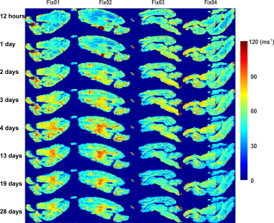 |
Influence of formalin-based fixatives on the MR properties of whole post-mortem pig brains
Azadeh Nazemorroaya1, Ali Aghaeifar1, Hildegard Schulz1, Thomas Shiozawa-Bayer2, Bernhard Hirt2, Klaus Scheffler3,4, and Gisela Hagberg4,5
1Max Planck Institute for Biological Cybernetics, Tuebingen, Germany, 2Clinical Anatomy, University of Tübingen, Tuebingen, Germany, 3High Field Magnetic Resonance,, Max Planck Institute for Biological Cybernetics, Tuebingen, Germany, 4Biomedical Magnetic Resonance, University of Tübingen, Tuebingen, Germany, 5High Field Magnetic Resonance, Max Planck Institute for Biological Cybernetics, Tuebingen, Germany
Post-mortem brain MRI can yield valuable information. However, tissue preservation and MR-compatibility of fixation agents are challenging. Here we investigated the effect of four MR-compatible formalin-based fixatives on the MR properties of pig brains at several timepoints after start of fixation up to one month. The inclusion agents known to improve the dielectric properties of the fixatives lead to greater R2* difference between GM-WM than conventional fixatives. Vice versa these agents lead to a decrease in T1 contrast.
|
|
4712.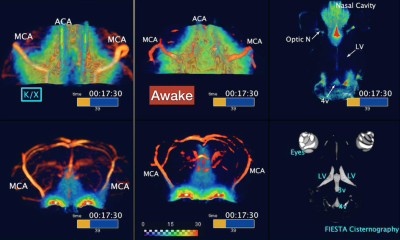 |
Glymphatic monitoring in awake state mouse brain with DCE-MRI imaging
Niklas Daniel Åke Persson1, Evan Hunter Stanton 1, Björn Sigurdsson1, Tuomas Lilius1, Humberto Mestre2, Maiken Nedergaard1,2, and Yuki Mori1,3
1Division of Glial Disease and Therapeutics, University of Copenhagen, Center for Translational Neuromedicine, Copenhagen, Denmark, 2Division of Glial Disease and Therapeutics, University of Rochester Medical Center, Center for Translational Neuromedicine, Rochester, NY, United States, 3Faculty of Health and Medical Sciences, University of Copenhagen, Panum NMR Core Facility, Copenhagen, Denmark
The glymphatic pathway is a novel pathway for interstitial solute clearance. Previous reports note that glymphatic patterns differ according to brain activity. Due to the motion-induced image blurring effects, DCE-MRI in the awake animal is difficult. We aimed to determine the feasibility of FISP-based fast DCE-MRI to assess the glymphatic pathway in awake mice. Head-fixed mice were habituated to restraint and received a cisterna magna cannulation. Awake DCE-MRI was performed 24 hours post surgery. Dynamic tracer distribution maps were calculated, and tracer distribution was measured in each brain segmentation. We successfully obtain DCE-MRI and demonstrate glymphatic dynamics in awake mice.
|
|
4713.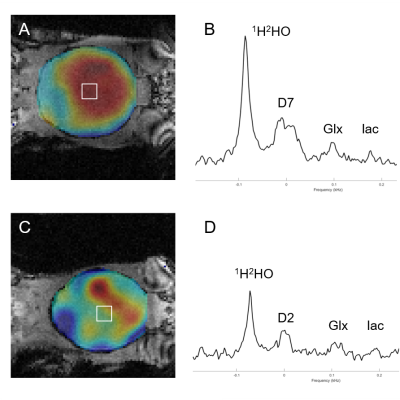 |
Deuterium Metabolic Imaging (DMI) of glucose metabolism in mouse brain.
Henk M. De Feyter1, Monique A. Thomas1, Peter B. Brown1, Akshay Khunte1, and Robin A. de Graaf1
1Dept. of Radiology and Biomedical Imaging, Yale University, New Haven, CT, United States
Deuterium Metabolic Imaging (DMI) is a novel approach providing high spatial resolution 3D metabolic data by combining 2H MRSI with administration of 2H-labeled substrates (1). Mapping of brain glucose metabolism has so far been focused on human and rat. Here we report the implementation of DMI for mapping of glucose metabolism in mouse brain. We described the dedicated radiofrequency coil, explore how different glucose administration approaches affect SNR, and illustrate mapping of glucose metabolism in a mouse model of glioblastoma.
|
|
4714.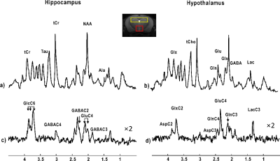 |
Comparison of metabolic fluxes in the mouse dorsal hippocampus and hypothalamus using indirect 1H-[13C] MRS upon [1,6-13C2] glucose infusion
Antoine Cherix1, Bernard Lanz1, and Hongxia Lei2
Video Permission Withheld
1Laboratory for Functional and Metabolic, Ecole Polytechnique Fédérale de Lausanne, Lausanne, Switzerland, 2Animal Imaging and Technology Core (AIT), Center for Biomedical Imaging (CIBM), Ecole Polytechnique Fédérale de Lausanne, Lausanne, Switzerland
With increased interests in understanding the hippocampal regulation of hypothalamic-pituitary-adrenocortical (HPA) axis, this study shows for the first time an in vivo comparison of metabolic fluxes in the mouse dorsal hippocampus and hypothalamus using indirect 1H-[13C] MRS upon [1,6-13C2] glucose infusion at 14.1T. This study provides foundation for investigating relevant evidence to the underlying mechanism of hippocampal regulation of HPA axis.
|
|
4715.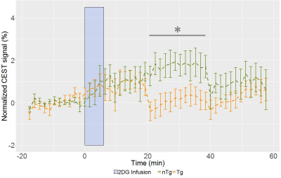 |
Characterization of cerebral metabolism in prodromal stage of disease progression in rat model of Alzheimer's disease
Illsung Joo1, Margaret Koletar1, Wilfred W Lam1, Wendy Oakden1, Greg J Stanisz1, JoAnne McLaurin1, and Bojana Stefanovic1
1Physical Sciences, University of Toronto, Toronto, ON, Canada
Alzheimer’s disease (AD) has complex underlying mechanism associated with progressive decline in multiple aspects of brain functions. This work aims to characterize cerebral metabolic alteration in presymptomatic AD in terms of glucose uptake and β-hydroxybutyrate (βHB) concentration in hippocampus (HC) and entorhinal cortex (EC) of rat model of AD using MR imaging and spectroscopic techniques. Glucose uptake was significantly reduced in HC and trending toward decrease in EC with signs of elevated HC βHB concentration in AD transgenic rat when compared to metabolic markers in healthy subjects, demonstrating that metabolic alteration precedes onset of AD cognitive symptoms.
|
|
4716.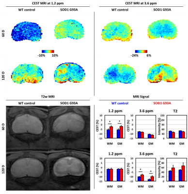 |
CEST MRI Detects Early Signal Changes in the Lumbar Spinal Cord of ALS SOD1 Mice
Aline M. Thomas1,2 and Jeff W.M. Bulte1,2
1Russell H. Morgan Department of Radiology and Radiological Science, Johns Hopkins University School of Medicine, Baltimore, MD, United States, 2Institute for Cell Engineering, Imaging Section and Vascular Biology Program, Johns Hopkins University School of Medicine, Baltimore, MD, United States
In amyotrophic lateral sclerosis (ALS), genetic abnormalities damage motor neurons, resulting in rapidly-progressing neurodegeneration, muscle wasting, and irreversible disability. Current imaging strategies (T2w, DTI, fMRI) monitor structural changes to motor neurons. Here, we investigated continuous wave chemical exchange saturation transfer (CEST) MRI to monitor molecular changes resulting from these abnormalities as a potentially complementary imaging biomarker for monitoring ALS disease progression.
|
|
4717.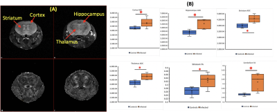 |
Microstructures and metabolites during HIV-1 infection of humanized microglia mice revealed by MRI and MRS
YUTONG LIU1, Saumi Mathews2, Ed Makarov2, Lili Guo2, Mariano Uberti1, Balasrinivasa Sajja1, Larisa Poluektova2, Howard Gendelman2, and Santhi Gorantla2
1Radiology, University of Nebraska Medical Center, Omaha, NE 68198, NE, United States, 2Pharmacology and Experimental Neuroscience, University of Nebraska Medical Center, Omaha, NE 68198, NE, United States
In this study, we performed DTI and MRS on a newly created mouse model of brain HIV infection. MRS results showed neuronal damage on hippocampus, and DTI showed diffuse neuroinflammation caused by HIV infection. White matter changes was also observed using DTI.
|
|
4718.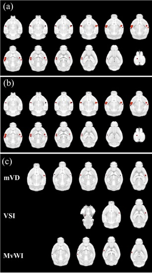 |
Microvascular Architecture Changes in the Brain of an Alzheimer’s disease Mouse
Suk-Ki Chang1, JeongYeong Kim2, DongKyu Lee3, Chang Hyun Yoo4, Jin San Lee5, Hak Young Rhee6, Chang-Woo Ryu7, HyungJoon Cho3, and Geon-Ho Jahng7
1Radiology, Hallym University medical center, Hwasung, Kyung-gi-Do, Republic of Korea, 2Department of physics, Kyung Hee University, Seoul, Republic of Korea, 3Department of Biomedical Engineering, Ulsan National Institute of Science and Technology, Ulsan, Republic of Korea, 4Department of Physics and Research Institute for Basic Sciences, Graduate School, Kyung Hee University, Seoul, Republic of Korea, 5Department of Neurology, Kyung Hee University Hospital, Seoul, Republic of Korea, 6Department of Neurology, Kyung Hee University Hospital at Gangdong, Seoul, Republic of Korea, 7Department of Radiology, Kyung Hee University Hospital at Gangdong, Seoul, Republic of Korea
To characterize and evaluate microvascular architectures presented by brain microvascular indices obtained with a 7T animal MRI system in the transgenic (Tg) AD-model mice and the non-Tg mice using monocrystalline iron oxide nanoparticle (MION) contrast agent, seven non-transgenic (Tg) mice and ten 5xFAD Tg mice were scanned to measure the R2 and R2* relaxation rates before and after injection of MION contrast agent. ΔR2, ΔR2*, BVf, mVD, VSI, and MvWI were greater in the Tg mouse group than in the non-Tg mouse group. ADC and mean vessel density Q were not significantly different between the two groups.
|
|
4719.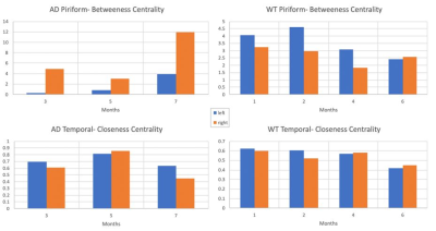 |
DTI-based Hemispheric Differences in Female 3xTgAD Mouse Model
David Hike1,2, Taylor Ariko1,2, Alina Stimmell3, Aaron Wilber3, and Samuel Colles Grant1,2
1Chemical & Biomedical Engineering, Florida State University, Tallahassee, FL, United States, 2CIMAR, National High Magnetic Field Laboratory, Tallahassee, FL, United States, 3Psychology, Florida State University, Tallahassee, FL, United States
This study utilizes DTI and graph theory as a method for early detection of structural changes inconnectivity related to Alzheimer’s Disease. As a function of phenotype and age, DTI analysis was implemented on 3xTgAD female mouse brains and wild type controls at 11.75-T. Current hemisphere dependent data shows differences between hemispheres within the age and phenotype for the parameters observed.
|
 Watch the Video
 View the Poster