
Digital Poster Session
Pediatric: Fetal & Developmental Brain
Pediatric
4570 -4585 Pediatric: Fetal & Developmental Brain - Pediatric Fetal
4586 -4599 Pediatric: Fetal & Developmental Brain - Pediatric Neuro: Development
4600 -4614 Pediatric: Fetal & Developmental Brain - Pediatric Neuro: Congenital Heart Disease & the Brain
4570.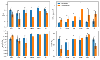 |
3D Fetal Brain Segmentation using an Optimized Deep Learning Approach
Li Zhao1, Xue Feng2, Josepheen Asis-Cruz 1, Yao Wu1, Kushal Kapse1, Axel Ludwig1, Dan Wu3, Kun Qing2, Carig H. Meyer2, and Catherine Limperopoulos1
1Childrens National Hospital, Washington, DC, United States, 2University of Virginia, Charlottesville, VA, United States, 3Zhejiang University, Hanzhou, China
An essential step to accurately quantify fetal brain development is to reliably segment brain regions and perform volumetric measurements. However, this task mainly relies on labor intensive manually contouring. In this work, a 3D U-Net method was optimized and evaluated for fetal brain segmentation. 3D U-Net and 4D atlas-based segmentation methods were compared on 46 fetal brain MRI scans with gestational age 26.4 to 39.1 weeks. The proposed method resulted in (1) higher consistency with the manual segmentation, (2) shorter processing time, and (3) more consistent results across gestational ages, compared to a 4D atlas-based method.
|
|
4571.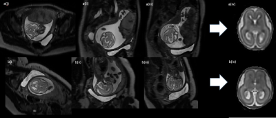 |
T2-Weighted Foetal Brain MRI: Utilising Slice-To-Volume Reconstruction to Reduce Repeat Acquisition Due to Fetal Motion
Elaine Felicity Green1, Emer Hughes1, Kathleen Colford1, Louise Dillon1, Joanna Allsop1, Laura McCabe1, Suzanne Hiscocks1, Lara Waite1, Anthony Price1, Jana Hutter1, Laurence Jackson1, Maria Murgasova-Kuklisova1, Joseph V Hajnal1,
and Mary A Rutherford1
1Centre for the Developing Brain, School of Biomedical Engineering and Imaging Sciences, Kings College London, London, United Kingdom
Single-shot turbo spin echo T2 (SSTSE) is a common sequence used in fetal brain MRI. It is typically acquired in stacks that are positioned according to fetal brain anatomy, producing images that are radiologically in-plane; axial, sagittal, and coronal. However, fetal motion can cause images to be out-of-plane and repeats are often necessary to rectify this. This study proposes a new, optimised method that acquires stacks in planes set to maternal anatomy, rather than fetal brain. We then utilise slice-volume reconstruction to re-register the volume to standard radiological planes.
|
|
4572.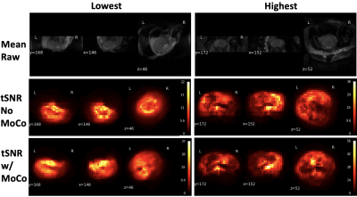 |
Quantifying motion and temporal SNR in fetal functional MRI within a large cohort
Saige Rutherford1 and Melissa Haskell2
1Psychiatry, University of Michigan, Ann Arbor, MI, United States, 2Electrical Engineering and Computer Science, University of Michigan, Ann Arbor, MI, United States
In this work we leverage a large dataset consisting of 535 fMRI sessions from 248 unique subjects to assess the data quality of fetal resting state functional MRI. Our main goal is to describe the quality of fetal resting state fMRI after image post-processing. We quantify how much motion is present, apply a range of motion censoring thresholds to show how much data remains post-censoring, and evaluate the temporal signal to noise ratio of the time-series pre and post motion correction. Our results show very low average tSNR (2.7-3.7) and high percentages (40-95%) of discarded data.
|
|
4573.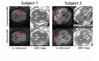 |
ZOOMit DWI of the Fetal Brain: A Preliminary Evaluation Comparing DWI at 3T
Liqi Yang1, Xiaoyong Zhang2, Zhenhui Huang3, and Xin Liu4
1BGI Company, ShenZhen, China, 2MR Collaborations, Siemens Healthcare Ltd, Shenzhen, China, 3Shenzhen Longgang District Central Hosptial, Shenzhen, China, 4Shenzhen Institutes of Advanced Technology, Shenzhen, China
This study evaluated the clinical utility of zoomed diffusion-weighted echo-planar imaging (ZOOMit DWI) of the fetal brain. The effectiveness of the method was compared with conventional echo-planar DWI. Based on imaging analysis of 10 cases, the results showed that ZOOMit DWI exhibits markedly reduced susceptibility and distortion artifacts relative to conventional DWI. ZOOMit-DWI may serve as a viable alternative for the fetal brain assessment and pathology diagnosis.
|
|
4574.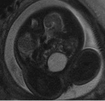 |
Determination of the decrease rate of the apparent diffusion coefficient in the fetal kidneys during the third trimester.
LORENA AZYADE MURRIETA GONZALEZ1, carla maria garcia moreno2, and MARIA BARRERA ESPARZA3
1RESONANCIA MAGNETICA, HOSPITAL ANGELES LOMAS, Huixquilucan Estado de Mexico, Mexico, 2RESONANCIA MAGNETICA, Hospital Angeles Lomas, Huixquilucan, Mexico, 3RESONANCIA MAGNETICA, HOSPITAL ANGELES LOMAS, Huixquilucan, Mexico
The main objective of the work was to determine the decrease rate of the apparent diffusion coefficient (ADC) in fetal kidneys using diffusion-weighted imaging (DWI) during the third trimester. We measured the renal ADC in 16 patients, and our findings exhibit an ADC decrease during the gestational age.
|
|
4575.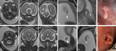 |
The importance of fetal MRI in the evaluation of external ear deformities
Tuantuan Wang1,2, Jinxia Zhu3, and Guangbin Wang2
1Shandong University, Jinan, China, 2Shandong Medical Imaging Research Institute, Jinan, China, 3MR Collaboration, Siemens Healthcare Ltd., Beijing, China Poster Permission Withheld
Fetal external ear deformities sometimes indicate that a chromosomal abnormality is present. This study aimed to demonstrate the feasibility of fetal MRI in the evaluation of auricular deformities, as well as the added value of external auditory canal atresia. We calculated the diagnostic sensitivity, specificity, and accuracy of fetal MRI and described the imaging features. Our study showed that fetal MRI can provide valuable information to assist in the diagnosis of external ear deformities.
|
|
4576.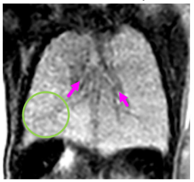 |
Fetal cardiac magnetic resonance imaging (FCMR); MR sequences, cardiac & non-cardiovascular findings with prenatal and postnatal correlation
Dianna M. E. Bardo1, Christopher Lindblade2, Fidaa Wishah1, Patricia Cornejo1, Jennifer Vaughn1, Mittun Patel1, and Luis Goncalves1
1Radiology, Phoenix Children's Hospital, Phoenix, AZ, United States, 2Cardiology, Phoenix Children's Hospital, Phoenix, AZ, United States Fetal MRI is a not uncommon imaging procedure, most often performed to diagnose neurologic abnormalities. Increasingly fetal cardiac MRI is performed to confirm CHD findings found on fetal echocardiography and associated non-cardiovascular anomalies, especially in mid-to-late gestation fetuses. This educational exhibit will provide guidance for image acquisition and show an extensive multi-modality case based library of fetal cardiac images, highlighting MRI findings, with correlation to postnatal imaging. |
|
 |
4577.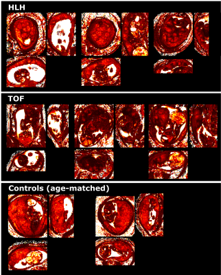 |
Qualitative and Quantitative analysis of placental function to characterize pathophysiology in congenital heart disease (CHD)
Johannes Klaus Steinweg1, Grace Tin Yan Hui2, Milou van Poppel1, Kathleen Colford2, David Lloyd1, Reza Razavi1, Kuberan Pushparajah1, Mary A Rutherford2, and Jana Hutter2
1Cardiovascular Imaging, King's College London, London, United Kingdom, 2Perinatal Imaging & Health, King's College London, London, United Kingdom
Congenital Heart Disease (CHD) is the most frequent congenital defect and varies widely in severity and subtype. The antenatal diagnosis of the severity is of key importance to plan delivery and treatment. The shared developmental pathways and close functional links with the fetal heart may lead to placental dysfunction that in turn may alter the in-utero environment to the detriment of mother and fetus. Hence, assessment of placental function delivers crucial complementary information to characterize pathophysiology in CHD. This study employs high-resolution whole-placental T2* relaxometry in 75 women (40 diagnosed with a fetus with CHD) to phenotype placental function.
|
4578.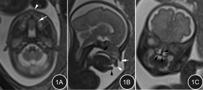 |
Evaluation of MRI in the Prenatal Dignosis of Cleft Lip and Palate
Mimi Tian1, Xiangtao Lin1,2, Shuwei Liu1, and Xiang Feng3
1Shandong Unversity, Jinan, China, 2Department of Radiology, Shandong Provincial Hosptial Affiliated to Shandong University, Jinan, China, 3MR Scientific Marketing, Siemens Healthcare Ltd, Beijing, China
This prospective study aimed to evaluate the value of MRI in prenatal diagnosis of cleft lip and palate. Pregnant females subject with fetal facial malformations were suspected by ultrasonography (US) and then underwent 1.5 T MRI scan. The diagnosis accuracy of the prenatal US and MRI was compared with the postnatal findings. A total of 44 fetuses were followed up with lip and/or cleft palate malformation. Compared to US, MRI provides more useful information in the diagnosis of fetal cleft lip and palate,which could be used as one of the effective alternative to prenatal ultrasound diagnosis.
|
|
|
4579.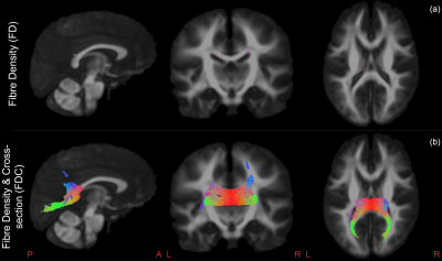 |
White matter development after prenatal chemotherapy exposure: a diffusion MRI fixel-based analysis.
Jeroen Blommaert1, Ahmed Radwan2, Charlotte Sleurs1, Ron Peeters2,3, Stefan Sunaert2,3, An-Sofie Gorissen1, Kristel Van Calsteren4,5, Frédéric Amant1,6,7, and Sabine Deprez2
1Oncology, KU Leuven, Leuven, Belgium, 2Imaging & Pathology, KU Leuven, Leuven, Belgium, 3Radiology, University Hospitals Leuven, Leuven, Belgium, 4Development and regeneration, KU Leuven, Leuven, Belgium, 5Gynaecology and Obstetrics, University Hospitals Leuven, Leuven, Belgium, 6Gynecological Oncology, The Netherlands Cancer Institute, Antoni van Leeuwenhoek, Amsterdam, Netherlands, 7Gynaecological oncology, Amsterdam university medical centers, Amsterdam, Netherlands
Currently, data are scarce on the long-term outcome of prenatal exposure to cancer treatment. In this preliminary analysis, we investigated white matter development in 9-year-old children who were prenatally exposed to chemotherapy, using multi-shell diffusion MRI and fixel-based analysis. We found indications of lower within-voxel (reflected by Fibre Density), macroscopic (reflected by Fibre Cross-section) and total (reflected by Fibre Density and Cross-section) intra-axonal volume of the splenium, isthmus and tapetal fibres of the corpus callosum in children with prenatal chemotherapy exposure compared to controls. This suggests prenatal chemotherapy exposure to potentially impact white matter development.
|
4580.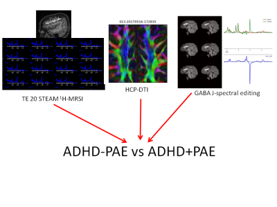 |
Neurochemical Correlates of Brain Abnormalities in Prenatal Alcohol Exposure: Interim Analysis
Jeffry R. Alger1,2,3, Joseph O'Neill4, Lisa Kilpatrick4, Katherine L. Narr1, Guldamla Kalender4, Ronald Ly4, Shantanu H. Joshi1, Sandy Loo4, Mary J. O'Connor4, and Jennifer G. Levitt4
1Neurology, University of California, Los Angeles, Los Angeles, CA, United States, 2Advanced Imaging Research Center, University of Texas Southwestern Medical Center, Dallas, TX, United States, 3NeuroSpectroScopics LLC, Sherman Oaks, CA, United States, 4Psychiatry and Biobehavioral Sciences, University of California, Los Angeles, Los Angeles, CA, United States
Multimodal MR neuroimaging was used to identify brain differences between attention deficit hyperactivity disorder (ADHD) associated with prenatal alcohol exposure (PAE) (ADHD+PAE), ADHD not associated PAE (ADHD-PAE) and normally-developing controls in an study of children aged 8-12 years. This is an interim report of findings obtained from an ongoing study after successful imaging of 73 subjects. Statistically-significant lateralized brain differences were identified in frontal white matter and gray matter. The findings provide preliminary support for the hypothesis that an objective diagnostic neuroimaging-based classifier can be developed.
|
|
4581.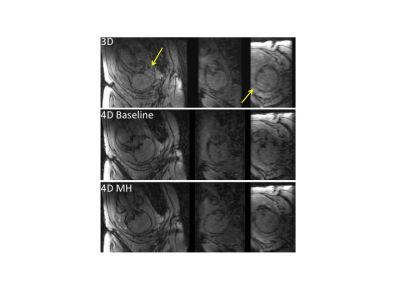 |
Whole Brain Cerebral Oxygenation Measurements in Fetuses Using 4D MRI
Jing Liu1, Yan Wang1, Duan Xu1, Patrick McQuillen1, and Shabnam Peyvandi1
1University of California San Francisco, San Francisco, CA, United States Poster Permission Withheld
We developed accelerated 4D MRI and efficient image processing tool for whole fetal brain oxygenation measurement. Fetuses with CHD have lower cerebral oxygenation compared with controls and exhibit an oxygenation increase with maternal hyperoxia testing while controls exhibit no change.
|
|
4582.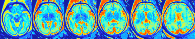 |
Preliminary Study on Quantitative Assessment of Fetal Brain Using MOLLI T1 Mapping Sequence
Haibo Qu1,2, Xuesheng Li3, Fenglin Jia3, Yi Liao3, Zhijun Ye3, Pei Li3, Sai Liu3, Xinmao Ma3, Xiaoyue Zhou4, Qing Li4, and Shaoyu Wang5
1West China Second University Hospital, Sichuan University, Chengdu, China, 2Key Laboratory of Birth Defects and Related Diseases of Women and Children (Sichuan University), Ministry of Education, Chengdu, China, 3West China Second University Hospital, Chengdu, China, 4MR Collaborations, Siemens Healthineers Ltd., Shanghai, China, 5MR Scientific Marketing, Siemens Healthineers Ltd., Xian, China
A modified Look-Locker inversion recovery T1 mapping technique was used to study the development of fetal brain. Our study found the MOLLI sequence with a short scan time could reduce the impact of fetal brain movements and perform robust T1 measurements. T1 values differ in brain regions and gestational weeks (from 24 to 36). A negative correlation was found between T1 values and gestational age in thalamus, posterior limb and brain stem. Therefore, T1 measured by the MOLLI sequence could be used as a biomarker for evaluating fetal brain development.
|
|
4583.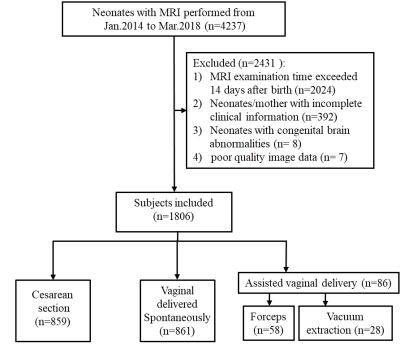 |
Assisted vaginal delivery increases the risk of punctate white matter lesions in neonatal brain found by MRI
Ting Liu1, Lingxia Zeng1, Xia Wang1, and Jian Yang1
1the First Afliated Hospital of Xi'an Jiaotong University, Xi'an, China
Punctate white matter lesion (PWML) was the common abnormal findings by MRI in neonates, which might predict poor motor and cognitive outcomes. Assisted vaginal delivery (AVD) increased brain injury. But the association between AVD and PWML is not clear. This study showed the mode of delivery is associated with PWML. AVD markedly increased the risk of PWML compared with cesarean delivery, especially vacuum extraction with cesarean section as a reference. The incidence of II and III-IV grade PWML of neonates with AVD was much higher than that with cesarean section.
|
|
4584.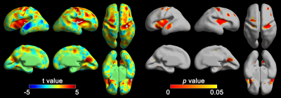 |
Altered Cortical Folding Depth in Fetuses with Down Syndrome
HyukJin Yun1, Juan David Perez1, Neel Madan2, Rie Kitano3, Shizuko Akiyama4, Diana W. Bianchi5, P. Ellen Grant1, Tomo Tarui4, and Kiho Im1
1Newborn Medicine, Boston Children's Hospital, Boston, MA, United States, 2Radiology, Tufts Medical Center, Boston, MA, United States, 3Obstetrics and Gynecology, Long Island Jewish Medical Center, New Hyde Park, NY, United States, 4Mother Infant Research Institute, Tufts Medical Center, Boston, MA, United States, 5Medical Genetics Branch, National Human Genome Research Institute, Bethesda, MD, United States
Analyzing fetal brains with Down syndrome is an interesting are because genetic factors affect brain development in early fetal stages. In addition to cortical volumetric and areal measures used in our previous studies, cortical folding in the fetal brain would be an important marker of altered neurodevelopment due to Down syndrome. Thus, we calculated sulcal depth to quantify cortical folding and compared sulcal depth between Down syndrome and typically developing fetuses. Down syndrome fetuses showed significantly altered sulcal depth compared to typically developing peers in the regions related to decreased neurogenesis in early fetal life.
|
|
4585.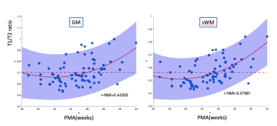 |
Mapping the spatiotemporal developmental pattern of the cortex and its adjacent white matter for preterm neonates using MRI
Shiyu Yuan1, Dr. Mengting Liu1, Jingda Yang 1, Dr. Arthur W. Toga1, Dr. A. James Barkovich2, Dr. Duan Xu2, and Dr. Hosung Kim 3
1USC Stevens Neuroimaging and Informatics Institute, Keck School of Medicine of USC, University of Southern California, Los Angeles, CA, United States, 2University of California, San Francisco, San Francisco, CA, United States, 3USC Stevens Neuroimaging and Informatics Institute as department, Keck School of Medicine of USC, University of Southern California, Los Angeles, CA, United States
Myelination is essential for normal brain function. The development of myelin begins in the third trimester of pregnancy and enables rapid synchronized communication between different brain regions. During the 3rd trimester of gestation overlapping with preterm birth, the neurodevelopment undergoes intensive cyto and myeloarchitecture changes. The T1/T2 ratio is a surrogate for the degree of myelination but has not been used to study early neurodevelopment. Using T1/T2 ratio, we show the unique spatiotemporal pattern of the cyto and myeloarchitecture changes in preterm neonates. This result may be useful in prediction of neurodevelopmental outcome and functional impairments in preterm survivors.
|
 View the Poster
 Watch the Video
4586.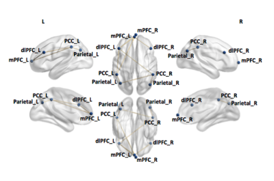 |
Functional Connectivity Alterations in Neural Networks Associated with Sustained Attention in Children with Epilepsy
Lynette Looi Ling1, Amanda Wood1, Elaine Foley1, and Stefano Seri 1,2
1School of Life and Health Sciences & Aston Neuroscience Institute, Aston University, Birmingham, West Midlands, United Kingdom, 2Children’s Epilepsy Surgery Service, Birmingham Children’s and Women’s Hospital NHS Foundation Trust, Birmingham, United Kingdom
Epilepsy is a common neurological disorder diagnosed in children that can be associated with impairments in sustained attention. Attentional deficits are linked to neural dysfunction within or between the default mode network and central executive network. Limited research has examined abnormalities in these networks in children with epilepsy. Using resting-state fMRI we found reduced connectivity within and between both networks in patients (n=18) compared to controls (n=16). The neural alterations found in patients could be potential predictors of attentional deficits, and subsequently aid in identifying children requiring intervention, however further studies are warranted to confirm this.
|
|
 |
4587.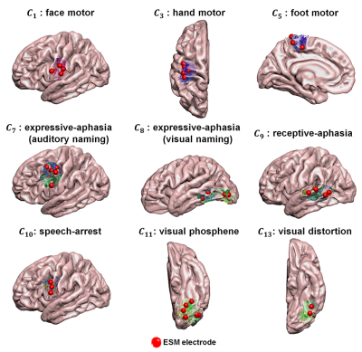 |
Deep learning of electrical stimulation mapping-driven DWI tractography to improve preoperative evaluation of pediatric epilepsy surgery
Min-Hee Lee1,2, Nolan O'Hara2,3, Csaba Juhasz1,2,3,4, Eishi Asano1,3,4, and Jeong-Won Jeong1,2,3,4
1Pediatrics, Wayne State University School of Medicine, Detroit, MI, United States, 2Translational Imaging Laboratory, Children's Hospital of Michigan, Detroit, MI, United States, 3Translational Neuroscience Program, Wayne State University School of Medicine, Detroit, MI, United States, 4Neurology, Wayne State University School of Medicine, Detroit, MI, United States
To investigate the clinical utility of deep convolutional neural network (DCNN)-tract-classification in the preoperative evaluation of children with focal epilepsy, DCNN-tract-classification deeply learned spatial trajectories of DWI tracts linking electrical stimulation mapping (ESM) findings, and then used to detect eloquent tracts. We found that the DCNN-tract-classification can achieve an excellent accuracy (98%) to detect eloquent areas. Also, the subsequent Kalman filter analysis showed that the preservation of detected areas predicts no postoperative deficits with a high mean accuracy across different functions (92%). Our findings demonstrate that DCNN-tract-classification may offer vital translational information in pediatric epilepsy surgery.
|
|
4588.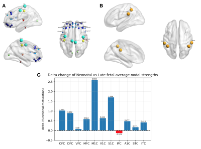 |
Resting State Functional Connectivity and Angiogenesis-related Gene Co-Expression Networks in early brain development
Serafeim Loukas1,2, Sandra Martin1, Joana Sa de Almeida1, Lana Vasung1,3, Dimitri Van De Ville2, Djalel Meskaldji1,4, and Petra S. Hüppi1
1Division of Development and Growth, Department of Pediatrics, University of Geneva, Geneva, Switzerland, 2Institute of Bioengineering, Ecole Polytechnique Fédérale de Lausanne (EPFL), Lausanne, Switzerland, 3Division of Newborn Medicine, Department of Medicine, Harvard Medical School, Boston, MA, United States, 4Institute of Mathematics, Ecole Polytechnique Fédérale de Lausanne (EPFL), Lausanne, Switzerland
During early brain development, rs-functional connectivity exhibits regional and age-specific activation patterns. We hypothesize that these rs-fMRI patterns might reflect maturity of intracerebral microvascular compartment linked to spatio-temporal genetic expression patterns. The genetic patterns were explored through postmortem human brain specimens and the rs-fMRI connectivity using a longitudinal preterm dataset. rs-functional connectivity and angiogenic genes expression show spatio-temporal differences during early brain development. We observe an increased role of the primary somatosensory and motor cortices from late-fetal to neonatal periods that might be driven by an increased expression of important angiogenic genes in these regions.
|
4589.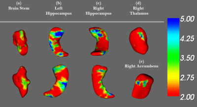 |
Investigating the brain volume and shape correlates of reading performance in children with reading difficulties
Yu-Chen Chuang1, Hsiao-Lan Sharon Wang2, and Jun-Cheng Weng1,3,4
1Department of Medical Imaging and Radiological Sciences, Chang Gung University, Taoyuan, Taiwan, 2Department of Special Education, National Taiwan Normal University, Taipei, Taiwan, 3Department of Psychiatry, Chang Gung Memorial Hospital, Chiayi, Taiwan, 4Medical Imaging Research Center, Radiological Research, Chang Gung University and Chang Gung Memorial Hospital, Linkou, Taoyuan, Taiwan
Reading disability is a special learning disorder based on the neurophysiological of the brain. Schooling children with reading disabilities (CRD) have different ways to process language from normal children (NCRD). Our study investigated the abnormality of brain volume and shape contributing to specific reading disabilities in schooling children. We found the significant difference in brain volume and shape of the parietal, temporal lobe and limbic system between CRD and NCRD. These brain areas are associated with the core phonological processing regions.
|
|
4590.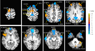 |
Altered functional connectivity of the primary auditory with cognitive networks in inattentive children and adolescents with hearing loss
Jianhong Li1, Weiwei Men2, David Zhu3, and Junfang Xian1
1Radiology, Beijing Tongren Hospital,Capital Medical University, Beijing, China, 2Center for MRI Research, Academy for Advanced Interdisciplinary Studies, Beijing City Key Lab for Medical Physics and Engineering, Institute of Heavy Ion Physics, School of Physics, Peking University, Beijing, China, 3Radiology and Cognitive Imaging Research Center, Michigan State University, East Lansing, MI, United States
Early profound deaf children and adolescents have been reported to suffer from cognitive decline and attentional deficit. Twenty-five inattentive children and adolescents with bilateral prelingual profound deafness and 30 age- and gender- matched normal controls were recruited in this study. The resting state functional connectivity differences were analyzed between two groups. The results indicated a direct impact of early auditory deprivation on high cognitive networks. The usage of hearing aid was also found to normalize the connectivity between primary auditory cortex and attention network.
|
|
4591.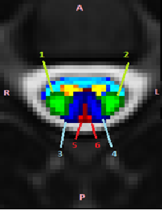 |
Development of Pediatric Spinal Cord White Matter Atlas: Preliminary Analysis
Shiva Shahrampour1, Benjamin De Leener2, Devon Middleton1, Mahdi Alizadeh1, Laura Krisa1, Adam Flanders3, Scott Faro3, Julien Cohen-Adad2, and Feroze Mohamed1
1Thomas Jefferson University, Philadelphia, PA, United States, 2Polytechnique Montreal, Montreal, QC, Canada, 3Thomas Jefferson University Hospital, Philadelphia, PA, United States
Spinal cord diffusion tensor imaging and atlas-based group analysis has potential for improving prognosis of spinal cord related diseases, aid clinical decision making and provide quantitative imaging biomarkers for spinal cord injury assessment.
|
|
4592.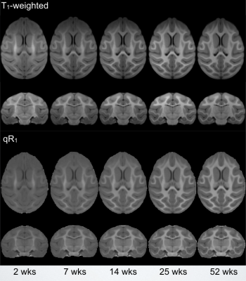 |
Investigation of Longitudinal Nonhuman Primate White Matter Maturation using Quantitative R1 Relaxometry
Douglas C Dean1,2,3, Jason F Moody2, Steve R Kecskemeti3, Nakul Aggarwal4, Jonathan A Oler4, Andrew L Alexander2,3,4, and Ned Kalin4
1Pediatrics, University of Wisconsin–Madison, Madison, WI, United States, 2Medical Physics, University of Wisconsin–Madison, Madison, WI, United States, 3Waisman Center, University of Wisconsin–Madison, Madison, WI, United States, 4Psychiatry, University of Wisconsin–Madison, Madison, WI, United States
Quantitative relaxometry affords a unique opportunity to map dynamic patterns of white matter development. Nonhuman primates offer a valuable model for investigating neurodevelopment, however, NHP studies utilizing relaxometry based approaches have been limited. Here, we utilize R1 relaxometry to map maturation in 35 rhesus macaques scanned across 5 timepoints during the first year of life. R1 is observed to rapidly increase during this period, with average and individual patterns exhibiting a logarithmic-like trajectory. Results highlight the onset of myelin during this period of development and demonstrate relaxometry to be a promising approach for characterizing patterns of early brain development.
|
|
4593.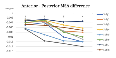 |
Improved quantitative accuracy using high-resolution DTI guided QSM to profile myelin maturation in pediatric brains
Lijia Zhang1, Chris Petty1, and Allen Song1
1Duke University Brain Imaging and Analysis Center, Durham, NC, United States
Quantitative susceptibility mapping (QSM) has the potential to help access brain development, in particular, the magnetic susceptibility changes during the myelination processes in the white matter. However, the quantitative accuracy of QSM is limited by its angle dependency to the fiber orientation. In this study, we propose a method utilizing ultra-high resolution diffusion tensor imaging (DTI) to delineate the major fiber bundles (i.e. corpus callosal fibers), followed by tract-based QSM, to largely remove the angle dependence and accurately quantify the magnetic susceptibility changes during brain development.
|
|
4594.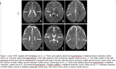 |
Prediction of spastic cerebral palsy based on radiographic feature in children with periventricular leukomalacia
Tingting Huang1,2, Zhe Liu1, Chao Jin1, Haoxiang Jiang3, Heng Liu1, Wei Xing2, Xiaocheng Wei4, Gang Zhang2, and Jian Yang5
1The First Affiliated Hospital of Xi’an Jiaotong University, Xi’an, China, 2The First Affiliated Hospital of Henan University of traditional Chinese Medicine, Zhengzhou, China, 3Xi’an Children Hospital, Xi’an, China, 4GE Healthcare, Peking, China, 5The First Affiliated Hospital of Xi’an Jiaotong University, Zhengzhou, China
Early identification of classification of cerebral palsy (CP) in children with periventricular leukomalacia (PVL) is crucial for specific rehabilitation treatment and improving the quality of life. In this study, we retrospectively analyzed the baseline clinical data and visually assessed PVL-associated MR signs in children with CP, found that T2WI abnormal hyperintensity in body of lateral ventricle, posterior limb of internal capsule (PLIC), lenticular nucleus and white matter volume reduction were independent predictors of spastic CP. Results indicated that the area under receiver operating characteristic curve of multivariate regression model was 0.85, sensitivity and specificity for this model were 74.4% and 85.7%, respectively.
|
|
4595.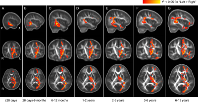 |
White Matter Left-lateralization from Birth through Childhood Revealed by Tract-based Spatial Statistics with Dynamic Age-specific Templates
Xianjun Li1, Yuli Zhang1, Mengxuan Li1, Miaomiao Wang1, Congcong Liu1, Chao Jin1, Xiaocheng Wei1, and Jian Yang1
1Department of Radiology, the First Affiliated Hospital of Xi’an Jiaotong University, Xi'an, China
Human brain exhibits structural and functional asymmetries. How the left-lateralization in white matter changes with age from newborns through childhood is not fully investigated. In this work, development of the left-lateralization from birth to 13 years is revealed by using tract-based spatial statistics with dynamic age-specific brain templates. Structural left-lateralization with larger fractional anisotropy in motor-related and language-related white matter is already present in newborns. The involvement of corticospinal tract, superior longitudinal fasciculus and uncinate fasciculus into regions with left-lateralization appears at different age. This may be associated with the development of fine motor and language during the early childhood.
|
|
4596.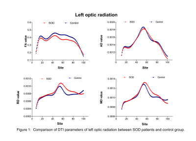 |
Investigating the change of optic radiation in Septo-optic dysplasia patients with DTI
Xiaoyu Wang1, Cong Tian1, Xinxin Tao1, Mengxuan Li1, Yuli Zhang1, Xiaocheng Wei2, and Jian Yang3
1The first affliated hospital of Xi'an Jiaotong University, Xi'an, China, 2MR Research China, GE Healthcare, Beijing, China, 3Radiology, The first affliated hospital of Xi'an Jiaotong University, Xi'an, China
Septo-optic dysplasia (SOD) is a rare congenital malformation disorder and have equally prevalent in males and females in early brain development. This study evaluated the micro-structure of optic radiation pathway using automatic fiber bundle quantization tracking (AFQ)in DT image to investigate the possible developmental impairment of optic radiation pathway that might cause by the abnormalanatomy structure of SOD patient. Result showed abnormal changes of optic radiation were found in SOD patients. The DTI tractography result could provides a non-invasive mapping of the optic radiation pathway and locate the injury.
|
|
4597.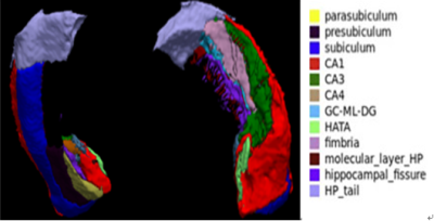 |
Subcortical abnormalities in adolescents with first-episode drug-naïve major depressive disorder
Ruohan Feng1, Lihua Zhuo1, Xinyue Hu2, Yingxue Gao2, Lianqing Zhang2, Yang Li3, Xinyu Hu2, Shi Tang2, Ming Zhou1, Guoping Huang3, and Xiaoqi Huang2
1Department of Radiology, the third hospital of Mianyang, Mianyang, China, Mianyang, China, 2Huaxi MR Research Center (HMRRC), Functional and Molecular Imaging Key Laboratory of Sichuan Province, Department of Radiology, West China Hospital, Sichuan University, Chengdu 610041, China, Chengdu, China, 3Department of Psychiatry, the third hospital of Mianyang, Mianyang, China, Mianyang, China
We analysis subcortical nucleus volume in 57 adolescents aged 13-18: 35 with MDD and 22 healthy controls (HC). Compared to HCs, the MDD group showed reduction in volume of the HATA nuclei of the left hippocampus and the subiculum nuclei of the right hippocampus, and increased volume in the LD、MDI、 MDm of the left thalamus and in the MV(Re) of the right thalamus. There were no significant difference in amygdala subregion between the MDD group and the HC group. We propose that these subregions will contribute to the affective and cognitive abnormities in adolescents with MDD.
|
|
4598.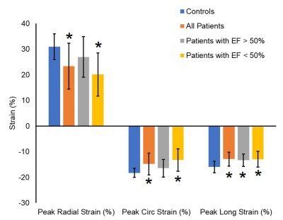 |
CHemotherapy-Induced cardiotoxicity: LongitudinaL imaging study (CHILL)
Nivedita K. Naresh1, Doaa Aly2, Michal Schafer2, Brian Fonseca3, Bruce Landeck2, Jenna Sopfe4, Lorna P Browne1, and Alex J Barker1,5
1Radiology, Children's Hospital Colorado, Aurora, CO, United States, 2Cardiology, Children's Hospital Colorado, Aurora, CO, United States, 3Pediatrics, Children's Hospital Colorado, Aurora, CO, United States, 4Oncology, Children's Hospital Colorado, Aurora, CO, United States, 5Bioengineering, University of Colorado Anschutz Medical Campus, Aurora, CO, United States
In this retrospective study, we explored the potential of cardiac MRI to identify functional imaging biomarkers of cardiotoxicity in pediatric patients exposed to anthracycline chemotherapy. Global and regional myocardial strain was reduced in patients with ejection fraction (EF)>50% indicating that myocardial strain may be an early imaging biomarker of cardiotoxicity in pediatric patients. Additionally, regional differences were found in strain measurements in this population. The results from this study will be used to perform prospective imaging studies to identify early functional imaging biomarkers of cardiotoxicity in pediatric population.
|
|
4599.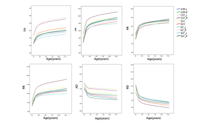 |
Depicting White Matter Developmental Trajectory Using Diffusional Kurtosis Imaging:from neonate through childhood
Yuli Zhang1, Xianjun Li1, Mengxuan Li1, Qinli Sun1, Miaomiao Wang1, Chao Jin1, Congcong Liu1, Fan Wu1, Xiaoyu Wang1, Huifang Zhao1, Yannan Chen1, Cong Tian1, Peiyao Chen1, Xiaocheng Wei2,
and Jian Yang1
1the First Affiliated Hospital, Xi'an Jiaotong University, Xi'an, China, 22. MR Research China, GE Healthcare, Xi'an, China, China
Brain development is a complex process linked with behavioral, emotional, cognitive, especially the maturation process of WM is both of great scientific and clinical importance1. However, the lack of continuous development of WM from neonate to childhood. Diffusion kurtosis imaging(DKI)is a diffuse imaging method that reflects non-Gaussian distribution information of biological water. This study aimed to use DKI parameters depicting developmental trajectory of WM. The fractional anisotropy(FA), mean kurtosis(MK),axial kurtosis(AK),radial kurtosis(RK)positively correlated with age; the axial diffusivity(AD) radial diffusivity(RD) negativly correlated with age. These parameters changed rapidly before the age of 2 years old, and then gradually slowed down.
|
4600.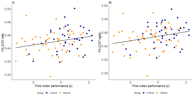 |
Altered corticospinal tract microstructure is associated with motor performance in adolescents with congenital heart disease
Melanie Ehrler1, Michael von Rhein1,2, Oliver Kretschmar3, Beatrice Latal1, and Ruth O'Gorman Tuura4
1Child Development Center, University Children's Hospital, Zurich, Switzerland, 2Developmental Pediatrics, Cantonal Hospital Winterthur, Winterthur, Switzerland, 3Cardiology, University Children's Hospital, Zurich, Switzerland, 4Center for MR Research, University Children's Hospital, Zurich, Switzerland
Children with congenital heart diseases (CHD) undergoing open-heart surgery are at an increased risk of motor impairment, but little is known about the neuroanatomical correlates of these deficits. The purpose of the present study was to examine the link between corticospinal tract (CST) microstructure, assessed with diffusion tensor MRI, and motor function in a cohort of adolescents with operated CHD. Compared to age-matched controls, adolescents with CHD showed lower CST anisotropy which correlated with worse fine motor function and poorer movement quality. CHD may therefore lead to alterations in motor tract development, increasing the risk of persistent motor impairments.
|
|
4601.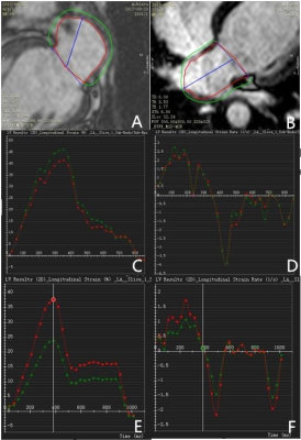 |
Abnormal Left Atrial Performance in Children with Repair Tetralogy of Fallot using CMR Feature Tracking
Liwei Hu1, Xiaodan Zhao2, Rongzhen Ouyang1, Shuang Leng2, WeiHui Xie1, Yafeng Peng1, Xiaofen Yao1, Yong Zhang3, Ru San Tan2,4, Liang Zhong2,4, and Yumin Zhong1
1Radiology, Shanghai Children's Medical Center, Shanghai, China, 2National Heart Centre Singapore, Singapore, Singapore, 3GE Healthcare, Shanghai, China, 4Duke-NUS Medical School, National University of Singapore, Singapore, Singapore
Right ventricular (RV) and atrial (RA) dilation has been clinically observed in patients after initial repair of tetralogy of Fallot (rTOF) and is associated with adverse long-term outcomes. Due to RA and left atrial (LA) interaction, we aimed to determine the effect of RA dilation on LA performance from feature tracking cardiovascular magnetic resonance (CMR) in pediatric rTOF patients. Results revealed that LA strains and strain rates were impaired in rTOF than age- and gender-matched healthy volunteers. RA volumes are negatively associated with LA strains. These findings may suggest LA diastolic dysfunction from chronic RV and RA dilations in rTOF, even at early stage after initial repair.
|
|
|
4602.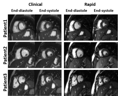 |
20-min Pediatric Cardiovascular Magnetic Resonance without Contrast or Anesthesia: Initial Evaluation in Kids with Congenital Heart Disease
Lexiaozi Fan1,2, Hassan Haji-Valizadeh3, Cynthia K Rigsby4, Joshua D. Robinson4, Paige Constance Nelson4, and Daniel Kim1,2
1Department of Radiology, Northwestern University Feinberg School of Medicine, Chicago, IL, United States, 2Department of Biomedical Engineering, Northwestern University, Evanston, IL, United States, 3Department of Medicine (Cardiovascular Division), Beth Israel Deaconess Medical Center & Harvard Medical School, Boston, MA, United States, 4Division of Pediatric Cardiology, Ann & Robert H. Lurie Children's Hospital of Chicago, Chicago, IL, United States
Cardiovascular MRI is an excellent diagnostic tool for children with congenital heart disease (CHD), but the long acquisition times, and need for sedation/anesthesia and the administration of a gadolinium-based contrast results it only accounts for only 2% of pediatric cardiac diagnostic tests. Our 20-min, free-breathing, non-contrast cardiovascular magnetic resonance (CMR) protocol is potentially a game-changer, because its total scan time (20 min) is considerably shorter than a standard protocol (90 min) and does not require anesthesia or gadolinium-based contrast agent.
|
4603.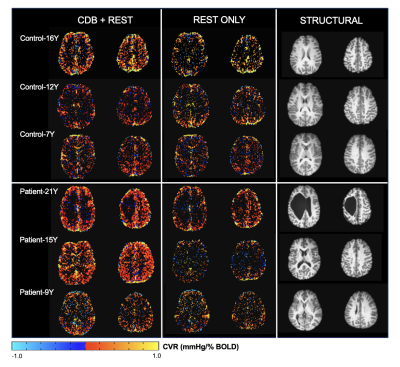 |
Feasibility of a cued deep breathing task to map cerebrovascular reactivity in healthy and clinical pediatric cohorts
Kristina Zvolanek1,2, Rachael Stickland1, and Molly Bright1,2
1Physical Therapy and Human Movement Sciences, Northwestern University, Chicago, IL, United States, 2Biomedical Engineering, McCormick School of Engineering, Northwestern University, Evanston, IL, United States
Cerebrovascular reactivity (CVR) mapping with MRI is emerging as a useful metric of vascular health. Tracking natural fluctuations in CO2 (a vasodilator) during a resting state fMRI scan has been proposed as a method to map CVR, but it is uncertain whether this method is reliable. We propose adding a two-minute breathing challenge prior to a resting state acquisition to transiently reduce CO2 levels. Here, we tested the feasibility of this method in a small pediatric cohort. The breathing challenge produced more robust CVR maps in both controls and children with cerebral palsy compared to resting state data alone.
|
|
4604.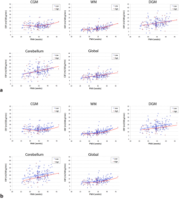 |
Longitudinal Assessment of Third Trimester Regional Cerebral Blood Flow in Very Preterm Infants
Zungho Zun1,2,3,4, Kushal Kapse1, Marni Jacobs3,5, Sudeepta Basu3,6, Mariam Said3,6, Nicole Andersen1, Jonathan Murnick1,3,4, Taeun Chang3,7,8, Adre du Plessis2,3, and Catherine Limperopoulos1,2,3,4
1Division of Diagnostic Imaging and Radiology, Children's National Hospital, Washington, DC, United States, 2Division of Fetal and Transitional Medicine, Children’s National Hospital, Washington, DC, United States, 3Department of Pediatrics, George Washington University, Washington, DC, United States, 4Department of Radiology, George Washington University, Washington, DC, United States, 5Division of Biostatistics and Study Methodology, Children's National Hospital, Washington, DC, United States, 6Division of Neonatology, Children's National Hospital, Washington, DC, United States, 7Division of Neurology, Children's National Hospital, Washington, DC, United States, 8Department of Neurology, George Washington University, Washington, DC, United States
Early newborn age is a crucial phase of brain development, which can be characterized by in-vivo measurement of cerebral blood flow (CBF). In this study we assessed regional CBF in very preterm infants longitudinally during the ex-utero third trimester using arterial spin labeling. CBF in preterm infants significantly increased with postmenstrual age in all regions with the most rapid increase and the highest mean of CBF in the cerebellum. Lower CBF was associated with intraventricular hemorrhage and patent ductus arteriosus. CBF in preterm infants was higher than that of healthy full-term controls at term equivalent age.
|
|
4605.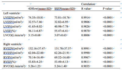 |
Right and Left Ventricular Volume Quantification in Children with repaired Tetralogy of Fallot Using 4D Flow MRI
Xiaofen Yao1, Liwei Hu1, Rongzhen Ouyang1, Yafeng Peng1, Weihui Xie1, Fei Feng2, and Yumin Zhong1
1Shanghai Children's Medical Center, Shanghai, China, 2GE Healthcare, Shanghai, China
4D flow CMR has been used to assess right and left ventricular function with increasing attention. We hypothesis that the results of right and left ventricular volume quantification using 4D flow has the same accuracy compared to that with 2D b-SSFP cine sequence in children with repaired Tetralogy of Fallot (r-TOF).
|
|
4606.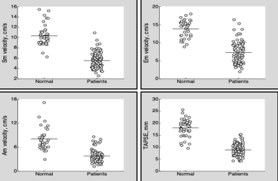 |
Assessment of Right Ventricular Systolic Abnormalities in Pediatric Repaired TOF Patients with Preserved EF: CMR Feature Tracking Analysis
Rongzhen Ouyang1, Shuang Leng2, Liwei Hu1, Aimin Sun1, Qian Wang1, Xiaofen Yao1, Xiaodan Zhao2, Ru-San Tan2, Yong Zhang3, Liang Zhong2,4, and Yumin Zhong1
1Shanghai Children's Medical Center, Shanghai Jiaotong University School of Medicine, Shanghai, China, 2National Heart Centre Singapore, Singapore, Singapore, 3GE Healthcare, Shanghai, China, 4Duke-NUS Medical School, National University of Singapore, Singapore, Singapore
Right ventricular (RV) ejection fraction (EF) <45% is insensitive for detecting systolic dysfunction due to pulmonary regurgitation after Tetralogy of Fallot repair (rTOF). In pediatric rTOF patients with RVEF≥45% and healthy controls, CMR feature tracking was used to assess RV systolic strains and displacement as well as systolic and diastolic myocardial velocities. We observed abnormal RV global longitudinal strain and myocardial velocities in rTOF despite normal RVEF. 78% and 75% of rTOF patients had tricuspid annular displacement systolic excursion <10.9 mm and peak systolic myocardial velocity <6.3cm/s (lower normal limits among controls), respectively, indicating high prevalence of systolic functional impairment.
|
|
|
4607.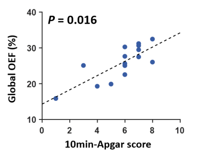 |
Brain oxygen extraction fraction is associated with 10minute Apgar score and severity of hypoxic ischemic encephalopathy
Dengrong Jiang1, W. Christopher Golden2, Charlamaine Parkinson2, Li Pan3, Zixuan Lin1, Hanzhang Lu1, Aylin Tekes1, Frances Northington2, and Peiying Liu1
1Department of Radiology, Johns Hopkins University School of Medicine, Baltimore, MD, United States, 2Department of Pediatrics, Johns Hopkins School of Medicine, Baltimore, MD, United States, 3Siemens Healthineers, Baltimore, MD, United States
Hypoxic-ischemic-encephalopathy (HIE) is a leading cause of neonatal mortality and severe neurological impairment in childhood. Quantification of cerebral oxygen utilization in HIE neonates may provide valuable information to guide the treatment and predict clinical outcomes. Oxygen-extraction-fraction (OEF) is an important index of the brain’s oxygen utilization. In this work, we used a rapid, non-invasive MRI technique to measure the global OEF in HIE neonates, and examined the association of OEF with Apgar scores and the severity of HIE. We demonstrated that global OEF was associated with 10minute Apgar score. Global OEF also decreased with the severity of HIE.
|
4608.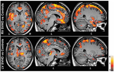 |
Wide-spread Reduced Cerebral Blood Flow in Patients with Right Ventricle Compared to Left Ventricle Single Ventricle Heart Disease
Rajesh Kumar1,2,3,4, Bhaswati Roy1, Xingfeng Shao5, Nancy J. Halnon6, Alan B. Lewis7, Mary A. Woo8, Danny JJ Wang5,9, and Nancy A. Pike8
1Anesthesiology, University of California at Los Angeles, Los Angeles, CA, United States, 2Radiology, University of California at Los Angeles, Los Angeles, CA, United States, 3Bioengineering, University of California at Los Angeles, Los Angeles, CA, United States, 4Brain Research Institute, University of California at Los Angeles, Los Angeles, CA, United States, 5Neurology, University of Southern California, Los Angeles, CA, United States, 6Division of Pediatric Cardiology, University of California at Los Angeles, Los Angeles, CA, United States, 7Division of Pediatric Cardiology, Children's Hospital of Los Angeles, Los Angeles, CA, United States, 8UCLA School of Nursing, University of California at Los Angeles, Los Angeles, CA, United States, 9Radiology, University of Southern California, Los Angeles, CA, United States
Single ventricle heart disease (SVHD) presents with either a dominant single right ventricle (RV) or left ventricle (LV). Individuals with RV dominant SVHD show worse outcomes, including worse cognition and quality of life, which may result from decreased cardiac output due to differences in ventricular size, shape, and function or other structural related sequela, contributing to regional cerebral blood flow (CBF) changes. We examined CBF changes between RV and LV over controls, and found more wide-spread changes in RV over LV. These findings indicate that worse outcomes in RV SVHD may result from compromised CBF over LV SVHD.
|
|
4609.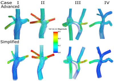 |
Comparison of advanced and simplified computational fluid dynamics simulations in the Glenn and Fontan circulations – an MRI validation study
Nicolas Aristokleous1, Petter Frieberg1, Johannes Töger 1, Petru Liuba2, Pia Sjöberg1, Einar Heiberg 1, and Marcus Carlsson1
1Department of Clinical Sciences Lund, Clinical Physiology, Skåne University Hospital, Lund University, Lund, Sweden, 2Department of Clinical Sciences Lund, Pediatric Heart Center, Skåne University Hospital, Lund University, Lund, Sweden Computational fluid dynamics (CFD) modelling may help patients with heart defects by predicting blood flow after interventions thus aiding surgical planning. Current CFD-techniques are not clinically feasible due to long computational times. We compared a simplified CFD technique to the state-of-the-art advanced CFD and validated the results with Magnetic Resonance Imaging. We could show that the simplified CFD method on a conventional laptop saved between 75%-99% computing time compared with advanced CFD on a dedicated computation server with comparable accuracy in results of flow distributions. This simplified CFD method may facilitate the implementation of CFD predictions in a clinical setting. |
|
4610.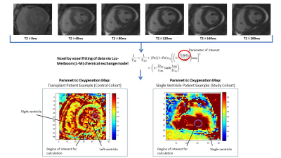 |
Non-invasive cardiac magnetic resonance blood oxygenation mapping in complex congenital heart disease
Lajja Desai1,2, Juliet Varghese3, Rizwan Ahmad3, Orlando Simonetti3, Cynthia Rigsby2, and Michael Markl1
1Northwestern University Feinberg School of Medicine, Chicago, IL, United States, 2Lurie Children's Hospital of Chicago, Chicago, IL, United States, 3Ohio State University, Columbus, OH, United States
Cardiac magnetic resonance (CMR) can be utilized for non-invasive estimation of blood oxygen (O2) saturation in complex pediatric heart disease (i.e., single ventricle physiology) with good correlation to current gold standard catheter-derived measurements. Oximetry maps derived from T2 CMR data can enhance visualization of regional O2 saturations and may guide optimization of image acquisition and analysis.
|
|
|
4611.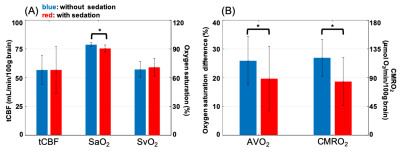 |
Effect of Sedation on Cerebral Metabolic Rate of Oxygen (CMRO2) in Children with Congenital Heart Disease
Pei-Hsin Wu1, Alexandra Batzdorf1, Arastoo Vossough2, Daniel J. Licht2, Mark A. Fogel2, and Felix W. Wehrli1
1Department of Radiology, University of Pennsylvania, Philadelphia, PA, United States, 2The Children's Hospital of Philadelphia, Philadelphia, PA, United States
CMRO2 was evaluated by MR susceptometry. The hypothesis that CMRO2 is lower in sedated young children with single ventricle heart defects (SVD) was investigated in this study. The results show that (1) lower CMRO2 was found in young subjects after administration of sevoflurane, compared to their unsedated peers, (2) brain volumes in patients with SVD were comparable to those reported in healthy children, (3) cerebral blood flow was smaller than the values reported in healthy subjects. The preliminary data indicate that results from awake and sedated subgroups cannot be pooled, at least not for the study of the neurometabolic consequences.
|
4612. |
Decreased fractional anisotropy in the diffusion tensor imaging of neonatal hypoxic ischemic encephalopathy with normal conventional MRI
Qiang Zheng1, Chenying Zhao2, Minhui Ouyang3, Arastoo Vossough4, Giulio Zuccoli3, Raymond Wang Sze4, Hao Huang4, and Misun Hwang4
1School of Computer and Control Engineering, Yantai University, Yantai, China, 2University of Pennsylvania, Philadelphia, PA, United States, 3Children’s Hospital of Philadelphia, Philadelphia, PA, United States, 4Children’s Hospital of Philadelphia; University of Pennsylvania, Philadelphia, PA, United States
Diffusion tensor imaging (DTI) plays an important role in the diagnosis and prognosis of neonatal hypoxic ischemic encephalopathy (HIE). However, studies on DTI in HIE neonates with normal DWI, T1/T2-weighted images (referred as conventional MRI) are rarely reported. Here, we compared fractional anisotropy (FA), mean diffusivity, radial diffusivity, axial diffusivity among controls, HIE neonates with normal conventional MRI, and HIE neonates with abnormal conventional MRI. The results demonstrate that FA is the most sensitive DTI measurement, and significantly decreased in HIE neonates with normal MRI, suggesting the importance of inclusion of DTI for evaluating HIE in clinical practice and research.
|
|
4613.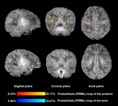 |
Quantitative assessment of the distribution of mild white matter injury in preterm and term neonates
Miaomiao Wang1, Xianjun Li1, Yuli Zhang1, Fan Wu1, Congcong Liu1, Xiaoyu Wang1, Cong Tian1, Peiyao Cheng1, Mengxuan Li1, Chao Jin1, XiaoCheng Wei2, and Jian Yang1
1The First Affiliated Hospital of Xi’an Jiaotong University, Xi’an, China, 2MR Research China, GE Healthcare, Beijing, China
White matter injury is common in neonates. The most common punctate white matter lesions (PWML) can disappear along with time and are easily missed diagnosis. Most current clinical scores account for the numbers and the size of PWML, but ignore the lesion location, which is important for the outcome prediction. This study aims to quantitatively assess the distribution of mild PWML in preterm and term neonates, and characterize its dynamic alterations with gestational age. Our study found that mild PWML is mainly located in the posterior of brain, with extensive injury in the preterm extending to the central region.
|
|
4614.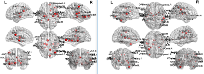 |
Differences in Functional Integration and Segregation in CHD Neonates: Interactions with Sex
Vincent Jerome Schmithorst1, Jodie Votava-Smith2, and Ashok Panigrahy1
1Radiology, Children's Hospital of Pittsburgh of UPMC, Pittsburgh, PA, United States, 2Children's Hospital of Los Angeles, Los Angeles, CA, United States
Functional network topology was compared between neonates with congenital heart disease (CHD) and normal controls and sex-X-CHD interactions were investigated. Using a cost-independent analysis, CHD neonates displayed reduced segregation globally (modularity, transitivity) and nodally (clustering coefficient, participation coefficient) mainly in frontal and subcortical regions; no significant sex-X-CHD interactions were found. Using a cost-dependent analysis, CHD neonates displayed reduced integration and segregation globally (global efficiency, transitivity) and nodally (nodal efficiency, clustering coefficient) in frontal, temporal, and subcortical regions. Significant sex-X-CHD interactions (M>F) were found in similar regions. Results may support a neurophysiological basis for differential neurodevelopmental outcomes related to sex.
|