
Digital Poster Session
Pediatric: Brain Structure & Function
Pediatric
4615 -4630 Pediatric: Brain Structure & Function - Pediatric Technical Advances
4631 -4645 Pediatric: Brain Structure & Function - Pediatric Neuro: Structural & Functional Networks
4646 -4659 Pediatric: Brain Structure & Function - Pediatric Neuro: Morphometry
4615.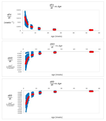 |
Mathematical Modeling of White Matter Development in Infant Rhesus Monkeys
Jason F. Moody1, Nakul Aggarwal2, Douglas C. Dean III1,3,4, Do Tromp2, Steve R. Kecskemeti4, Jonathan Oler2, Ned H. Kalin2, and Andrew L. Alexander1,2,3,4
1Department of Medical Physics, University of Wisconsin-Madison, Madison, WI, United States, 2Department of Psychiatry, University of Wisconsin-Madison, Madison, WI, United States, 3Department of Pediatrics, University of Wisconsin-Madison, Madison, WI, United States, 4Waisman Center, University of Wisconsin-Madison, Madison, WI, United States In this study, we use prototypical DTI metrics to assess longitudinal changes in white matter (WM) across the postnatal rhesus macaque brain, over the first year of life. DTI trajectories extracted from 37 ROIs conform to a logarithmic model and illustrate an initial 10-week period of exceedingly rapid WM development, followed by a prominent plateau in alterations at approximately 6 months of age. K-means clustering of model parameters suggests distinct regional differences in WM maturation. Our analysis provides an early quantitative framework for attaining insights into healthy postnatal WM development, and eventually, establishing connections between WM deterioration and human psychopathology. |
|
4616.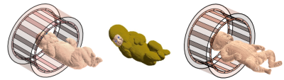 |
SAR and temperature in neonate models resulting from exposure to 7T head coil
Shaihan J Malik1,2, Jeff W Hand1,2, Ryan Satnarine1, and Joseph V Hajnal1,2
1Biomedical Engineering Department, School of Biomedical Engineering and Imaging Sciences, King's College London, London, United Kingdom, 2Centre for the Developing Brain, School of Biomedical Engineering and Imaging Sciences, King's College London, London, United Kingdom
A safety study for neonatal MRI at 7T using an generic head transmit coil was performed using two baby voxel models, one of which was created as part of this study. Both adult and age-adjusted tissue properties were used in performing RF simulations. Simulations showed that SAR/W is increased for neonatal imaging, but the efficiency per B1+2 is comparable to adult imaging. The new neonate model additionally had a 'blanket' layer that could be used to simulate thermal insulation as is often used in practice. Thermal simulations suggest that scanning within IEC SAR limits does not lead to excessive heating.
|
|
4617.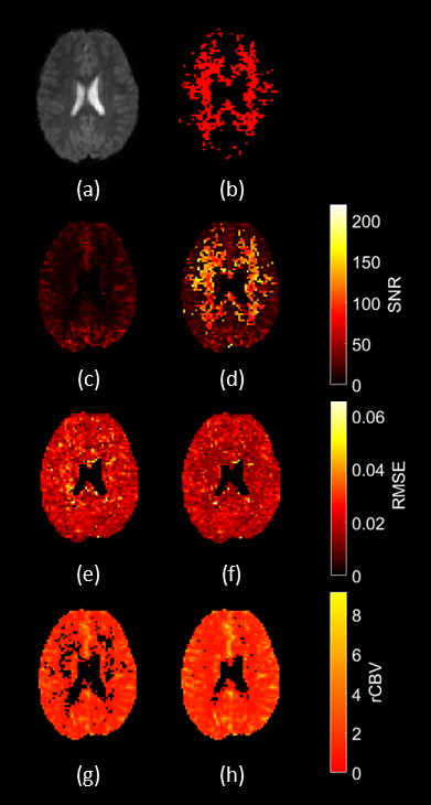 |
An Automated Processing Pipeline to Assess and Improve Data Quality for Multicentre Pediatric Dynamic Susceptibility Contrast Imaging
Stephen Powell1,2, Stephanie Withey2,3,4, Yu Sun2,5, James Grist2, Lesley MacPherson6, Laurence Abernathy7, Barry Pizer8, Richard Grundy9, Simon Bailey10, Dipayan Mitra11, Dorothee Auer12, Shivaram Avula7, Theodoros N. Arvanitis2,3,13,
and Andrew Peet2,3
1Physical Sciences for Health CDT, University of Birmingham, Birmingham, United Kingdom, 2Institute of Cancer and Genomic Sciences, University of Birmingham, Birmingham, United Kingdom, 3Department of Oncology, Birmingham Children's Hospital, Birmingham, United Kingdom, 4RRPPS, University Hospitals Birmingham NHS Foundation Trust, Birmingham, United Kingdom, 5School of Biological Sciences and Medical Engineering, Southeast University, Nanjing, China, 6Radiology, Birmingham Children's Hospital, Birmingham, United Kingdom, 7Radiology, Alder Hey Children's NHS Foundation Trust, Liverpool, United Kingdom, 8Oncology, Alder Hey Children's NHS Foundation Trust, Liverpool, United Kingdom, 9The Children’s Brain Tumour Research Centre, University of Nottingham, Nottingham, United Kingdom, 10Sir James Spence Institute of Child Health, Royal Victoria Infirmary, Newcastle, United Kingdom, 11Neuroradiology, The Newcastle upon Tyne Hospitals NHS Foundation Trust, Newcastle, United Kingdom, 12Sir Peter Mansfield Imaging Centre, University of Nottingham, Nottingham, United Kingdom, 13Institute of Digital Healthcare, University of Warwick, Coventry, United Kingdom
Obtaining robust perfusion measures from pediatric Dynamic Susceptibility Contrast (DSC-) MRI, such as cerebral blood volume (CBV), is challenging due to variability in acquisition protocols between centres and a heterogeneous patient population. Quality control (QC) is currently carried out by expert qualitative review. An automated QC pipeline was developed which used denoising to salvage data, and assessed data quality using logistic regression classification, with signal-to-noise ratio (SNR) and root mean square error (RMSE) in a gamma variate fit to the first pass as predictors. SNR was the key factor in data quality and denoising is important in assuring appropriate analysis.
|
|
4618.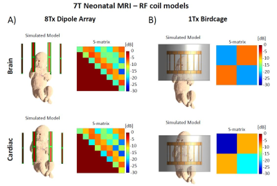 |
8-channel dipole array for 7 Tesla neonatal brain and cardiac MRI
Jérémie Daniel Clément1, Raphael Tomi-Tricot2, Ryan Satnarine1, Jeffrey Hand1, Shaihan Malik1,3, Irena Zivkovic4, Andrew Webb4, Jo Hajnal1, and Özlem Ipek1
1Biomedical Engineering Department, School of Biomedical Engineering and Imaging Sciences, King's College London, London, United Kingdom, 2MR Research Collaborations, Siemens Healthcare Limited, Frimley, United Kingdom, 3Centre for the Developing Brain, School of Biomedical Engineering and Imaging Sciences, King's College London, London, United Kingdom, 4Department of Radiology, Leiden University Medical Center, Leiden, Netherlands
An 8-channel dipole array was investigated for neonatal brain and cardiac MR imaging at 7T. The coil array was compared using electromagnetic field simulations to a single-channel birdcage coil on a realistic baby model for brain and cardiac imaging. B1+-efficiency and SAR10g were evaluated. The dipole coil array demonstrated significant lower SAR10g levels for both imaging positions. The working of the constructed dipole array was assessed on adult head. We conclude that dipole arrays are a valuable approach for neonatal brain and cardiac MR imaging at 7T, and could be part of future ethics approval of a neonatal MRI application.
|
|
4619.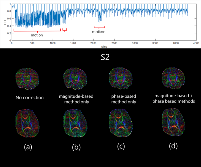 |
Application of phase-based motion outlier detection to infant dMRI
Nahla M H Elsaid1, Jiachen Zhuo2, Jerry L Prince3, Yu-Chien Wu1, and Rupa Radhakrishnan1
1Department of Radiology and Imaging Sciences, Indiana University, Indianapolis, IN, United States, 2Department of Diagnostic Radiology and Nuclear Medicine, University of Maryland School of Medicine, Baltimore, MD, United States, 3Department of Electrical and Computer Engineering, Johns Hopkins University, Baltimore, MD, United States Detecting and eliminating motion-corrupted slices is crucial in diffusion MRI (dMRI), and particularly essential in imaging neonates. Conventional magnitude-based outlier rejection methods are intensity-based and can usually detect and correct intra-volume movement but can miss outliers in cases of small continuous motions. Phase-based methods can be used to detect motion independently, regardless of the slice-to-volume location. The phase-based method is reasonably accurate and computationally fast, and may be better suited for real-time detection of motion in dMRI. Combining magnitude and phase methods could produce the best results. Here, we evaluate the phase-based method versus the magnitude-based method in neonatal data.
|
|
4620.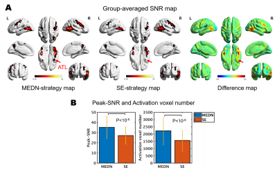 |
An integrated multi‐echo denoising strategy improves the performance of the FPS-fMRI approach on a face localizer
Ziyang Chen1, Minjie Wen2, Xiaoqing Gao3, Yi-Cheng Hsu4, Hongjian He1, and Jianhui Zhong1,5
1Center for Brain Imaging Science and Technology, Key Laboratory for Biomedical Engineering of Ministry of Education, College of Biomedical Engineering and Instrumental Science, Zhejiang University, Hangzhou, Zhejiang, China, Hangzhou, China, 2Department of Psychology and Behavioral Sciences, Zhejiang University, Hangzhou, China, Hangzhou, China, 3Center for Psychological Sciences, Zhejiang University, China, Hangzhou, China, 4MR Collaboration NE Asia, Siemens Healthcare, Shanghai, China, Shanghai, China, 5Department of Imaging Sciences, University of Rochester, Rochester, NY, USA, Rochester, NY, United States
The fast periodic stimulation (FPS) fMRI approach can be influenced by noise with its frequency spectrum close to the specific task frequency. In this study, we investigated the effect of an integrated multi-echo denoising strategy compared with a single-echo strategy on a face localizer task. The group-averaged SNR increased on a typical face processing network due to a significant decrease in the noise frequency standard deviation. False-positive error introduced by the denoising process could be well tolerated in the presence of a positive activation increase.
|
|
4621.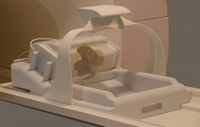 |
Size-Adaptable 32-Channel Receive Array for Pediatric Brain Imaging at 3 Tesla
Anpreet Ghotra1, Heather Kosakowski2, Atsushi Takahashi2, Markus May1, Alina Scholz1, Nicolas Kutscha1, Mirsad Mahmutovic1, Lawrence L Wald3, Nancy Kanwisher2, Rebecca Saxe2, and Boris Keil1
1Institute of Medical Physics and Radiation Protection, TH Mittelhessen University of Applied Science, Giessen, Germany, 2McGovern Institute for Brain Research, Massachusetts Institute of Technology, Cambridge, MA, United States, 3Martinos Center for Biomedical Imaging, Dept. of Radiology, Massachusetts General Hospital, Harvard Medical School, Boston, MA, United States
The aim of this study is a hardware related approach to increase the subject’s compliance for fMRI with awake infants. An infant-friendly head coil array was developed to improve sensitivity, spatial resolution, accelerated encoding, motion insensitivity, and subject tolerance in pediatric MR-imaging. The coil was characterized with both bench and image metrics and compared to a 32-channel adult head coil. The size-optimized 32-channel infant array coil provides increased reception sensitivity and is well-suited for highly accelerated fMRI data acquisitions in awake infants.
|
|
4622.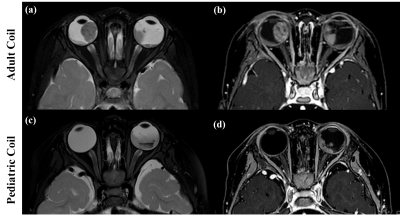 |
High-resolution MRI of retinoblastoma achieved by the dedicated pediatric coil at 3T
Nan Li1, Hui Zheng2, Ting Gui2, Qiufeng Yin2, Yang Xin3, Shuheng Zhang3, Qiang He3, Xiaoliang Zhang4, Xin Liu5, Hairong Zheng5, Dengbin Wang2, and Ye Li5
1Shenzhen Institutes of Advanced Technology, Chinese Academy of Sciences, Shenzhen, China, shenzhen, China, 2Department of Radiology, Xinhua Hospital, Shanghai Jiao Tong University School of Medicine, Shanghai, China,, Shanghai, China, 3Shanghai United Imaging, Shanghai, China, Shanghai, China, 4Department of Biomedical Engineering, State University of New York at Buffalo, NY, United States., Buffalo, NY, United States, 5Lauterbur Imaging Research Center, Shenzhen Institutes of Advanced Technology, Chinese Academy of Sciences, Shenzhen, China, shenzhen, China
High resolution MRI has become a very useful diagnostic tool in evaluation children with retinoblastoma. In this paper, higher spatial resolution images of retinoblastoma in 3T MRI were acquired by using the dedicated pediatric coil with optimized imaging protocol. 8 examinations in this study were analyzed and assessed. The results show that all were achieved the good image quality, which is very significance for the accurate diagnosis in retinoblastoma and evaluation of therapeutic effect in clinically.
|
|
4623.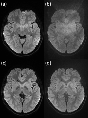 |
Quiet Diffusion-weighted Imaging in Pediatric Patients with Moyamoya Disease
Satoshi Nakajima1, Yasutaka Fushimi1, Tomohisa Okada2, Gosuke Okubo1, Yusuke Yokota1, Sonoko Oshima1, Sayo Otani1, Azusa Sakurama1, Krishna Pandu Wicaksono1, and Kaori Togashi1
1Department of Diagnostic Imaging and Nuclear Medicine, Kyoto University Graduate School of Medicine, Kyoto, Japan, 2Human Brain Research Center, Kyoto University Graduate School of Medicine, Kyoto, Japan Poster Permission Withheld
Diffusion-weighted imaging (DWI) is an essential MR sequence for evaluating pediatric patients with moyamoya disease (MMD). Acoustic noise associated with DWI may lead to motion artifact. Compared with conventional DWI (cDWI), quiet DWI (qDWI) is considered less noisy and able to keep children more relaxed and stable. We evaluated the advantages of qDWI compared with cDWI in pediatric MMD patients. Compared with cDWI, qDWI induced fewer artifacts in sedated pediatric MMD patients, whereas in unsedated patients, the frequencies of qDWI- and cDWI-induced artifacts were similar. qDWI and cDWI had the same performance for detecting restricted diffusion.
|
|
4624.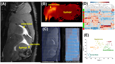 |
Machine-Learning Segmentation and Classification of Pediatric Brain Tumors Based on Preclinical Multiparametric Advanced Fast Imaging (MAFI)
Marina Stukova1, Samuel Henehan2, Jenna Steiner2, Alaina Marquardt2, and Natalie Julie Serkova3
1Radiology, University of Colorado Anschutz Medical Campus, Aurora, CO, United States, 2University of Colorado Anschutz, Aurora, CO, United States, 3Radiology, University of Colorado Anschutz, Denver, CO, United States
Brain tumors are the second most common malignancy in childhood (exceeded only by leukemia). Clinically, multiparametric MRI is now considered to be the neuroimaging standard for detecting brain tumors. Pediatric brain tumors have a diverse array of clinical manifestations, cellular and molecular phenotypes, and tumor habitats. Previously, we reported on the initial development of a non-gadolinium, Multiparametric Advanced Fast Imaging (MAFI) protocol at the 9.4 Tesla in patient derived xenografts models (PDX, ISMRM 2019). Here, we expanded our study to include all existing pediatrics PDX mouse models for machine learning based segmentation and classification of four major brain tumor subtypes.
|
|
4625.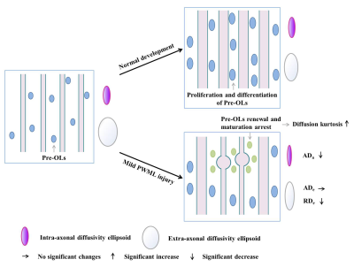 |
DKI is a sensitive method for detect the white matter patho-morphological variations in neonates with mild white matter injury
Miaomiao Wang1, Mengxuan Li1, Xianjun Li1, Congcong Liu1, Yuli Zhang1, Fan Wu1, Xiaoyu Wang1, Cong Tian1, Peiyao Cheng1, XiaoCheng Wei2, and Jian Yang1
1The First Affiliated Hospital of Xi’an Jiaotong University, Xi’an, China, 2MR Research China, GE Healthcare, Beijing, China
White matter injury is common in neonates. The most common punctate white matter lesions (PWML) can disappear along with time and are easily missed diagnosis. Since DTI is not sensitive to detect the white matter (WM) microstructural changes in mild PWMLs, DKI may provide more details for capture the WM alterations. This study aims to investigate the utility of DKI in characterizing the WM microstructural variations following mild PWMLs. Our study found that diffusion kurtosis and two-compartment metrics are sensitive imaging markers for characterize the WM microstructural alterations of mild PWMLs, which is associated with reactive gliosis and axonal swelling.
|
|
4626.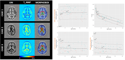 |
Simultaneous micro- and macrostructural assessment of normal brain development
Bénédicte Maréchal1,2,3, Gian Franco Piredda1,2,3, Tom Hilbert1,2,3, Clovis Tauber4, Jean-Philippe Cottier4, Jean-Philippe Thiran3, Tobias Kober1,2,3, and Baptiste Morel4,5
1Advanced Clinical Imaging Technology, Siemens Healthcare AG, Lausanne, Switzerland, 2Department of Radiology, Lausanne University Hospital and University of Lausanne, Lausanne, Switzerland, 3LTS5, École Polytechnique Fédérale de Lausanne (EPFL), Lausanne, Switzerland, 4UMR 1253, iBrain, Université de Tours, Inserm, Tours, France, 5Pediatric Radiology Department, Clocheville Hospital, CHRU of Tours, Tours, France
MRI has become a routine diagnostic modality for evaluating normal structural and metabolic development of the brain in children. In this work, MP2RAGE sequence parameters and consecutive automated brain morphometry are optimized to overcome cohort-specific challenges due to wide variations in head size, head shape, and rapid changes in tissue contrast associated with myelination. Reference ranges for both volumetric and T1-relaxometry measures are built from a pediatric population with normal MRI findings. We also provide an initial assessment of potential clinical applications of these micro- and macrostructural features and corresponding normative ranges using selected cases with abnormal findings.
|
|
4627.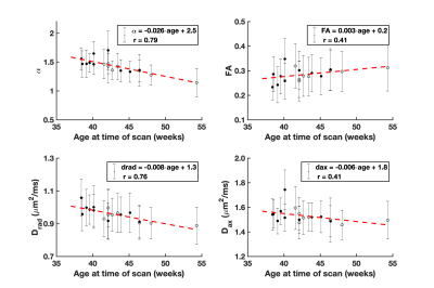 |
Power-law fits for the direction-averaged diffusion MRI signal: a potential marker for white matter maturation in non-feeding infants
Emilie T McKinnon1,2,3, Hunter G Moss1,3, Bashar W Badran4, Dorothea D Jenkins5, and Jens H Jensen1,3
1Department of Neuroscience, Medical University of South Carolina, Charleston, SC, United States, 2Department of Neurology, Medical University of South Carolina, Charleston, SC, United States, 3Center for Biomedical Imaging, Medical University of South Carolina, Charleston, SC, United States, 4Brain Stimulation Laboratory, Department of Psychiatry, Medical University of South Carolina, Charleston, SC, United States, 5Department of Pediatrics and Neonatology, Medical University of South Carolina, Charleston, SC, United States
Preterm or hypoxic ischemic birth is associated with postpartum feeding problems and white matter dysmaturation. Noninvasive vagus nerve stimulation has been proposed as therapy, and here we explore the application of high b-value diffusion MRI to assess changes in white matter following stimulation. Our hypothesis is that using high b-values will improve sensitivity to alterations in white matter as compared with traditional diffusion measures. Power-law fits for high b-value diffusion MRI data were investigated in 12 neonates. We find that the power-law exponent decreases in early development and give preliminary results for its application as a marker for treatment response.
|
|
4628.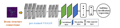 |
A deep transfer learning model for early prediction of cognitive deficits using brain structural connectome data in very preterm infants
Ming Chen1,2, Hailong Li1, Jinghua Wang3, Weihong Yuan3,4, Adebayo Brainmah4, Mekibib Altaye5, Nehal Parikh1,6, and Lili He1,4,6
1The Perinatal Institute and Section of Neonatology, Perinatal and Pulmonary Biology, Cincinnati Children's Hospital Medical Center, Cincinnati, OH, United States, 2Department of Electronic Engineering and Computing Science, University of Cincinnati, Cincinnati, OH, United States, 3Department of Radiology, University of Cincinnati College of Medicine, Cincinnati, OH, United States, 4Imaging Research Center, Cincinnati Children's Hospital Medical Center, Cincinnati, OH, United States, 5Department of Biostatistics, Cincinnati Children's Hospital Medical Center, Cincinnati, OH, United States, 6Department of Pediatrics, University of Cincinnati College of Medicine, Cincinnati, OH, United States
The high risk of cognitive deficits is a major concern for parents and clinicians caring for premature babies. Early and accurate identification of children at risk is urgently needed for early treatment decision. We propose a deep transfer learning model to predict cognitive deficits at 2 years corrected age using brain structural connectome data obtained at term. The proposed model was able to identify infants at high-risk of later cognitive deficit with an accuracy of 78.5% and an AUC of 0.75. The predicted cognitive scores were significantly correlated with corresponding Bayley-III cognitive scores, with a Pearson’s correlation coefficient of 0.48.
|
|
4629.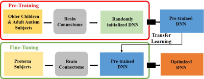 |
Early prediction of neurodevelopmental deficits in very preterm infants using a multi-task deep transfer learning model
Lili He1,2,3, Hailong Li1,3, Jinghua Wang4, Ming Chen1,5, Jonathan R. Dillman4,6, and Nehal Parikh1,2
1The Perinatal Institute and Section of Neonatology, Perinatal and Pulmonary Biology, Cincinnati Children's Hospital Medical Center, Cincinnati, OH, United States, 2Department of Pediatrics, University of Cincinnati College of Medicine, Cincinnati, OH, United States, 3Imaging Research Center, Cincinnati Children's Hospital Medical Center, Cincinnati, OH, United States, 4Department of Radiology, University of Cincinnati College of Medicine, Cincinnati, OH, United States, 5Department of Electronic Engineering and Computing Science, University of Cincinnati, Cincinnati, OH, United States, 6Department of Radiology, Cincinnati Children's Hospital Medical Center, Cincinnati, OH, United States
We proposed a deep transfer learning model using the fusion of clinical and brain functional connectome data obtained at term for early neurodevelopmental deficits prediction at two years corrected age in very preterm infants. The proposed model was first trained in an unsupervised fashion using 884 subjects from publicly available ABIDE repository, then fine-tuned and cross-validated on 33 very preterm infants. Our model achieved an AUC of 0.77, 0.63 and 0.74 on the risk stratification of cognitive, language and motor deficits, respectively. Our findings demonstrated the feasibility of using deep transfer learning on connectome data for abnormal neurodevelopment prediction.
|
|
4630.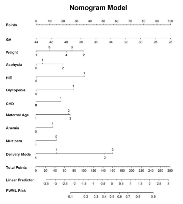 |
Predictive Model of MRI Screening for Neonatal Punctate White Matter Lesion Based on Multi-Center Data
Congcong Liu1, Qinli Sun1, Miaomiao Wang1, Xiaoyu Wang 1, Yannan Cheng1, Ting Liu1, Zhe Liu1, Pan Cao1, Fan Wu1, Yuli Zhang1, Huifang Zhao1, Chao Jin1, Xianjun Li1, Xingxing Tao1,
Xiaocheng Wei2, and Jian Yang1
1Department of Radiology, The First Affiliated Hospital of Xi’an Jiaotong University, Xi'an, China, 2MR Research China, GE Healthcare, Xi'an, China
PWML with high incidence (~20%) was correlated with adverse neurodevelopment outcomes and lesions disappeared as time goes on. Although MRI can accurately evaluate PWML, currently it’s not a universal screening method. This study aimed to develop a predictive model of MRI screening for PWML based on clinical factors, and help clinicians identify which neonates should undergo MRI. 5899 neonates were recruited. Logistic regression analysis were used to develop the model. Ten independent factors were identified and the model was presented as nomogram. Current model showed good discrimination and clinically use. It will help clinicians conveniently and early make decision.
|
 View the Poster
 Watch the Video
4631.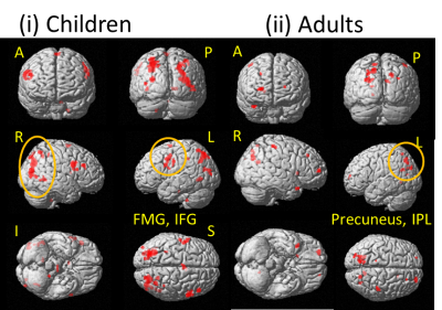 |
Dorsal-ventral stream development for visual word processing
Sunita Gudwani1,2, S. Senthil Senthil Kumaran3, Rajesh Sagar4, Madhuri Behari5, Manju Mehta6, Vaishna Narang7, SN Dwivedi8, and NR Jagannathan3
1Department of ENT, Escorts Heart Institute and Research Center, New Delhi, India, 2Former Department of NMR and MRI Facility, Former ALL INSTITUTE OF MEDICAL SCIENCES, NEW DELHI, India, 3Department of NMR and MRI Facility, All India Institute of Medical Sciences, New Delhi, India, 4Department of Psychiatry, All India Institute of Medical Sciences, New Delhi, India, 5Department of Neurology, Fortis Hospital, New Delhi, India, 6Department of Psychiatry (Psychology Unit), All India Institute of Medical Sciences, New Delhi, India, 7Department of Linguistics, School of Language, Jawahar Lal Nehru University, New Delhi, India, 8Department of Biostatistics, All India Institute of Medical Sciences, New Delhi, India
Reading skill is window to the world that we acquire with development. Dual-route model predicts our proficiency of grapheme (text) to phoneme (sound) conversion and semantic decoding (understanding content) for visual words. Fast processing by frontoparietal pathway while meaningful word reading but lexical decision to pseudowords proceeds slowly and exhaustively. Pseudoword successful orthographic mapping recruits ventral and then dorsal areas. This phonological proficiency and exhaustive mental lexicon search during reading is automatized with skill development. Current study evidences recruitment of inferior frontal gyrus (BA44 and 45), insula, thalamus, caudate nucleus and cortical reorganizations of skill developmental with age
|
|
 |
4632.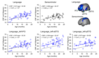 |
Delineation of language network maturation during infancy with multi-modal perfusion and functional MRI
Minhui Ouyang1, Qinlin Yu1, Huiying Kang2, Yun Peng2, Bo Hong3, and Hao Huang1,4
1Department of Radiology, The Children's Hospital of Philadelphia, Philadelphia, PA, United States, 2Department of Radiology, Beijing Children’s Hospital, Capital Medical University, Beijing, China, 3Department of Biomedical Engineering, School of Medicine, Tsinghua University, Beijing, China, 4Department of Radiology, Perelman School of Medicine, University of Pennsylvania, Philadelphia, PA, United States
Human language capabilities are acquired during infancy. We hypothesized that language brain circuit emergence is supported by rapidly increasing regional cerebral blood flow (rCBF) to meet the metabolic demands. However, the spatiotemporal distribution of rCBF in language network during infancy is unknown. In this study, we acquired resting-state fMRI and pCASL perfusion MRI from 48 infants aged 0-24months for identifying language network and quantifying rCBF, respectively. Heterogeneous and significant age-dependent rCBF increases were found at specific functional regions of language network during this critical period.
|
4633.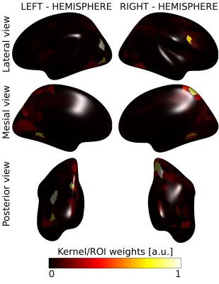 |
Visual processing activation in the magnocellular pathway is associated with Developmental Dyslexia
Denis Peruzzo1, Sara Mascheretti2, Chiara Andreola2,3, Gabriele Amorosino1,4, Stefania Tirelli1, Daniela Redaelli1, and Filippo Arrigoni1
1Neuroimaging Unit, IRCCS Eugenio Medea, Bosisio Parini, Italy, 2Child Psychopathology Unit, IRCCS Eugenio Medea, Bosisio Parini, Italy, 3Laboratory for the Psychology of Child Development and Education (LaPsyDE), University Paris Descartes, Paris, France, 4NeuroInformatics Laboratory (NILab), Bruno Kessler Foundation, Trento, Italy According to the magnocellular (M) deficit theory, deficits in the visual M functions predict reading skills and are associated with Developmental Dyslexia (DD). We acquired two well-established visual tasks (i.e. a sinusoidal grating and a coherent dots motion detection) in 22 Normal Readers (NR) and 22 children with DD. We applied a multi-variate pattern analysis approach to combine the two tasks. The combination of their activation patterns significantly discriminated between NR and children with DD, suggesting that differences in the M functions are present between the two populations and supporting the M deficit theory |
|
4634.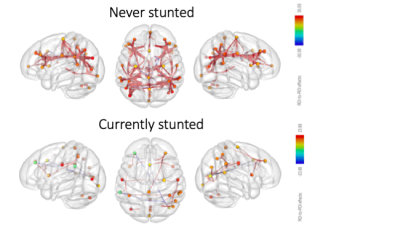 |
Functional Brain Network Connectivity Patterns in Never and Currently Stunted Young Children in India
Muriel Marisa Katharina Bruchhage1,2, Giang-Chau Ngo1,2, Madhuri Tiwari3, Aarti Kumar3, Vishwajeet Kumar3, Viren D'Sa1,2, and Sean C. L. Deoni1,2,4
1Warren Alpert Medical School, Brown University, Providence, RI, United States, 2Advanced Baby Imaging Lab, Hasbro Children's Hospital, Providence, RI, United States, 3Community Empowerment Lab, Lucknow, India, 4Maternal, Newborn, and Child Health Discovery & Tools, Bill & Melinda Gates Foundation, Seattle, WA, United States
Growth stunting is an indicator of poor child development, negatively impacting cognitive performance and overall health until adulthood. While previous studies have investigated links between neurocognitive functioning and resting state network functional connectivity (fc), most have been conducted in developed countries. In our study, we investigate brain network fc at rest in young children (<2 years) living in an Indian region with one of the worst human development indicators. When currently stunted, fewer fc networks were present than in never stunted children, possibly indicating an effect of stunting on brain fc already at a very young age.
|
|
|
4635.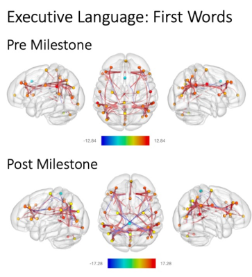 |
Functional Correlates to Achieving Early Motor and Language Milestones
Muriel Marisa Katharina Bruchhage1,2, Giang-Chau Ngo1,2, Viren D'Sa1,2, and Sean C. L. Deoni1,2,3
1Warren Alpert Medical School, Brown University, Providence, RI, United States, 2Advanced Baby Imaging Lab, Hasbro Children's Hospital, Providence, RI, United States, 3Maternal, Newborn, and Child Health Discovery & Tools, Bill & Melinda Gates Foundation, Seattle, WA, United States
Developmental milestones are essential for a child’s development, with early milestones building a stepping-stone for more complex skills later. In this study, we investigate resting state functional connectivity (fc) before and after major developmental milestones (walking, pincer grasp, first words) were reached in a large cohort of typically developing children (3 to 30 months). Reaching a milestone showed a different fc pattern across all skill domains. Opposite to more complex skills where fc strengthened with skill advancement, walking recruited fewer fc networks when the milestone was reached, possibly underlining the importance of automaticity and complexity demand for the milestone achieved.
|
4636.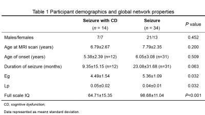 |
Alterations in brain white matter structural network in children with seizure and cognitive dysfunction
Yannan Cheng1, Chao Jin2, Xianjun Li2, Congcong Liu2, Miaomiao Wang2, Huifang Zhao2, Xiaoyu Wang2, Yuli Zhang2, Fan Wu2, Mengxuan Li2, Cong Tian2, Peiyao Chen2, Xiaocheng Wei3, Jianxin Guo2,
and Jian Yang2
1Department of Radiology, the First Affiliated Hospital, Xi’an Jiaotong University, Xi'an, China, Xi'an, China, 2the First Affiliated Hospital, Xi’an Jiaotong University, Xi'an, China, Xi'an, China, 3MR Research China, GE Healthcare, Bei Jing, People's Republic of China, Xi'an, China
Seizures are the most common disease in children and some types like epilepsy usually cause many serious co-morbidities such as cognitive dysfunction. We detailed the alterations of brain white matter network in children with seizure and cognitive dysfunction by comparing the difference of network properties and connectivity strength between the patient and control groups. Here, we found that children with seizure and cognitive dysfunction show a suboptimal brain organization with reduced information integration ability, and abnormalities in multiple intrinsic functional connectivity may be the structural basis for cognitive impairment in patients with seizures.
|
|
4637.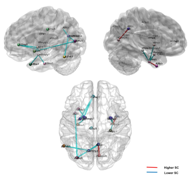 |
The subregion scheme of the precuneus in children with autism spectrum disorder: a multimodal network analysis based on the Brainnetome Atlas
Xin Xu1,2 and Qiyong Gong1,2
1Huaxi MR Research Center (HMRRC), Department of Radiology, West China Hospital of Sichuan Uinversity, Chengdu, China, 2Psychoradiology Research Unit of the Chinese Academy of Medical Sciences (2018RU011)), West China Hospital of Sichuan Uinversity, Chengdu, China
To explore the brain network characteristics in children with ASD, we calculated the whole-brain structural and functional connectivity and its network topological attributes between ASD children and typical development (TD) by using whole-brain multimodal connectivity network analysis based on the Brainnetome Atlas. This study found that the left dorsomedial parieto-occipital sulcus was the area of overlap within both structural and functional connectivity abnormalities in ASD children compared with TD, implicated that this subregion of the precuneus was a key node in ASD brain network during childhood.
|
|
4638.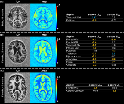 |
Detecting and characterizing abnormal brain lateralization using quantitative MRI in clinical practice
Gian Franco Piredda1,2,3, Baptiste Morel4,5, Maximilien Perivier4,5, Clovis Tauber4, Jean Philippe Cottier4, Jean-Philippe Thiran2,3, Bénédicte Maréchal1,2,3, Tom Hilbert1,2,3, and Tobias Kober1,2,3
1Advanced Clinical Imaging Technology, Siemens Healthcare AG, Lausanne, Switzerland, 2Department of Radiology, Lausanne University Hospital and University of Lausanne, Lausanne, Switzerland, 3LTS5, École Polytechnique Fédérale de Lausanne (EPFL), Lausanne, Switzerland, 4UMR 1253, iBrain, Université de Tours, Inserm, Tours, France, 5Pediatric Radiology Department, Clocheville Hospital, CHRU of Tours, Tours, France
The right-left lateralization of cognitive abilities in the human brain is reflected in hemispheric asymmetries. Since abnormal deviations of these asymmetries were linked to neurological disorders, establishing reference norms of hemispheric lateralization has clinical relevance. To this end, the normal evolution of brain asymmetries during development was modelled in a cohort of healthy young subjects. Normative ranges for brain asymmetries in volumes and T1 were established using a linear model while accounting for sex differences and age. Initial results in data of epileptic patients demonstrate the potential of the established norms for detecting abnormal brain lateralization on a single-subject basis.
|
|
4639.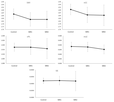 |
Less Integrated and Less Efficient Structural Brain Networks in Children Treated for ALL
Wilburn E Reddick1, John O Glass1, Ruitian O Song1, Ching-Hon Pui2, and Sima Jeha2
1Diagnostic Imaging, St. Jude Children's Research Hospital, Memphis, TN, United States, 2Oncology, St. Jude Children's Research Hospital, Memphis, TN, United States
This study assessed whole-brain structural connectomes in 252 children treated for ALL during early phase of treatment and at end of therapy relative to 89 normal healthy age-similar controls. Both small worldness index and clustering coefficient were significantly lower in patients early and late in therapy relative to the controls but did not change during therapy. However, both characteristic path length and local efficiency significantly decreased during therapy. Decreased network integration and less efficient information transfer in patients treated for ALL is likely to result in decreased performance on neurocognitive testing by end of therapy.
|
|
|
4640.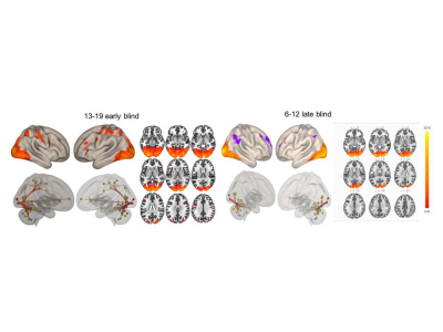 |
Structural and Functional changes in ventral and dorsal stream during phonological and haptic language in early and late blind
A Ankeeta1, s Senthil Kumaran1, and Rohit Saxena2
1NMR and MRI facility, All India Institute of Medical Sciences, New Delhi, India, 2RP centre, Opthalmology, All India Institute of Medical Sciences, New Delhi, India
Anatomical and functional MRI data for Braille and phonological noun pair assessment test was acquired in 40 early blind (EB), forty late blind (LB) subjects and thirty sighted controls (SC) in two age groups 6-12 years and 13-19 years (all right-handed). Visual impairment induces structural and functional modification in the visual cortex. Late blind subjects exhibited differences in the V1 13-19 years age range, but not in the 6-12 years age group. Duration of blindness and education influence the extent of plasticity of the V1 and language network.
|
4641.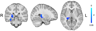 |
Alterations in brain functional connectivity in pediatric migraine
Tiffany Bell1,2,3, Akashroop Khaira1,2,3, Mahak Sandhu1,2,3, Megan Webb1,2,3, Melanie Noel3,4, Farnaz Amoozegar2,5, and Ashley D Harris1,2,3
1Department of Radiology, University of Calgary, Calgary, AB, Canada, 2Hotchkiss Brain Institute, Calgary, AB, Canada, 3Alberta Children's Hospital Research Institute, Calgary, AB, Canada, 4Department of Psychology, University of Calgary, Calgary, AB, Canada, 5Department of Clinical Neurosciences, University of Calgary, Calgary, AB, Canada
Though migraine is a common childhood disease, there has been relatively little investigation into migraine in children. Here we use resting-state functional connectivity analyses to investigate functional alterations in brain activity in children with migraine. In agreement with the adult literature, we show altered resting-state connectivity in children with migraine compared to healthy controls in areas related to sensory processing.
|
|
4642.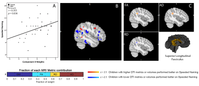 |
Linked signatures of brain structure and microstructure at preschool age predict reading readiness in early childhood
Jess E Reynolds1,2,3, Kathryn Y Manning1,2,3, Dmitrii Paniukov1,2,3,4, Deborah Dewey2,3,4,5, and Catherine Lebel1,2,3
1Radiology, University of Calgary, Calgary, AB, Canada, 2Owerko Centre, Alberta Children Hospital Research Institute, University of Calgary, Calgary, AB, Canada, 3Hotchkiss Brain Institute, University of Calgary, Calgary, AB, Canada, 4Pediatrics, University of Calgary, Calgary, AB, Canada, 5Community Health Sciences, University of Calgary, Calgary, AB, Canada
We aimed to identify whether variations in grey and white matter brain structure during early childhood predict future pre-reading skills. We examined anatomical (T1-weighted) and diffusion tensor (DTI) images from 35 children at 3.5years(±3months). Children were assessed for their pre-reading abilities using NEPSY-II subtests one year later (4.5years±3months). A data-driven linked independent component analysis was used to identify components of DTI and morphometry measures with shared variability across subjects that related to pre-reading ability approximately a year later. Our results suggest the co-development of grey and white matter brain structures in early life predicts future pre-reading capabilities in preschool children.
|
|
4643.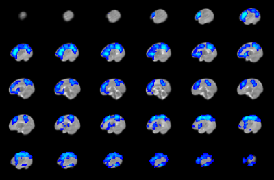 |
Greater reduction in functional connectivity strength in term neonates with congenital heart disease born earlier in gestation
Vincent Jerome Schmithorst1, Cecilia Lo2, Philip Adams2, Jodie Votava-Smith3, and Ashok Panigrahy1
1Radiology, Children's Hospital of Pittsburgh of UPMC, Pittsburgh, PA, United States, 2University of Pittsburgh, Pittsburgh, PA, United States, 3Children's Hospital of Los Angeles, Los Angeles, CA, United States
Functional connectivity strength (FCS) differences are investigated in term neonates with congenital heart disease (CHD) as compared to normal controls. In CHD neonates, FCS is reduced in default mode network (DMN), salience network (SN), and central executive network (CEN) regions. However, widespread positive post-conceptional age (PCA) at birth – X – CHD interactions were found, indicating this effect is more pronounced for CHD neonates born earlier in gestation, and that prolonged in utero exposure may be beneficial for development of brain functional connectivity. Negative correlations of nasal nitric oxide (nNO) and FCS were also found, suggesting a partly vascular etiology.
|
|
4644. |
Altered brain activation during cognitive control in children with enuresis
Mengxing Wang1, Xiangyu Zheng2, Yue Liu1, Jun Ma*2,3, and Xiaoxia Du*4
1College of Medical Imaging, Shanghai University of Medicine&Health Science, Shanghai, China, 2Department of Developmental and Behavioral Pediatrics, Shanghai Institute of Pediatric Translational Medicine, Shanghai Children’s Medical Center, Shanghai Jiao Tong University School of Medicine, Shanghai, China, 3MOE‑Shanghai Key Laboratory of Children’s Environmental Health, Shanghai Jiao Tong University School of Medicine, Shanghai, China, 4Shanghai Key Laboratory of Magnetic Resonance and Department of Physics, School of Physics and Materials Science, East China Normal University, Shanghai, China
This study was to investigate the potential disturbance in cognitive control in enuretic children using functional MRI. Two groups including enuretic and healthy children were scanned during a color-word Stroop task to evaluate their task performance and neural response. Compared to the healthy subjects, the children with enuresis showed stronger Stroop effect and increased activation for the incongruent > congruent condition contrast in the anterior cingulate and the precentral/postcentral gyri. Our results suggested that the enuretic children showed altered neural responses to conflict events and tended to compensate by greater activation to sustain normal cognitive control.
|
|
4645.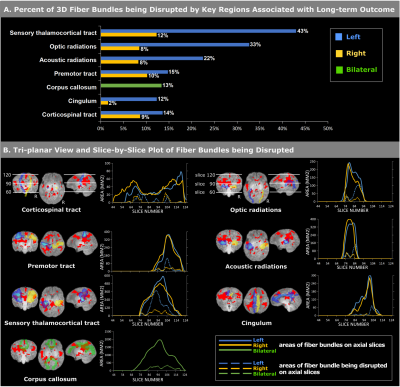 |
Fibers and Neuroanatomy in the Left Hemisphere Are More Associated with Neonatal Death and 2-Year Outcome after Hypoxic Ischemic Encephalopathy
Yanan Song1,2, Sara V. Bates3,4, Rebecca J. Weiss3,4, Randy L. Gollub4,5, Sheng He1,4, Camilo Jaimes4,6, Susan Sotardi4,7, Yue Zhang1,4, Tonghua Liu2, P. Ellen Grant1,4,6, and Yangming Ou1,4,6
1Fetal-Neonatal Neuroimaging Data Science Center, Boston Children's Hospital, Boston, MA, United States, 2Beijing University of Chinese Medicine, Beijing, China, 3Department of Pediatrics, Massachusetts General Hospital, Charlestown, MA, United States, 4Harvard Medical School, Boston, MA, United States, 5Department of Psychiatry, Massachusetts General Hospital, Charlestown, MA, United States, 6Department of Radiology, Boston Children's Hospital, Boston, MA, United States, 7Department of Radiology, Massachusetts General Hospital, Charlestown, MA, United States
Hypoxic ischemic encephalopathy affects ~5/1000 neonates and causes neonatal death and 2-year disability. Using neonatal MRI to predict neonatal and 2-year outcomes remains a key open question. However, expert scoring of neonatal MRI, the current norm, only focuses on a few key brain regions based on subjective clinical experience. We objectively and comprehensively examined the associations with neonatal and 2-year outcomes throughout the brain. Our new findings – many voxels, subregions and fibers that are not considered in expert scoring systems, with a left hemisphere dominance, were associated with outcomes – provide new knowledge for future individualized outcome prediction.
|
4646.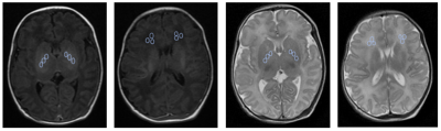 |
The ratio of T1 and T2 signals of MRI improves the diagnostic value of early hyperbilirubinemia brain damage in neonates
Jinhong Yu1, Yanwei Miao1, Lizhi Xie2, Bingbing Gao1, Yu Bing1, Li Yang1, and Ailian Liu1
1The First Affiliated Hospital of Dalian Medical University, Dalian, China, 2GE Healthcare, Beijing, China
Exposure to high levels of bilirubin can cause severe motor symptoms and cerebral palsy. The aim of this study was to analyze the diagnostic value for early brain damage caused by hyperbilirubinemia (HB) in neonates using T1-weighted imaging (T1WI) and T2-weighted imaging (T2WI).
|
|
4647.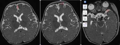 |
Age-related changes of the extracerebral space in normal infants observed with 3D T2 MR images
Wang Xiaohu1, Ren huipeng1, Fan qing1, Wei Xiaocheng2, and Ren Zhuanqin1
1Baoji Central Hospital, Baoji, China, 2MR Research China, GE Healthcare, Beijing, China
External hydrocephalus (EH) can cause poor prognosis, but there is no consensus exists on diagnostic criteria for pathological EH in infants and young children. In this study, we measured extracerebral space of 212 healthy subjects on different anatomical slices and their age correlation were analyzed. The results demonstrated that extra cerebral space measured at different position on axial plane features similar age-related changes. The results of this study may be a valuable reference in diagnosis of external hydrocephalus.
|
|
4648.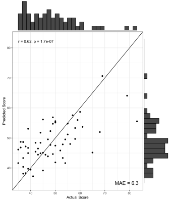 |
Morphometric Similarity Better Predicts Later Cognition in Paediatric Traumatic Brain Injury than Single Structural Features
Daniel J. King1, Stefano Seri1,2, Cathy Catroppa3,4, Vicki A. Anderson3,4, and Amanda G. Wood1,5
1School of Life and Health Sciences & Aston Neuroscience Institute, Aston University, Birmingham, United Kingdom, 2Department of Clinical Neurophysiology, Birmingham Women’s and Children’s Hospital NHS Foundation Trust, Birmingham, United Kingdom, 3Brain and Mind Research, Clinical Sciences, Murdoch Children’s Research Institute, Melbourne, Australia, 4Department of Psychology, Royal Children’s Hospital, Melbourne, Australia, 5School of Psychology, Faculty of Health, Melbourne Burwood Campus, Deakin University, Geelong, Victoria, Australia Neuroanatomical correlates of long-term cognitive impairment after a paediatric traumatic brain injury are not established. Acutely acquired T1w MPRAGE MRI scans were used to calculate morphometric similarity between cortical areas. A supervised learning approach showed that, after cross-validation, morphometric similarity explained 12% variance in cognitive functioning two-years post-injury, beyond that of individual structural features. Thus, morphometric similarity is a useful approach to understand the diffuse effects of neurological insult on the still-developing brain and how this may predict later neuropsychological functioning. |
|
4649.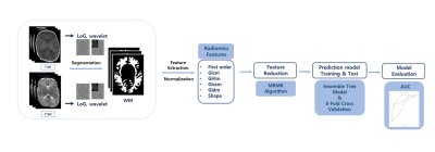 |
Application of radiomics for predicting poor psychomotor outcome in preterm neonates using brain MRI
You Won Shin1, Taehoon Shin1, Yoon Ho Nam2,3, and Hyun Gi Kim2
1Division of Mechanical and Biomedical Engineering, Ewha Womans University, Seoul, Korea, Republic of, 2Department of Radiology, Eunpyeong St. Mary’s Hospital, College of Medicine, The Catholic University of Korea, Seoul, Korea, Republic of, 3Department of Radiology, Seoul St. Mary's Hospital, College of Medicine, The Catholic University of Korea, Seoul, Korea, Republic of
To predict poor psychomotor development in preterm neonates who underwent MRI at term-equivalent age, we implemented radiomics feature analysis of white matter on T1-and T2-weighted images. A total 1920 features were derived, and optimal number of features were selected. The area under the ROC curve (AUC) for the diagnostic abilities of the radiomics analysis were 0.657, 0.814, and 0.690, using T1-weighted images, T2-weighted images, and both T1- and T2-weighted images, respectively. In conclusion, radiomics for term-equivalent age brain MRI can be useful for predicting poor psychomotor outcome in preterm neonates.
|
|
4650.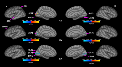 |
Grey matter hypertrophy and atrophy in early-blind adolescents: a surface-based morphometric study
Zhifeng Zhou1, Xia Liu2, Long Qian3, Yang Fan4, Hai Li4, Gangqiang Hou2, Wentao Jiang2, and Hengguo Li5
1Radiology, Shenzhen Mental Health Center/Shenzhen Kangning Hospital, Shenzhen, China, 2Shenzhen Mental Health Center/Shenzhen Kangning Hospital, Shenzhen, China, 3GE Healthcare, Beijing, China, 4Beijing Intelligent Brain Cloud, Inc, Beijing, China, 5The First Affiliated Hospital of Jinan University, Guangzhou, China
Numerous studies have explored brain structural abnormalities in blind people. However, most of them were based on volumetric measures, using voxel-based morphology, or in a specific region of interest, which may conceal fine anatomic details in other features. This study investigated alterations in cortical morphologies (thickness, volume and surface area) in early-blind adolescents (EBAs) using a surface-based morphometric method. Our results showed that there was disuse atrophy, compensatory and experience-related neural plasticity in the cerebral cortex of EBAs due to the visual deprivation.
|
|
|
4651.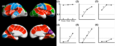 |
Spatiotemporal patterns of sulcal pits in the fetal brain
HyukJin Yun1, Lana Vasung1, Tomo Tarui2, Caitlin K. Rollins3, Cynthia M. Ortinau4, P. Ellen Grant1, and Kiho Im1
1Newborn Medicine, Boston Children's Hospital, Boston, MA, United States, 2Pediatric Neurology, Tufts Medical Center, Boston, MA, United States, 3Neurology, Boston Children's Hospital, Boston, MA, United States, 4Pediatrics, Washington University in St. Louis, St. Louis, MO, United States
Sulcal pits is known to represent the first cortical folds emerging in early fetal life. However, little is known about sulcal pits in the fetal brain. Since fetal brain showed dynamic changes of cortical folding during the second half of gestation, it is necessary to investigate spatial and temporal patterns of sulcal pits in the fetal brain. We analyzed spatial distribution and quantified emergence timing of pits using 48 typically developing fetuses. Sulcal pits were uniformly distributed in the fetal brain, and their timing of emergence are regionally different. It demonstrates sulcal pits are important landmarks of human brain development.
|
4652.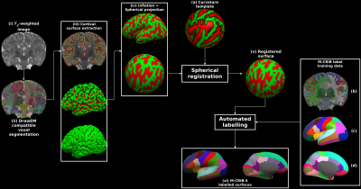 |
Parcellation of the neonatal cortex using Surface-based Melbourne Children's Regional Infant Brain atlases (M-CRIB-S)
Christopher Leslie Adamson1, Bonnie Alexander1, Gareth Ball1, Richard Beare1,2, Jeanie Chong3,4,5, Alicia Spittle1,4,5, Lex Doyle1,4,5, Peter Anderson1,5,6, Marc Seal1,5, and Deanne Thompson1,5
1Developmental Imaging, Murdoch Childrens Research Institute, Parkville, Australia, 2Department of Medicine, Monash Unviersity, Melbourne, Australia, 3Murdoch Childrens Research Institute, Parkville, Australia, 4Royal Womens Hospital, Parkville, Australia, 5University of Melbourne, Parkville, Australia, 6Monash Unviersity, Clayton, Australia Longitudinal studies measuring changes in cortical morphology over time are best facilitated by parcellation schemes compatible across all life stages. The Melbourne Children's Regional Infant Brain (M-CRIB) and M-CRIB 2.0 atlases provide voxel-based parcellations of the cerebral cortex compatible with the Desikan-Killiany (DK) and the Desikan-Killiany-Tourville (DKT) cortical labelling schemes. The curvature template registration targets, average surfaces, labelling training data, and pipeline execution scripts are available at (https://www.github.com/DevelopmentalImagingMCRI/MCRIBS). |
|
4653.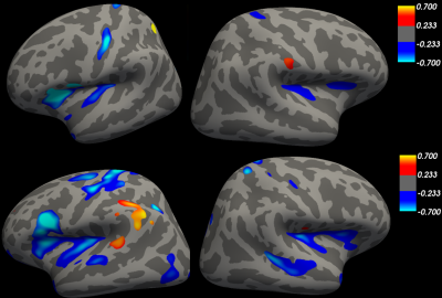 |
Brain region scaling differences between children and adults
Huangyuan Chen1 and Qing Cai1,2
1Institute of Cognitive Neuroscience, East China Normal University, Shanghai, China, 2NYU-ECNU Institute of Brain and Cognitive Science, New York University Shanghai, Shanghai, China
Individual brain size can vary as much as 1.5-fold. Furthermore, brain regions might not expand linearly proportional relative to total brain size. Scaling is a measure of such expansion. However, only few studies have investigated the differences in scaling pattern between childhood and adulthood. Here we analyzed structural T1 weighted images from children and adults datasets. We found that nonlinear scaling regions are more widely distributed in adults than in children. Therefore, we propose that individual brain regional growth might be influenced by “initial” brain size.
|
|
4654.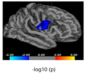 |
Characterization of cortical abnormalities in adolescents with first-episode drug-naïve major depressive disorder
Xinyue Hu1, Ruohan Feng2, Lianqing Zhang1, Yang Li3, Xinyu Hu1, Shi Tang1, Yingxue Gao1, Lihua Zhuo2, Guoping Huang3, and Xiaoqi Huang1
1Huaxi MR Research Center (HMRRC), Functional and molecular imaging Key Laboratory of Sichuan Province, Department of Radiology, West China Hospital, Sichuan University, Chengdu 610041, China, Chengdu, China, 2Department of Radiology, the third hospital of Mianyang, Mianyang, China, Mianyang, China, 3Department of Psychiatry, the third hospital of Mianyang, Mianyang, China, Mianyang, China
The detailed information about neuroanatomical alterations in adolescents with first-episode drug-naïve major depressive disorder (MDD) is still unknown. We measured cortical thickness, surface area and volume in 35 MDD patients and 22 healthy controls (HC). The MDD group, compared to HCs, showed reduction in cortical volume of the right precentral gyrus. We also found that MDD severity significantly correlated with cortical abnormalities in a number of regions, primarily in frontal and parietal lobes. These regions are implicated in emotion processing whose abnormities will contribute to the affective and cognitive dysfunction in adolescents with MDD.
|
|
4655.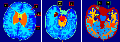 |
Sex Differences across Brain Regions in Neonates
Jianliang Gao1, Ahmed Fetit1, John Cupitt1, Jonathan Passerat-Palmbach1, Amir M Y F M Alansary1, Antonios Makropoulos1, Emma Robinson2, Sean Fitzgibbon3, Matteo Bastiani3,4, Nickolas Harper2, Lucilio Cordero Grande2, Anthony N Price2, Eugene Duff3,
Steve Smith3, Joseph V Hajnal2, David Edwards2, and Daniel Rueckert1
1BioMedIA, Department of Computing, Imperial College London, London, United Kingdom, 2Centre for the Developing Brain, Division of Imaging Sciences and Biomedical Engineering, King's College London, London, United Kingdom, 3Wellcome Centre for Integrative Neuroimaging, FMRIB, Nuffield Department of Clinical Neurosciences, University of Oxford, Oxford, United Kingdom, 4Sir Peter Mansfield Imaging Centre, School of Medicine, University of Nottingham, Nottingham, United Kingdom
We present for the first time a large cohort of neonatal brain MR images were acquired. Based on 505 subjects' MRI brain imaging data were acquired as part of the developing Human Connectome Project (dHCP) , we investigated the differences of 92 brain regions and brain volume without and with normalization of the volumes. Using multivariate analysis and adaptive step-down false discovery rate (FDR) control methods to examine term born neonates, we found 90 out of 92 regions and the brain volume statistically significant between sexes. But selected data normalization methods also suggested less statistical significance in neonatal brain regions.
|
|
4656.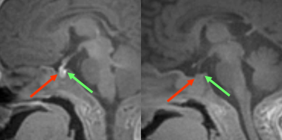 |
The Signal Intensity of Anterior and Posterior Lobe of Pituitary and Thyroid Gland in Preterm and Term Infants
Sayo Otani1, Yasutaka Fushimi1, Satoshi Nakajima1, Yusuke Yokota1, Sonoko Oshima1, Azusa Sakurama1, Krishna Pandu Wicaksono1, Tomohisa Okada2, and Kaori Togashi1
1Department of Diagnostic Imaging and Nuclear Medicine, Kyoto University Graduate School of Medicine, Kyoto, Japan, 2Human Brain Research Center, Kyoto University Graduate School of Medicine, Kyoto, Japan Poster Permission Withheld
We have evaluated the signal and the volume of anterior pituitary, posterior pituitary and thyroid gland in preterm and term infants on MRI by using 3D T1-PETRA sequence. Our study showed that the intensity of anterior pituitary was positively correlated with gestational age (GA), negatively correlated with chronological age (CA). We showed the intensity of posterior pituitary was also positively correlated with GA, negatively correlated with CA. We also demonstrated that the intensity of thyroid was positively correlated with GA, negatively correlated with CA. Our results may have relation to the maturity of hypothalamic-pituitary-thyroid (HPT) axis.
|
|
4657.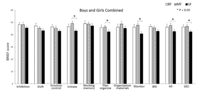 |
Infant Diets, Brain Cortical Development, and Executive Functions in Children
Ting Li1,2, Thomas M Badger3, Betty Jayne Bellando3, Seth Sorensen3, and Xiawei Ou1,3,4
1Arkansas Children's Research Institute, LITTLE ROCK, AR, United States, 2University of Arkansas at Little Rock, LITTLE ROCK, AR, United States, 3University of Arkansas for Medical Sciences, LITTLE ROCK, AR, United States, 4Arkansas Children's Nutrition Center, LITTLE ROCK, AR, United States
While it is known that breastfeeding promotes healthy brain development in children, the comparative effects of formulas feeding substantially differing in composition (i.e., milk-based vs. soy-based) on brain development are unclear. In this study, we recruited healthy 8-year-old children who were predominately breastfed, cow’s milk formula fed, or soy-based formula fed during infancy, and evaluated their brain cortical development using MRI. We also assessed their executive functions using parental-reported Behavior Rating Inventory of Executive Function (BRIEF) assessment. Differences in cortical thickness in multiple brain regions and differences in BRIEF scores were both observed for children in different infant diet groups.
|
|
4658.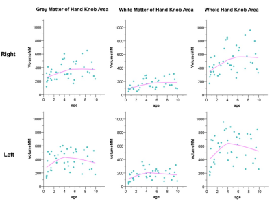 |
The Morphological Development of Hand Knob Areas in Children Aged 6m to 10 years
Fan Wu1, Xianjun Li1, Yuli Zhang1, Miaomiao Wang1, Congcong Liu1, Xiaoyu Wang1, Chao Jin1, Mengxuan Li1, Cong Tian1, Peiyao Chen1, Xiaocheng Wei2, and Jian Yang1
1Department of Diagnostic Radiology, The First Affiliated Hospital of Xi’an Jiaotong University, Xi’an, China, 2MR Research China, GE Healthcare, Xi'an, China
The cortical representation of hand motor function, hand knob region, is localized in the precentral gyrus. We found that the age-related rapid development of hand knob areas peaks at 4-5 years, and the left side increases faster than right. And the different increase rate between the left and right hand knob regions of normal children may be related to dominant hemisphere of handedness. This study will help to understand the process of hand function development in children and provide the research basis for the study of the upper limb pathway of the nerve center.
|
|
4659. |
Tailored rehabilitation in developmental dyslexia based on neurobiological model
Sunita Gudwani1,2, S.Senthil Kumaran3, Rajesh Sagar4, Madhuri Behari5, Manju Mehta6, Vaishna Narang7, SN Dwivedi8, and NR Jagannathan3
1Department of ENT, Escorts Heart Institute and Research Center, New Delhi, India, 2Former Department of NMR and MRI Facility, Former ALL INSTITUTE OF MEDICAL SCIENCES, NEW DELHI, India, 3Department of NMR and MRI Facility, All India Institute of Medical Sciences, New Delhi, India, 4Department of Psychiatry, All India Institute of Medical Sciences, New Delhi, India, 5Department of Neurology, Fortis Hospital, New Delhi, India, 6Department of Psychiatry (Psychology Unit), All India Institute of Medical Sciences, New Delhi, India, 7Department of Linguistics, School of Language, Jawahar Lal Nehru University, New Delhi, India, 8Department of Biostatistics, All India Institute of Medical Sciences, New Delhi, India
Reading a cultural invention by human species is complex cognitive skill. The ventral regions contribute as integrating bottom-up (feed-forward) generic visual processing with top-down influences from phonological and semantic areas. Developmental dyslexia a neurodevelopmental disorder, despite its description since century ago is yet unclear. The heterogeneous deficits, persists and may have emotional-socio-economic consequences. The study shows that if domain specific remediation, tailored according to subject’s neurobiological positive and negative signs along with behavioral measures, it benefits the individuals. Post-training improvement in behavioral performance and reorganizations in neural processing towards normalization, rather than compensatory regions attributed to optimal outcome
|