
Digital Poster Session
Pediatric: Brain Metabolism & Acquired Brain Disease
Pediatric
4660 -4675 Pediatric: Brain Metabolism & Acquired Brain Disease - Pediatric Neuro: Diffusion
4676 -4688 Pediatric: Brain Metabolism & Acquired Brain Disease - Pediatric Neuro: Metabolic Disease & Metabolism
4689 -4704 Pediatric: Brain Metabolism & Acquired Brain Disease - Pediatric Neuro: Acquired Brain Disease
Session Topic: Pediatric: Brain Metabolism & Acquired Brain Disease
Session Sub-Topic: Pediatric Neuro: Diffusion
Digital Poster
Pediatric
4660.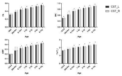 |
Hemispheric asymmetry of brain motor-related white matter:a diffusion kurtosis imaging study
Yuli Zhang1, Xianjun Li1, Mengxuan Li1, Chao Jin1, Miaomiao Wang1, Qinli Sun1, Fan Wu1, Congcong Liu1, Yannan Chen1, Xiaoyu Wang1, Huifang Zhao1, Cong Tian1, Peiyao Chen1, Xiaocheng Wei2,
and Jian Yang1
1the First Affiliated Hospital, Xi'an Jiaotong University, Xi'an, China, 22. MR Research China, GE Healthcare, Xi'an, China, China
Motor function development is a necessary condition for later life. It is closely related to cognitive psychological development1. As an important white matter reflecting the motor function, clarify the asymmetry of corticospinal tract (CST) development, which is of great significance for revealing the developmental of behavior and exploring the mechanism of disease2. This study aims to use DKI parameters investigate the asymmetry of CST development. Our results suggest that from 0-13 years old, there is hemispheric asymmetry in the development of CST, and they show left-sided dominance ;the asymmetry of CST appears at 6 months.
|
|
4661. |
Low Frequency Fluctuations in White Matter of Perinatally HIV-Infected Adult Youths: Glial Cycling for Neuroplasticity and Repair?
Manoj Kumar Sarma1, Amrita Pal2, Margaret A. Keller3,4, Tamara Welikson5, Joseph Ventura6, David E. Michalik7, Karin Nielsen-Saines8, Jaime Deville8, Andrea Kovacs9, Eva Operskalski9, Joseph A. Church9,10, Irwin Walot11, Paul M. Macey2,
Bharat Biswal12, and M. Albert Thomas1
1Radiological Sciences, David Geffen School of Medicine at UCLA, Los Angeles, CA, United States, 2UCLA School of Nursing, David Geffen School of Medicine at UCLA, Los Angeles, CA, United States, 3Pediatrics, Harbor-UCLA Medical Center, Torrance, CA, United States, 4The Lundquist Institute for Biomedical Innovation at Harbor-UCLA Medical Center, Torrance, CA, United States, 5Semel Institute for Neuroscience and Human Behavior, David Geffen School of Medicine at UCLA, Los Angeles, CA, United States, 6Psychiatry and Biobehavioral Sciences, David Geffen School of Medicine at UCLA, Los Angeles, CA, United States, 7Infectious Diseases-Pediatrics, Miller Children’s Hospital of Long Beach, Long Beach, CA, United States, 8Pediatrics, David Geffen School of Medicine at UCLA, Los Angeles, CA, United States, 9Pediatric, Keck School of Medicine of University of Southern California, Los Angeles, CA, United States, 10Children’s Hospital Los Angeles, Los Angeles, CA, United States, 11Radiology, Los Angeles County Harbor- UCLA Medical Center, Torrance, CA, United States, 12Biomedical Engineering, New Jersey Institute of Technology, Newark, NJ, United States
We evaluated the functional brain activity in perinatally HIV-infected youth (PHIVY) on cART by quantifying the amplitude of low frequency fluctuations (ALFF) and correlated with clinical and cognitive measures. We observed higher neural activity for ALFF in PHIVY compared to control. ALFF values in the cerebral white matter were positively correlated with viral load. Higher neural activity was associated with poorer performance in psychomotor function, abstract thinking, and social cognition. The findings suggest that long-term consequence of higher neuroinflammation and associated neurorepair in PHIVY may have a significant impact on regional spontaneous neuronal firing consequently impacting neurodevelopment and cognitive functioning.
|
|
4662.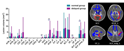 |
Left inferior fronto-occipital fasciculus is key anatomical location of cognitive delay in preterm infants with mild white matter injury
Miaomiao Wang1, Xianjun Li1, Congcong Liu1, Xiaoyu Wang1, Yannan Cheng1, Huifang Zhao1, Xingxing Tao1, Fan Wu1, Yuli Zhang1, Mengxuan Li1, Cong Tian1, Peiyao Chen1, Chao Jin1, XiaoCheng Wei2,
and Jian Yang3
1Department of Diagnostic Radiology, The First Affiliated Hospital of Xi’an Jiaotong University, Xi’an, China, 2MR Research China, GE Healthcare, Beijing, China, 3Department of Diagnostic Radiology, the First Affiliated Hospital of Xi’an Jiaotong University, Xi’an, China
Punctate white matter lesions (PWMLs) are common in the preterm. The mild PWMLs may result in cognitive impairments and lesion location is closely associated with neurodevelopmental outcomes. Accurate assessment of lesions location on qualitative MRI is difficult; therefore, this study aims to investigate the lesion-symptom relationship between locations of mild PWMLs and cognitive functioning in preterm infants at a corrected age of 3-6 months by diffusion tensor imaging. Lesion volume on IFOF_L is significantly larger in the cognitive delayed group than those in the normal group, and this is an independent factor (OR: 1.26) associated with the adverse cognitive development.
|
|
4663.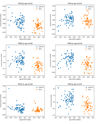 |
Peak width of skeletonized water diffusion MRI in the neonatal brain
Manuel Blesa Cabez1, Paola Galdi1, Gemma Sullivan1, Emily N. Wheater1, David Q. Stoye1, Gillian J. Lamb1, Alan J Quigley2, Michael J. Thrippleton1, Mark E Bastin1, and James P Boardman1
1University of Edinburgh, Edinburgh, United Kingdom, 2Royal Hospital for Sick Children, Edinburgh, United Kingdom
Preterm birth is closely associated with cognitive impairment and generalised dysconnectivity of developing white matter. Peak width of skeletonised DTI (MD, RD, AD, FA) and NODDI (NDI, ODI) metrics were used for characterising global connectivity during brain development. PSNDI was an excellent predictor for prematurity with an accuracy of 81 ± 10 %, followed by PSMD that achieved an accuracy of 77 ± 9 %. We conclude that the high accuracy in prediction and the ease of computation of these biomarkers make them useful new metrics of diffuse brain connectivity in neonatal populations.
|
|
 |
4664.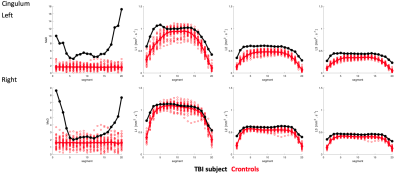 |
Mahalanobis Distance Tractometry (MaD-Tract) for Multivariate Analyses
Jose Manuel Guerrero-Gonzalez1, Peter Ferrazzano2, and Andrew Alexander1
1Medical Physics, University of Wisconsin - Madison, Madison, WI, United States, 2Pediatrics, University of Wisconsin - Madison, Madison, WI, United States
We present a multi-parameter tract profiling approach which allows for comparing diffusion imaging quantities between a single subject and a reference group along specific white matter path ways using the Mahalanobis distance (MaD). Implementation of MaD-Tract on a pediatric TBI-affected brain revealed several deviant tract segments in terms of elevated MaD values along multiple tracts compared to a group of healthy individuals.
|
4665. |
Early developmental differences of white matter tracts in preterm infants with brain injury
Tao Guo1, Dawn Gano2, Donna Ferriero2, James barkovich1, and Duan Xu1
1Department of Radiology and Biomedical Imaging, University of California, San Francisco, CA, United States, 2Department of Neurology and Pediatrics, University of California, San Francisco, CA, United States
We detected early developmental differences of white matter tracts in preterm infants with brain injury (BI). Nineteen preterm infants underwent two MRI scans. Microstructural development was analyzed combining conventional diffusion parameters and advanced NODDI parameters . We found BI preterm infants showed higher ODI in internal capsule, which was positively correlated with brain injuries severity; during the longitudinal period, most of the tracts in non-injured preterm infants had increased FA and Vic, decreased MD and ODI, while in BI preterm infants, only a few tracts developed. This study indicated white matter development was different in preterm infants with and without BI.
|
|
4666. |
Unveiling early cortical and white-matter maturation and the effect of music in preterm brain development: a longitudinal fixel-based analysis
Joana S. de Almeida1, Lara Lordier1, Sebastien Courvoisier2, François Lazeyras2, and Petra Hüppi1
1Department of Women-Children-Teenagers, Hôpitaux Universitaires de Genève, Geneva, Switzerland, 2Department of Radiology and Medical Informatics, CIBM, University of Geneva, Geneva, Switzerland
Dynamic brain macrostructural and microstructural changes occur from mid-fetal stage to birth. Prematurity disrupts brain maturation during this critical period and music might enhance activity-dependent-plasticity during early brain development. Using a longitudinal fixel-based analysis, we evaluated preterm brain macro and microscopic fiber changes from 33 weeks to term-age and the impact of music, during neonatal unit stay, on these changes. We show that fiber density (FD) and fiber-bundle cross-section (FC) increase in all major white-matter fibers over time, while in the cortex FD decreases and FC increases. Music intervention lead to a significant cortical FC increase in comparison to standard-of-care.
|
|
| 4667. | Relationships between myelin development and neurodevelopmental outcomes in very preterm and typically developing children
Deanne K Thompson1,2,3,4, Joseph YM Yang2,3,5,6, Jian Chen2, Claire E Kelly1,2, Chris L Adamson2, Bonnie Alexander1,2, Lillian G Matthews7, Katherine J Lee1,3,8, Rod W Hunt1,3,9, Jeanie LY Cheong1,10,11, Megan Spencer-Smith1,12, Marc L Seal2,3, Terrie E Inder1,7,
Lex W Doyle1,3,10,11, and Peter J Anderson1,12
1Victorian Infant Brain Studies, Murdoch Children's Research Institute, Parkville, Australia, 2Developmental Imaging, Murdoch Children's Research Institute, Parkville, Australia, 3Department of Paediatrics, University of Melbourne, Parkville, Australia, 4Florey Institute of Neuroscience and Mental Health, Parkville, Australia, 5Neuroscience Research, Murdoch Children’s Research Institute, Parkville, Australia, 6Department of Neurosurgery, the Royal Children’s Hospital, Parkville, Australia, 7Department of Pediatric Newborn Medicine, Brigham and Women’s Hospital, Harvard Medical School, Boston, MA, United States, 8Clinical Epidemiology & Biostatistics Unit, Murdoch Children’s Research Institute, Parkville, Australia, 9Neonatal Medicine, Royal Children’s Hospital, Parkville, Australia, 10Royal Women's Hospital, Parkville, Australia, 11Department of Obstetrics and Gynaecology, University of Melbourne, Parkville, Australia, 12Turner Institute for Brain and Mental Health, School of Psychological Sciences, Monash University, Clayton, Australia
Children born very preterm have altered myelination compared with full-term controls, but whether the T1-T2 ratio is a sensitive measure for understanding neurodevelopmental functioning remains unknown. This study found that the T1-T2 ratio trajectory between 7 and 13 years of age in the uncinate fasciculus was related to IQ scores, and 13-year T1-T2 ratios in almost all white matter regions were associated with motor functioning in both birth groups. Myelin development assessed using the T1-T2 ratio appears to be sensitive to predicting some neurodevelopmental outcomes.
|
|
4668.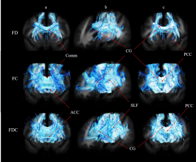 |
Fixel-based analysis of white matter differences between patients with cerebral palsy and typically developing children
Chih-Chien Tsai1, Chia-Ling Chen2,3, Yao-Liang Chen4, Jur-Shan Cheng5, Sung-han Lin6, and Jiun-Jie Wang1,6,7
1Healthy Aging Research Center, Chang-Gung University, TaoYuan, Taiwan, 2Department of Physical Medicine and Rehabilitation, Chang Gung Memorial Hospital, Linkou, TaoYuan, Taiwan, 3Graduate Institute of Early Intervention, College of Medicine, Chang Gung University, TaoYuan, Taiwan, 4Department of Medical Imaging and Intervention, Chang Gung Memorial Hospital, Keelung Branch, Keelung, Taiwan, 5Clinical Informatics and Medical Statistics Research Center, Chang-Gung University, TaoYuan, Taiwan, 6Department of Medical Imaging and Radiological Sciences, Chang Gung University, TaoYuan, Taiwan, 7Medical Imaging Research Center, Institute for Radiological Research, Chang Gung University/Chang Gung Memorial Hospital, TaoYuan, Taiwan
Disruption to white matter pathways is an important contributor to the pathogenesis of cerebral palsy but fail to delineate white matter tracts with lesions precisely with conventional magnetic resonance imaging. Fixel-based analysis, which has recently emerged as a useful fiber-specific tool for examining white matter structure, was used in this study. Reductions of fixel-based metrics in patients with cerebral palsy are represented in the corpus callosum, superior/posterior thalamic radiation, optic radiation, superior longitudinal fasciculus, and cingulum with corresponding direction of fiber tract. By using fixel-based analysis, this study described the white matter differences during development in patients with cerebral palsy.
|
|
4669. |
Age-specific DTI templates for human brain from 28 days to children
Mengxuan Li1, Xianjun Li1, Yuli zhang1, Miaomiao Wang1, Chao Jin1, Congcong Liu1, Yannan Cheng1, Xiaoyu Wang1, Huifang Zhao1, Fan Wu1, Cong Tian1, Peiyao Chen1, Xiaocheng Wei2, and Jian Yang1
1Radiology, the First Affiliated Hospital, Xi'an Jiaotong University, Xi'an, China, Xi'an, China, 2MR Research China, GE Healthcare, Bei Jing, China
The human brain develops rapidly from 28 days to children,there are distinct developmental trends at different ages.This period is important and difficult to acquire the images because of motion artifacts.Diffusion Tensor Imaging(DTI)was used to study brain structure.When analyzing DTI images of different individuals,a common standard space is needed to ensure the tissue diffusion information used for different individual images corresponds to the same spatial location.To understand how the human brain develops between 28 days and children,it is essential to make age-specific DTI templates for human brain.We built DTI templates for human brain of six periods from 28 days to children.
|
|
4670. |
How does Microstructural in lntra-axonal and Extra-axonl Space Change in Preterm Infants with Punctate White Matter Lesions?
Mengxuan Li1, Xianjun Li1, Yuli zhang1, Miaomiao Wang1, Yannan Cheng1, Congcong Liu1, Chao Jin1, Fan Wu1, Xiaoyu Wang1, Huifang Zhao1, Cong Tian1, Peiyao Chen1, Xiaocheng Wei2, and Jian Yang1
1Radiology, the First Affiliated Hospital, Xi'an Jiaotong University, Xi'an, China, Xi'an, China, 2MR Research China, GE Healthcare, Bei Jing, China
Punctate white matter lesions (PWML)are common in preterm infants.Extensive microstructural changes were observed previously for different PWML grades by using diffusion tensor imaging (DTI).However, the changes of intra-axon and extra-axon remain to be investigated. White matter tract integrity (WMTI)metrics derived from diffusion kurtosis imaging (DKI)provide information of intra-axonal or extra-axonal spaces. Our study aimed to use WMTI metrics to detect the Microstructural Variations.Compared to DTI metrics, the change trends of fractional anisotropy (FA),axial diffusivity (AD) and radial diffusivity (RD)are in agreement with previous findings.Furthermore, intra-axonal diffusivity (Da)unchange or reduce in lntra-axonal space.There were increased RDe, reduced/unchanged ADe in extra-axonal space.
|
|
4671. |
Maternal Anxiety and Depression during Pregnancy and Newborn’s Brain White Matter Development
Rachel M Graham1, Betty Jayne Bellando1, Seth Sorensen1, Li Jiang2, Charles M Glasier1, Raghu H Ramakrishnaiah1, Fang Lu1, Amy C Rowell1, and Xiawei Ou1,2,3
1University of Arkansas for Medical Sciences, LITTLE ROCK, AR, United States, 2Arkansas Children's Research Institute, LITTLE ROCK, AR, United States, 3Arkansas Children's Nutrition Center, LITTLE ROCK, AR, United States
This prospective study examined the relationships between maternal depression and anxiety during pregnancy and newborn’s brain white matter development. Healthy pregnant women were recruited at the 3rd trimester. Depression was assessed using the Beck Depression Inventory, Second Edition (BDI-II), and anxiety was assessed using the State-Trait Anxiety Inventory(STAI). Their newborns underwent an MRI examination of the brain at 2 weeks of age, which included diffusion tensor imaging to evaluate white matter development. Fractional anisotropy (FA) maps were generated and correlated with the BDI and STAI scores using tract-based spatial statistics. Negative correlations between FA values and BDI/STAI scores were found in multiple white matter regions, suggesting that depression and anxiety during pregnancy may impact the in utero brain white matter development.
|
|
4672. |
Magnitude and timing of major white matter along-tract maturation with NODDI
Kirsten M Lynch1, Ryan P Cabeen2, Kristi A Clark2, and Arthur W Toga2
1Mark and Mary Stevens Neuroimaging and Informatics Institute, University of Southern California, Los Angeles, CA, United States, 2Mark and Mary Stevens Institute for Neuroimaging and Informatics, University of Southern California, Los Angeles, CA, United States
White matter maturation is a heterogeneous phenomenon that can be probed by biophysical models. The purpose of this study was to characterize the development of white matter tracts with NODDI from infancy through adolescence. To probe the regional nature of white matter development, we use an along-tract approach to enable more fine-grained analysis. White matter tracts showed exponential age-related changes in NDI with spatially distinct maturational patterns. Our along-tract analyses elucidate hemispheric asymmetries within tracts which may be reflective of their functional specialization. Together, these results help to disentangle the processes that define the trajectory of white matter maturation.
|
|
4673.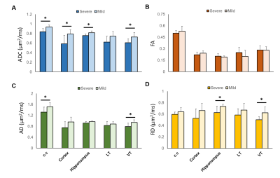 |
Early Changes in Diffusion Tensor Metrics between Different Final Damage Outcomes after Experimental Neonatal Hypoxic Ischemia
Yu-Chieh Jill Kao1,2,3, Chia-Feng Lu4, Bao-Yu Hsieh5, Chao-Ching Huang6, and Cheng-Yu Chen1,2,3,7
1Neuroscience Research Center, Taipei Medical University, Taipei, Taiwan, 2Department of Radiology, School of Medicine, College of Medicine, Taipei Medical University, Taipei, Taiwan, 3Translational Imaging Research Center, Taipei Medical University Hospital, Taipei, Taiwan, 4Department of Biomedical Imaging and Radiological Sciences, National Yang-Ming University, Taipei, Taiwan, 5Department of Biomedical Imaging and Radiological Science, China Medical University, Taichung, Taiwan, 6Department of Pediatrics, College of Medicine, National Cheng Kung University, Tainan, Taiwan, 7Department of Medical Imaging, Taipei Medical University Hospital, Taipei, Taiwan
Changes in diffusion tensor metrics within 6 h after hypoxic ischemia (HI) in different brain regions in neonatal rats is associated with the final lesion severity after hypoxic ischemia. We also demonstrated that the early changes within 6 h after HI may also correlate to the alteration in ultra-structure in the neurons and axons following the injury.
|
|
4674.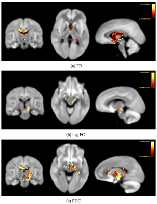 |
Fixel-based analysis reveals white matter changes following balance training in young brain injured patients: A longitudinal study
Xiaoyun Liang1,2, Chun-Hung Yeh2, Juan F Domínguez D1, Govinda Poudel1, and Karen Caeyenberghs1
1Mary Mackillop Institute for Health Research, Australian Catholic University, Melbourne, Australia, 2Florey Institute of Neuroscience and Mental Health, University of Melbourne, Melbourne, Australia
Traumatic brain injury (TBI) is one of the leading causes of death and disability in children and adolescents. Young TBI patients suffer from gross motor deficits, such as postural control deficits, which can severely compromise their daily life activities. Training programs have shown behavioral improvement; evidence of changes in WM morphology, however, has not been clear. We employ a fixel-based analysis (FBA) to investigate whether balance training results in significant changes of WM organization across whole brain in young TBI patients over time. Our results have shown that balance training induced signigicant macrostructural white matter changes (i.e. log-FC & FDC).
|
|
4675.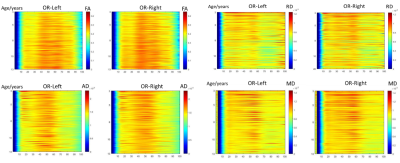 |
Spatiotemporal changes of optic radiation from 3-13 years: a diffusion tensor imaging study
Cong Tian1, Chao Jin1, Xingxing Tao1, Xianjun Li1, Miaomiao Wang1, Congcong Liu1, Yannan Cheng1, Fan Wu1, Yuli Zhang1, Mengxuan Li1, Xiaoyu Wang1, Peiyao Chen1, Huifang Zhao1, Xiaocheng Wei2,
and Jian Yang1
1the First Affiliated Hospital of Xi’an Jiaotong University, Xi’an, China, 2MR Research China, GE Healthcare, Xi’an, China
Optic radiations(OR), which connecting the lateral geniculate nuclei and visual cortex, plays a critical role in visual function. Detailing the maturation process of OR is helpful in understanding the visual development and identifying the abnormalities. Diffusion tensor imaging can quantify the brain white matter development. Using DTI-based automating fiber-tract quantification, this study explored the spatiotemporal OR changes in children aged 3-13 years. Results indicated that the anterior, middle, and posterior segments of OR showed asynchronous developmental patterns. Specifically, the mid-segment of OR presented more mature than the anterior segment, while right OR showed more extended mature than left OR.
|
 Watch the Video
 View the Poster
Session Topic: Pediatric: Brain Metabolism & Acquired Brain Disease
Session Sub-Topic: Pediatric Neuro: Metabolic Disease & Metabolism
Digital Poster
Pediatric
4676.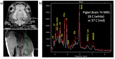 |
1H MRS of brain metabolism during cardiopulmonary bypass surgery in a neonatal pig model
Daniel Spielman1, Meng Gu1, Ralph Hurd1, Kirk Riemer2, Hiroki Ito2, and Frank Hanley2
1Radiology, Stanford University, Stanford, CA, United States, 2Cardiothoracic Surgery, Stanford University, Stanford, CA, United States
Although cardiopulmonary bypass (CPB) surgery in the neonate carriers significant risk for long-term neuronal deficits, details of brain metabolism during varying flow conditions for CPB, such as deep hypothermic circulatory arrest (DHCA) versus antegrade cerebral perfusion (ACP), remain incompletely understood. Metabolic changes in the brain have been observed under different CPB conditions, but which are truly causative of neurotoxicity and injury remains unclear. In this preliminary preclinical study, brain metabolism, as assessed using 1H MRS, during DHCA is active and abnormal, resulting in a buildup of lactate and loss of energy substrates. ACP may prevent these abnormalities.
|
|
4677.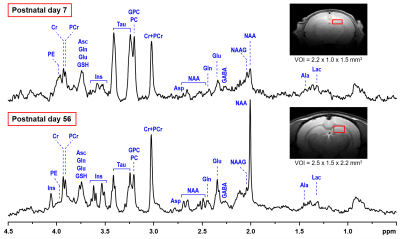 |
Intranasal insulin administration in a rat model of neonatal hypoinsulinemic hyperglycemia
Ivan Tkac1, Kathleen Ennis2, William H Frey II3, and Raghavendra Rao2
1Center for Magnetic Resonance Research, University of Minnesota, Minneapolis, MN, United States, 2Department of Pediatrics, University of Minnesota, Minneapolis, MN, United States, 3Center for Memory & Aging, HealthPartners Institute, St Paul, MN, United States
Hyperglycemia (HG) is common in the neonatal period in extremely low gestational age infants. The purpose of this study was to investigate acute and long-term effects of intranasal insulin administration on hippocampal neurochemical profile in a rat model of human perinatal HG. Hypoinsulinemic HG was induced in neonatal rats by injecting streptozotocin on postnatal day P2. Hippocampal neurochemical profiles were assessed at P7 and P56. Preliminary in vivo 1H MRS data demonstrate the feasibility and efficacy of intranasal insulin administration for normalizing neurochemical homeostasis in the hippocampus exposed to transient hypoinsulinemic HG in the neonatal period.
|
|
4678.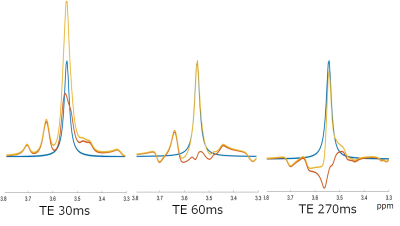 |
Quantification of brain glycine in an infant with nonketotic hyperglycemia: a serial proton magnetic resonance spectroscopy study
Moyoko Tomiyasu1,2, Jun Shibasaki3, Yasuhiro Kawai4, Tatsuya Higashi1, Takayuki Obata1, and Noriko Aida1,2
1Molecular Imaging and Theranostics, National Institutes for Quantum and Radiological Science and Technology, Chiba, Japan, 2Radiology, Kanagawa Children's Medical Center, Yokohama, Japan, 3Neonatology, Kanagawa Children's Medical Center, Yokohama, Japan, 4Neurology, Kanagawa Children's Medical Center, Yokohama, Japan
We serially examined in vivo brain glycine levels in an infant with nonketotic hyperglycemia (NKH) using single-voxel proton magnetic resonance spectroscopy (MRS) with different point-resolved spectroscopic localization sequence echo times (TEs). The time-course changes of brain glycine obtained with TEs of 60 and 270 ms corresponded with glycine concentrations in cerebrospinal fluid and with the clinical condition of the infant. By appropriately combing a pulse sequence and acquisition parameters, proton MRS can evaluate brain glycine levels to help diagnose NKH.
|
|
4679.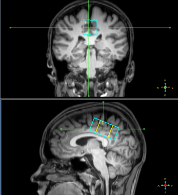 |
Pediatric acute mTBI study of GABA concentration in posterior cingulate cortex
Andrei Manzhurtsev1,2, Maxim Ublinskiy1,3, Petr Menshchikov1, Alexey Yakovlev2,4, Olga Bozhko3, Tolib Akhadov3, and Natalia Semenova1,3,4
1Emanuel Institute of Biochemical Physics of the Russian Academy of Sciences, Moscow, Russian Federation, 2Clinical and Research Institute of Emergency Surgery and Trauma, Moscow, Russian Federation, 3Clinical and Research Institute of Emergency Pediatric Surgery and Traumatology, Moscow, Russian Federation, 4Semenov Institute of Chemical Physics of the Russian Academy of Science, Moscow, Russian Federation
This is the first measurement of pure GABA concentration in the PCC of children with mild TBI in the acute phase. MEGA-PRESS pulse sequence was used in order to obtain GABA signal not contaminated by macromolecules. The result obtained disagrees with the previous study, where [GABA] was increased in the anterior cingulate cortex of children with mTBI when comparing to healthy controls. The findings may signify that ACC is more sensitive to mTBI than PCC.
|
|
|
4680.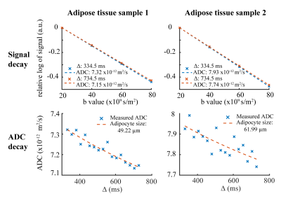 |
Comparison of adipocyte size measurements with histology and high b-value diffusion-weighted spectroscopy in human white adipose tissue at 3 T
Dominik Weidlich1, Julius Honecker2, Stefan Ruschke1, Lisa Patzelt1, Cora Held1, MingMing Wu1, Daniela Franz1, Stefanie Winkler2, Hans Hauner2, and Dimitrios C. Karampinos1
1Department of Diagnostic and Interventional Radiology, Technical University of Munich, Munich, Germany, 2Else Kröner Fresenius Center for Nutritional Medicine, Technical University of Munich, Freising, Germany
Despite its high relevance in metabolic research, the non-invasive measurement of adipocyte size remains an unmet need. DW-MRS has been previously applied to probe diffusion restriction effects in-vivo to measure lipid droplets in animals up to diameters of 10 µm and in humans up to diameters of 50 µm. However, probing diffusion restriction in white adipose tissue, consisting of adipocytes up to a size of 150 µm, on a clinical system remains a major challenge. This work proposes to measure adipocyte sizes with high b-value long diffusion time DW-MRS. In a preliminary analysis, the presented method matches with histology.
|
4681.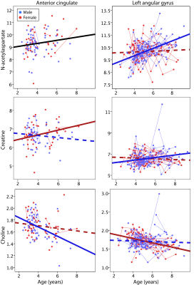 |
Characterization of brain metabolite changes in early childhood
Ashley D Harris1,2, Tiffany Bell1,2, Mercedes Bagshawe1,2, Elodie Boudes1,2, and Catherine Lebel1,2
1Radiology, University of Calgary, Calgary, AB, Canada, 2Alberta Children's Hospital Research Institute and the Hotchkiss Brain Institute, University of Calgary, Calgary, AB, Canada
Using proton MRS, we examine longitudinal changes in tNAA, tCr, tCho, Ins and Glx in early childhood in a typically developing children ages 2.4-9.3 years. We show NAA increases in the anterior cingulate across both sexes while age-relate changes in NAA in the left angular gyrus are only seen in boys. In the anterior cingulate tCr in girls increases and tCho decreases in boys. By contrast in the left angular gyrus tCr increases in boys and tCho decreases in girls. These different metabolite trajectories in different brain regions may be associated with different development seen between boys and girls.
|
|
4682.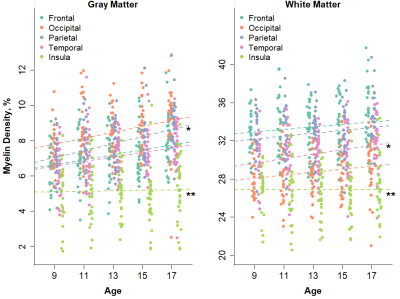 |
Characterization of Gray and White Matter Myelination Across Adolescence Using Macromolecular Proton Fraction Mapping
Neva M. Corrigan1, Vasily L. Yarnykh2, Daniel S. Hippe2, Julia P. Owen2, Christina Zhao1, and Patricia K. Kuhl1
1Institute for Learning and Brain Sciences, University of Washington, Seattle, WA, United States, 2Radiology, University of Washington, Seattle, WA, United States
Although previous studies have investigated brain structural changes across adolescent development, little attention has been paid to the myelination that occurs in the gray matter during this period. We utilized macromolecular proton fraction mapping to investigate myelination of gray and white matter in a cross-sectional sample of 146 adolescents at 9, 11, 13, 15 and 17 years of age. Throughout most of the brain, gray matter myelin density was found to increase at a faster rate with age than white matter myelin density. Our findings suggest that gray matter myelination is a significant component of brain maturation during adolescent development.
|
|
4683.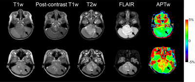 |
Differentiation of Low- and High-Grade Pediatric Brain Tumors with Amide Proton Transfer Imaging
Xingwang Yong1, Hongxi Zhang2, Zhipeng Shen3, Xinchun Chen2, Weibo Chen4, Dan Wu1, and Yi Zhang1
1Key Laboratory for Biomedical Engineering of Ministry of Education, Department of Biomedical Engineering, College of Biomedical Engineering & Instrument Science, Zhejiang University, Hangzhou, Zhejiang, China, 2Department of Radiology, Children’s Hospital, Zhejiang University School of Medicine, Hangzhou, Zhejiang, China, 3Department of Neurosurgery, Children’s Hospital, Zhejiang University School of Medicine, Hangzhou, Zhejiang, China, 4Philips Healthcare, Shanghai, China
Amide proton transfer (APT) imaging was applied to the grading of pediatric brain tumors for the first time with its performance evaluated by the Receiver Operating Characteristic (ROC) curve. Ninety-two patients were enrolled in this study, of whom 48 cases were high-grade tumors and 44 cases were low-grade tumors. An experienced radiologist delineated the initial ROIs, which were then automatically masked and shrunk to avoid artifacts and subjective bias. APT MRI was able to differentiate low-grade pediatric brain tumors from high-grade ones, with a maximum area under the ROC curve of 0.95 in conjunction with quantitative T1 and T2.
|
|
4684.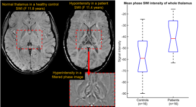 |
Advanced permutation test of children’s thalami reveals correlation between age and iron accumulation in aspartylglucosaminuria (AGU) disease
Viljami Sairanen1, Anna Tokola1, Ritva Tikkanen2, Minna Laine3, and Taina Autti1
1HUS Medical Imaging Center, University of Helsinki and Helsinki University Hospital, Helsinki, Finland, 2Institute of Biochemistry, University of Giessen, Giessen, Germany, 3Department of Child Neurology, University of Helsinki and Helsinki University Hospital, Helsinki, Finland
We report an original finding of a strong linear correlation (F=26, p=1.5e-4, R2=0.65) between iron accumulation within specific thalamic structures and the age of children with aspartylglucosaminuria (AGU). AGU is a rare lysosomal storage disorder which has no cure, causes a negative effect on the development of a child, and leads to a premature death. We used affine image registration and implemented a voxel-wise permutation test to locate where AGU patients have higher filtered phase SWI intensities (i.e. more iron) than controls. Furthermore, we demonstrated that permutation test was crucial for discovering the linear correlation between iron accumulation and age.
|
|
4685.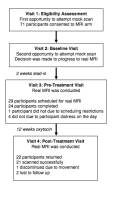 |
Uptake of MRI and associated data quality in an inclusive clinical trial in children with Autism Spectrum Disorder
Marilena M DeMayo1, Izabella M Pokorski1, Ian B Hickie2, and Adam J Guastella1
1University of Sydney, Camperdown, Australia, 2Brain and Mind Centre, University of Sydney, Camperdown, Australia
Individuals with Autism Spectrum Disorder (ASD) show high rates of comorbidities, including intellectual disability. Often, individuals with comorbid conditions are excluded from clinical trials and neuroimaging studies, potentially biasing the development of treatments and subtypes. In an inclusive trial (without functioning constraints) for children with ASD (aged 3-12 years), 71 participants consented to MRI and 24 (34%) completed an MRI scan, following a familiarization procedure. Twenty-one participants had a successful post-treatment MRI. This study reports on the resultant data quality and shows the potential to include MRI in trials of complex populations who, typically, are excluded.
|
|
4686.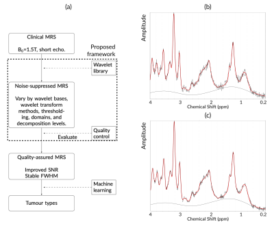 |
Optimized pediatric brain tumor diagnosis by using in vivo magnetic resonance spectroscopy and machine learning
Dadi Zhao1,2, James T. Grist1,2, Yu Sun1,2, Vijay Sawlani1,3, and Andrew C. Peet1,2
1Institute of Cancer and Genomic Sciences, University of Birmingham, Birmingham, United Kingdom, 2Birmingham Children's Hospital NHS Foundation Trust, Birmingham, United Kingdom, 3Queen Elizabeth Hospital Birmingham NHS Foundation Trust, Birmingham, United Kingdom Poster Permission Withheld
Previous studies have shown that magnetic resonance spectroscopy (MRS) can provide diagnostic classifiers for childhood brain tumors. Here we investigate the effect of noise suppression on classification and build it into a pipeline aimed at optimizing the diagnosis. Although people have proposed several algorithms to suppress noise in MRS, the clinical application is still largely restricted to apodisation. We propose a wavelet-based framework to suppress the noise of in vivo MRS, thereby the signal quality of in vivo MRS and the classification accuracy of tumor cases was improved. The framework could be used in the clinical diagnosis through MRS.
|
|
4687.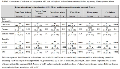 |
Associations of body size and composition with regional brain volumes and white matter microstructure in very preterm infants
Katherine A Bell1,2, Lillian G Matthews1,2, Anna K Prohl2,3, Sara Cherkerzian1,2, Terrie E Inder1,2, Simon K Warfield2,3, Shun Onishi4, and Mandy B Belfort1,2
1Department of Pediatric Newborn Medicine, Brigham & Women's Hospital, Boston, MA, United States, 2Harvard Medical School, Boston, MA, United States, 3Computational Radiology Laboratory, Department of Radiology, Boston Children's Hospital, Boston, MA, United States, 4Department of Pediatric Surgery, Research Field in Medical and Health Sciences, Medical and Dental Area, Research and Education Assembly, Kagoshima University, Kagoshima, Japan
For very preterm infants, body size and composition (lean versus fat mass) may index brain growth and microstructural development. Among 85 very preterm infants at term equivalent age, we studied associations of body size/composition with brain magnetic resonance imaging (MRI) outcomes including total and regional brain volumes, and fractional anisotropy of white matter tracts. Larger body size and more lean--but not fat--mass were associated with larger brain volumes and higher fractional anisotropy of multiple white matter tracts. Lean mass accrual may index brain growth and development. MRI may be useful for studying effects of nutritional exposures on the preterm brain.
|
|
4688.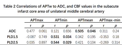 |
Correlation analysis between amide proton transfer weighted and blood flow status in infarct core of patients with subacute ischemic stroke
Yuhan Jiang1, Yanwei Miao1, Liangjie Lin2, Zhiwei Shen2, Peipei Chang1, Yiwei Che1, Ailian Liu1, Qingwei Song1, and Jiazheng Wang2
1the First Affiliated Hospital of Dalian Medical University, Dalian, China, Dalian, China, 2Philips Healthcare, Beijing, China, Beijing, China
Amide proton transfer weighted (APTw) MR imaging enables detections of metabolite and pH value changes, while the arterial spin label (ASL) imaging can assess hemodynamic changes in brain tissue after cerebral infarction. This study aimed to evaluate correlation between APT-related metabolic changes and blood flow status in the infarct core of patients with subacute ischemic stroke by APTw and ASL MR techniques. Significant correlations of APT to apparent diffusion coefficient (ADC) and cerebral blood flow (CBF) values in the infarct region were observed.
|
Session Topic: Pediatric: Brain Metabolism & Acquired Brain Disease
Session Sub-Topic: Pediatric Neuro: Acquired Brain Disease
Digital Poster
Pediatric
4689.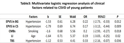 |
Investigation of cerebral small vessel disease and impacting clinical factors in young patients with symptomatic cerebral infarction
Yuhan Jiang1, Yanwei Miao1, Peipei Chang1, Yiwei Che1, and Lizhi Xie2
1the First Affiliated Hospital of Dalian Medical University, Dalian, China, Dalian, China, 2GE Healthcare, MR Research China, Beijing, Beijing, China, Beijing, China
In this study, we selected the first infarction of young patients as a study object, retrospective analysis of young cerebral infarction, senile cerebral infarction and healthy young people's brain cerebral small vessel diseases (CSVD) imaging findings, and further analysis of its clinical influencing factors to further explore the etiology of infarction in young patients. We found that CSVD often occurs in young and elderly patients of cerebral infarction, while it is more frequent and serious in elderly patients. Hypertension, smoking, and age may be factors influencing cerebral small vessel disease in young patients with cerebral infarction.
|
|
4690. |
Utilising multi-parametric MRI to non-invasively predict tumour type in paediatric neuro-oncological disease: a multi-centre study.
James Timothy Grist1, Stephanie Timothy Withey2, Lesley MacPherson3, Adam Oates4, Stephen Timothy Powell2, Jan Novak5, Laurence Abernethy6, Barry Pizer7, Ricahrd Grundy8, Simon Bailey9, Dipayan Mitra9, Theodoros N Arvantis10, Dorothee P. Auer8,
and Andrew C Peet2
1University of Birmingham, BIRMINGHAM, United Kingdom, 2University of Birmingham, Birmingham, United Kingdom, 3Birmingham Women's and CHildren's NHS foundation trust, Birmingham, United Kingdom, 4Birmingham Women's and Children's NHS foundation trust, Birmingham, United Kingdom, 5Aston University, Birmingham, United Kingdom, 6Alder Hey Children's NHS foundation trust, Liverpool, United Kingdom, 7Institute of Translation Medicine, University of Liverpool, Liverpool, United Kingdom, 8University of Nottingham, Nottingham, United Kingdom, 9Royal Victoria Infirmary, Newcastle, United Kingdom, 10University of Warwick, Warwick, United Kingdom
This study focuses on utilising supervised Machine Learning to combine both diffusion and perfusion weighted imaging to discriminate between the three most common pediatric brain tumour types: Pilocytic Astrocytoma, Ependymoma, and Medulloblastoma.
|
|
4691. |
Combining different high resolution MR vessel imaging sequences in pediatric medulloblastoma survivors: Be faster than the stroke event
Yasemin Tanyildizi1, Marie-Astrid Neu2, Arthur Wingerter2, Alexandra Russo2, Marc Alexander Brockmann1, and Joerg Faber2
1Neuroradiology, University Medical Center Mainz, Mainz, Germany, 2Pediatric Hematology/ Oncology/ Hemostaseology, University Medical Center Mainz, Mainz, Germany
Imaging cerebro- vasculopathy after radio-chemotherapy in pediatric Medulloblastoma survivors, by assessing internal media thickening of the external carotid artery through ultrasound and comparing black blood high resolution vessel wall imaging sequences with TOF-MR-Angiography, Volume Interpolated GRE/±CE and T2 tse fat saturated sequences. The underlying study indicates a superiority of black blood sequences in early detection of vascular changes, which might lead to vasculopathy, in comparison to the commonly used sequences. Thus follow-up MRI protocols might include black blood vessel wall images.
|
|
4692. |
Evaluating the Alternation of Brain Structure in Deaf Children by using Multimodal Structural Analysis
Hang Qu1, Wei Wang1, and Weiqiang Dou2
1Radiology Department, The Affiliated Hospital of Yangzhou University, Yangzhou, China, 2MR Research China, GE Healthcare, Beijing, China
In this study, we used Surface Based Morphometry (SBM) and Tract-Based Spatial Statistics (TBSS) analysis to investigate the alternation of brain structure in deaf children. The cortical thickness of the deaf group significantly decreased in the left postcentral gyrus, superior parietal lobule, paracentral lobule, precuneus, the right transverse temporal gyri, and the middle temporal gyrus. The left precuneus local gyrification index increased. The fractional anisotropy value of the white matter in deaf children is lower than control group. These findings demonstrated that structural changes in deaf children occurred in both the grey and white matter, and revealed that the neuroplastic changes associated with cross-modal reorganization in the precuneus.
|
|
4693.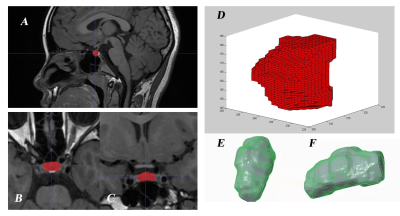 |
3D automatic segmentation analysis of pituitary in children with precocious puberty
Hang Qu1, Yu Pan1, RuiHong Chen1, Weiqiang Dou2, Weiyin Vivian Liu2, and Wei Wang1
1The Affiliated Hospital of Yangzhou University, Yangzhou, China, 2GE Healthcare, MR Research China, Beijing, P.R. China, Beijing, China
To explore changes of pituitary morphology in precocious children.An in-house software based on Matlab platform was used to obtain pituitary morphological measurements. In this research the height of the pituitary in the CPP group was higher than that in the PPP group and the normal group. The pituitary volume of the CPP group was larger than that of the normal group.Pituitary volume and surface area of CCP group were positively correlated with hormone metabolism levels. The height of pituitary gland is still the index of differential diagnosis, however pituitary volume and surface area have better correlation with hormone level.
|
|
4694.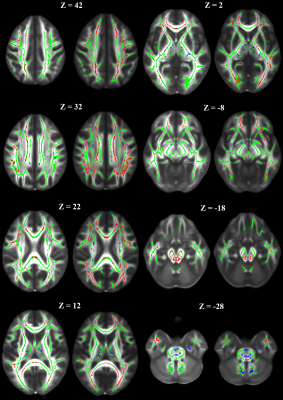 |
Confirming outcomes of disrupted white matter integrity in an independent cohort of children treated for medulloblastoma
John O. Glass1, Jared J. Sullivan1, Yian Guo2, Julie H. Harreld1, Yimei Li2, Giles W. Robinson3, Amar Gajjar3, and Wilburn E. Reddick1
1Diagnostic Imaging, St. Jude Children's Research Hospital, Memphis, TN, United States, 2Biostatistics, St. Jude Children's Research Hospital, Memphis, TN, United States, 3Oncology, St. Jude Children's Research Hospital, Memphis, TN, United States
This study confirmed changes seen in a cohort of 146 patients and 92 normal healthy age-similar controls in an independent cohort of 141 patients. Fractional anisotropy (FA) was analyzed using the TBSS tools in FSL. After surgery but before any additional therapy, FA was significantly reduced in both cohorts. Additionally, a large increase in the percentage of skeleton voxels where the patients at least reach the level of controls two years from diagnosis was observed. This suggest the possibility that the newer therapy is having a less toxic impact on the microstructural integrity in this pediatric medulloblastoma population.
|
|
4695.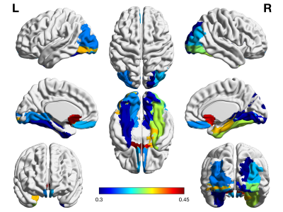 |
R2* mapping in children with attention deficit hyperactivity disorder: a whole-brain analysis
Yingqian Chen1, Shu Su1, Long Qian2, Yan Dai1, Miao Fan1, Hongyu Zhang3, and Zhiyun Yang1
1Department of Radiology, First Affiliated Hospital, Sun Yat-sen University, Guangzhou, China, 2MR Research, GE Healthcare, Beijing, China, 3Department of Pediatric, First Affiliated Hospital, Sun Yat-sen University, Guangzhou, China
Attention deficit/hyperactivity disorder (ADHD) might associate with the iron deficiency, but few studies reported on the correlation between brain iron level and the severity of illness. To address whether the changes of iron deposition are correlated with the cognitive function deficits in ADHD, the R2* mapping were applied in current study. The results indicated that the correlations between the R2* value and the symptom severity of ADHD were found in in brain regions within the basal ganglia region, temporal lobe and occipital lobe, which also indicated that R2* mapping might have the potential efficacy in the auxiliary diagnosis of ADHD.
|
|
4696.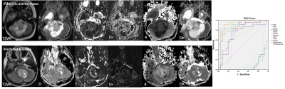 |
Differentiation of Low- and High-grade Pediatric Brain Tumors by Using intravoxel incoherent motion imaging and diffusion kurtosis imaging
Dejun She1
1Radiology, The First Affiliated Hospital of Fujian Medical University, Fuzhou, China
To demonstrate the quantitative parameters derived from intravoxel incoherent motion imaging (IVIM) and diffusion kurtosis imaging (DKI) models can be used to improve the accuracy of MR imaging for differentiating among low- and high-grade pediatric brain tumors.
|
|
4697.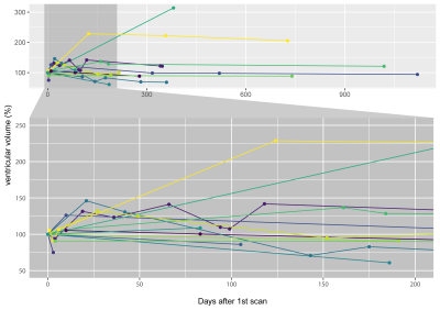 |
MRI reveals brain ventricle expansion in pediatric patients with acute disseminated encephalomyelitis
Jason Michael Millward1, Luca Bramé1, Kevin Rostásy2, Matthias Baumann3, Thoralf Niendorf1,4, and Sonia Waiczies1
1Berlin Ultrahigh Field Facility, Max Delbrück Center for Molecular Medicine, Berlin, Germany, 2Department of Paediatric Neurology, Children's Hospital Datteln, Witten/Herdecke University, Datteln, Germany, 3Division of Paediatric Neurology, Department of Paediatrics I, Medical University of Innsbruck, Innsbruck, Austria, 4Experimental and Clinical Research Center, Charité - Universitätsmedizin Berlin, Berlin, Germany
We show dynamic variations in brain ventricle volume (VV) in longitudinal MRI scans of pediatric patients with acute disseminated encephalomyelitis (ADEM). A majority of patients showed decreases in VV directly following the initial clinical event, or following subsequent VV expansion. This suggests that the VV expansion in these patients was not due to irreversible brain atrophy, but rather likely reflected processes associated with acute disease. Calculations of VV (and volumes of other brain structures) done using the automated tool FreeSurfer were significantly affected by gadolinium-based contrast agents; comparing pre- and post-contrast scans should be avoided in longitudinal studies.
|
|
4698.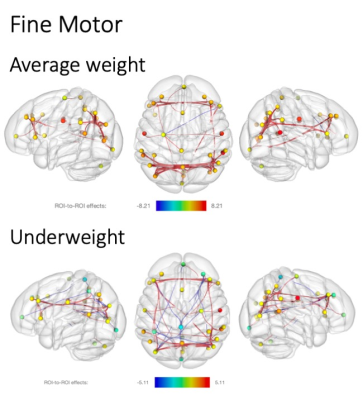 |
Motor Ability Network Connectivity in Undernourished Young Children in India
Muriel Marisa Katharina Bruchhage1,2, Giang-Chau Ngo1,2, Madhuri Tiwari3, Aarti Kumar3, Vishwajeet Kumar3, Viren D'Sa1,2, and Sean C. L. Deoni1,2,4
1Warren Alpert Medical School, Brown University, Providence, RI, United States, 2Advanced Baby Imaging Lab, Hasbro Children's Hospital, Providence, RI, United States, 3Community Empowerment Lab, Lucknow, India, 4Maternal, Newborn, and Child Health Discovery & Tools, Bill & Melinda Gates Foundation, Seattle, WA, United States
Early motor skill acquisition is an important developmental stepping stone for more complex cognitive tasks. While cognitive performance and overall health has shown to be negatively impacted by malnutrition, few studies have been conducted to investigate its influence on neurodevelopment. Here, we investigate the relationship of motor ability and brain functional connectivity (fc) in currently underweight and average weight young children (< 2 years) living in an Indian region with one of the worst human development indicators. With increase in motor complexity, fewer fc were available in the undernourished cohort only, possibly indicating an under-recruitment of brain networks.
|
|
4699.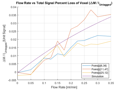 |
Measuring CSF shunt flow with MRI using flow enhancement of signal intensity (Shunt-FENSI)
Mingxiao Zhang1,2, Natalie Aw2,3, Mark Doose1, Paul M. Arnold4, Jason Huston4, William C. Olivero2, and Bradley P. Sutton2
1Department of Bioengineering, University of Illinois at Urbana-Champaign, Urbana, IL, United States, 2Beckman Institute, University of Illinois at Urbana-Champaign, Urbana, IL, United States, 3Department of Electrical & Computer Engineering, University of Illinois at Urbana-Champaign, Urbana, IL, United States, 4Carle Foundation Hospital, Urbana, IL, United States
Ventriculo-peritoneal shunts are used for the treatment of hydrocephalus in pediatric patients. Monitoring the condition of CSF flow through the shunt is vital for identifying possible shunt failure. We propose an MRI-based monitoring technique, Shunt-FENSI, for accurate, quantitative and non-invasive shunt flow measurement. The Shunt-FENSI technique uses pseudocontinuous sub-voxel labeling to build up signal due to flow within a larger imaging voxel. We demonstrate high accuracy in phantom scans, where we match our tagged signal measurement with a simulation which includes tagging, flow, and T1 recovery.
|
|
4700.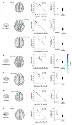 |
The Neurobiology of Placebo Response in Adolescents with Generalized Anxiety Disorder
Lu Lu1,2, Hailong Li1, Jeffrey Mills3, Heidi Schroeder2, Sarah Mossman2, Sara Varney2, Kim Cecil4, Melissa DelBello2, Amir Levine5, Xiaoqi Huang1, Qiyong Gong1, John Sweeny1,2, and Jeffrey Strawn2
1Huaxi MR Research Center (HMRRC), Functional and molecular imaging Key Laboratory of Sichuan Province,Department of Radiology, West China Hospital of Sichuan University, Chengdu, China, 2Department of Psychiatry and Behavior Neuroscience, College of Medicine, University of Cincinnati, Cincinnati, OH, United States, 3Carl H. Lindner College of Business, University of Cincinnati, Cincinnati, OH, United States, 4Cincinnati Children's Hospital Medical Center, Cincinnati, OH, United States, 5Division of Child and Adolescent Psychiatry, Department of Psychiatry, College of Physicians and Surgeons, Columbia University, New York, NY, United States
Increasing placebo response rates represent a significant barrier to detecting treatment effects in pediatric psychiatry clinical trials. To identify biomarkers for it in adolescents with generalized anxiety disorder prior to their entering a clinical trial, whole brain dynamic and static functional connectivity (FC) were used. Dynamic and static FC between the amygdala, dorsal anterior cingulate cortex, ventrolateral prefrontal cortex and regions that subserve emotion control and inhibition were significantly associated with degree of placebo response and differed between placebo responders and non-responders. These findings may be used to decrease placebo response in clinical trials to more effectively evaluate novel treatments.
|
|
4701.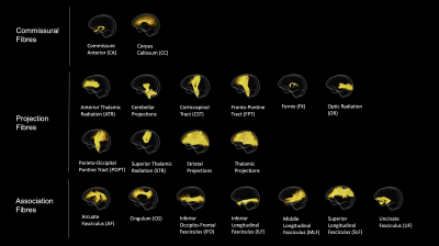 |
Fibre specific tract profiles of children with ADHD: Evidence from fixel-based analysis
Ian Fuelscher1, Emma Sciberras1,2,3, Daryl Efron2,3,4, Vicki Anderson4,5, Christian Hyde1, and Timothy J Silk1,4,5
1School of Psychology, Deakin University, Geelong, Australia, 2Population Health, Murdoch Children's Research Institute, Melbourne, Australia, 3Department of Paediatrics, University of Melbourne, Melbourne, Australia, 4The Royal Children's Hospital, Melbourne, Australia, 5Clinical Sciences, Murdoch Children's Research Institute, Melbourne, Australia
Diffusion tensor imaging (DTI) studies examining white matter microstructure in children with attention-deficit/hyperactivity disorder (ADHD) have provided conflicting results, possibly reflecting limitations of the DTI framework. We leverage fixel-based analysis (FBA), a fibre specific assessment of white matter microstructure, to comprehensively evaluate white matter tract profiles of children with ADHD. Relative to controls, children with ADHD show both reduced fibre density and reduced fibre cross section across association, projection and commissural fibres. FBA metrics further correlate with ADHD symptom severity. We conclude that ADHD symptomatology in childhood may be subserved by altered communication pathways across the brain.
|
|
4702.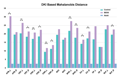 |
DKI-Based Mahalanobis Distance Assessing Alterations of Brain White Matter in Infants With Benign Enlargement of Subarachnoid Space
Congcong Liu1, Xianjun Li1, Miaomiao Wang1, Chao Jin1, Fan wu1, Cong Tian1, Mengxuan Li1, Xiaocheng Wei2, and Jian Yang1
1Department of Radiology, The First Affiliated Hospital of Xi’an Jiaotong University, Xi'an, China, 2MR Research China, GE Healthcare, Xi'an, China
Some infants with BESS were accompanied with mildly motor and language delay. White matter (WM) development is important to neurodevelopmen, but relationships between BESS and WM maturation are not very clear. Mahalanobis distance is a feasible multivariate approach to evaluate white matter maturation. This study aims to quantitative assess the WM microstructures of in infant with BESS aged 4-6 months via DKI-based Mahalanobis distance. Larger Mahalanobis distance was found in infants with BESS among most WM tracts. All these changes of WM tracts suggested underlying alterations and prematuration of white matter. It may provide additional information for the neurodevelopment outcomes.
|
|
4703.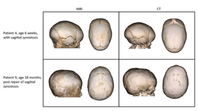 |
Replacing CT with high-resolution MRI for 3D pediatric cranial bone imaging
Kamlesh B. Patel1, Cihat Eldeniz2, Gary B. Skolnick3, Udayabhanu Jammalamadaka2, Paul K. Commean2, Manu S. Goyal2, Matthew D. Smyth4, and Hongyu An2
1Plastic and Reconstructive Surgery, Washington University in St. Louis, Saint Louis, MO, United States, 2Radiology, Washington University in St. Louis, Saint Louis, MO, United States, 3Surgery, Washington University in St. Louis, Saint Louis, MO, United States, 4Neurosurgery, Washington University in St. Louis, Saint Louis, MO, United States
Ionizing radiation from computed tomography (CT) imaging increases the risk of cancer. Patients with craniosynostosis often undergo repeated head CT scans, exacerbating the cumulative risk. Magnetic Resonance Imaging (MRI) has the potential to be a radiation-free safe alternative. Previously proposed methods in this area are not widely utilized because of suboptimal osseous/soft tissue contrast, vulnerability to motion, and the need for manual post-processing. In this study, we propose a high-resolution radial MRI protocol with improved tissue contrast and less sensitivity to motion. Moreover, we seek to evaluate its feasibility by using a blinded clinical evaluation.
|
|
4704. |
An In-vivo Assessment of Regional Brain Temperature during Whole-body Cooling for Neonatal Encephalopathy
Tai-Wei Wu1, Jessica Lee Wisnowski1, Rachel Chapman1, Marvin D Nelson2, Benita Tamrazi2, and Stefan Bluml2,3
1Neonatology, Children's Hospital Los Angeles/USC, Los Angeles, CA, United States, 2Radiology, Children's Hospital Los Angeles/USC, Los Angeles, CA, United States, 3Rudi Schulte Research Institute, Santa Barbara, CA, United States
Regional brain temperatures in newborns hypoxic-ischemic encephalopathy (HIE) were measured during whole-body hypothermia (TH) to test the hypothesis that brain temperature profile is non-homogenous and is related to pattern or severity of brain injury. We found that whole body hypothermia was effective in cooling deep brain structures, while superficial structures were warmer and significantly higher than rectal temperature. It was also observed that infants with more severe brain injury exhibited higher brain temperatures without regional temperature differences.
|