
Scientific Session
CSF flow & Glymphatic Imaging
| Tuesday Parallel 2 Live Q&A | Tuesday, 11 August 2020, 15:15 - 16:00 UTC | Moderators: Nivedita Agarwal & Olivier Baledent |
0643.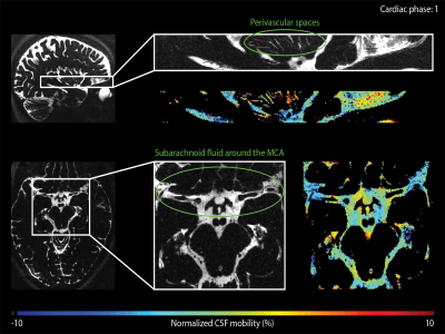 |
The driving force of glymphatics: influence of the cardiac cycle on CSF-mobility in perivascular spaces in humans
Lydiane Hirschler1, Bobby A Runderkamp2, Suzanne L Franklin1,3, Thijs van Harten1, Aart Nederveen2, Matthan WA Caan4, and Matthias JP van Osch1
1Leiden University Medical Center, Leiden, Netherlands, 2Radiology and Nuclear Medicine, Amsterdam UMC, University of Amsterdam, Amsterdam, Netherlands, 3University Medical Centre Utrecht, Utrecht, Netherlands, 4Department of Biomedical Engineering & Physics, Amsterdam University Medical Center, Amsterdam, Netherlands
Recently, flow of cerebrospinal fluid (CSF) has been shown to play an important role in the waste clearance of the brain, ushering in the concepts of glymphatics and intramural periarterial drainage. Despite the importance of brain waste removal, the exact clearance mechanisms such as its driving force are still poorly understood and remain highly controversial. In this study, we image how the cardiac cycle influences the CSF-mobility in the human brain, in the perivascular spaces of the basal ganglia as well as in large CSF-filled spaces, i.e. ventricles and subarachnoid space around large arteries.
|
|
 |
0644.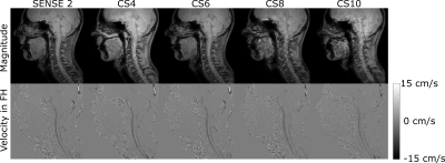 |
Compressed sensing accelerated 4D flow magnetic resonance imaging of the cerebrospinal fluid.
Kristina Sonnabend1, Elena Jäger1, David Maintz1, Kilian Weiss1,2, and Alexander Bunck1
1University of Cologne, Faculty of Medicine and University Hospital Cologne, Institute for Diagnostic and Interventional Radiology, Cologne, Germany, 2Philips GmbH, Hamburg, Germany
Cerebrospinal fluid (CSF) flow dynamics are relevant parameters in the diagnosis of neurological diseases and can be accessed by three-dimensional time-resolved phase-contrast MRI (4D flow MRI). However, these measurements are accompanied by long scan times making acquisition acceleration necessary to accomplish clinical feasibility. The aim of this study was to evaluate the feasibility of compressed sensing (CS) acceleration in 4D flow MRI of the CSF. CS factors 4 to 10 were compared against the conventional SENSE in 16 healthy subjects. Preliminary results show feasibility of CS factor 6 with comparable image and velocity data quality.
|
0645.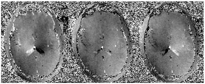 |
Cyclic Intracerebral Coherent Motion on Peripheral-Pulse-Gated Ultra-Low VENC MRI: Noninvasive Depiction of Glymphatic Flow?
Robert Y Shih1,2, J Kevin DeMarco1,2, J Kent Werner1,2, Justin E Costello1,2, Isabelle Heukensfeldt Jansen3, Luca Marinelli3, Thomas K Foo3, and Vincent B Ho1,2
1Uniformed Services University of the Health Sciences, Bethesda, MD, United States, 2Walter Reed National Military Medical Center, Bethesda, MD, United States, 3GE Global Research, Niskayuna, NY, United States
A combination of pressure gradients from arterial pulsatility, respiratory cycles, and resistance changes is thought to drive convective influx of CSF into paraarterial spaces for rapid exchange with ISF, followed by efflux into paravenous spaces toward arachnoid granulations, meningeal lymphatics, or cranial nerves. Visualization of this phenomenon was attempted with peripheral-pulse-gated phase contrast sequences at VENC = 5 mm/s (gradient echo) and 0.24 mm/s (spin echo) in four healthy adults using an ultra-high-performance MAGNUS gradient coil. Very slow intracerebral coherent motion was depicted, cerebropetal during systole, cerebrofugal during diastole, possibly reflecting bulk flow in paravascular spaces of the glymphatic system.
|
|
0646. |
Blood flow in the internal carotid arteries is correlated with CSF outflow from the ventricular system
Karin Markenroth Bloch1, Tekla M. Kylkilahti2,3, Olle Haglund1, Linn C. Lingehall2,3, Nils Fregne2,3, Johannes Töger4, and Iben Lundgaard2,3
1National 7T facility, LBIC, Lund University, Lund, Sweden, 2Department of Experimental Medical Science, Lund University, Lund, Sweden, 3Wallenberg Centre for Molecular Medicine, Lund University, Lund, Sweden, 4Department of Clinical Sciences, Lund University, Lund, Sweden
Little is known about which physiological parameters regulate CSF production. In this work, we tested the hypothesis that cerebral blood flow and heart rate play roles in CSF regulation. We used 7T MR to quantify CSF flow in the cerebral aqueduct and blood flow in the carotid arteries of healthy volunteers. We found that CSF outflow from the ventricular system correlated with blood flow in the internal carotid arteries, whereas there was no significant effect of heart rate on CSF outflow. This suggests that cerebral blood flow affects CSF flow and production.
|
|
 |
0647.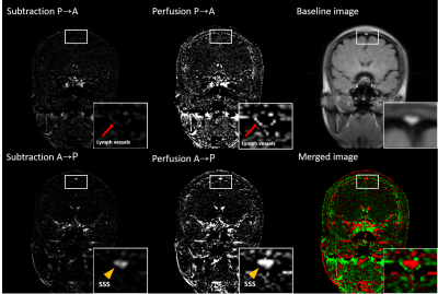 |
Human meningeal lymphatic vessels can be imaged by inversion recovery alternate ascending/descending directional navigation (ALADDIN)
Jun-Hee Kim1 and Sung-Hong Park1
1Department of Bio and Brain engineering, Korea Advanced Institute of Science and Technology, Daejeon, Korea, Republic of
Recent studies showed meningeal lymphatic vessels significantly contribute to the clearance mechanisms of cerebrospinal fluid (CSF) and the immune system in central nervous system. In this study, we tried to image human dural meningeal lymphatic vessels (mLVs) using inversion recovery alternate ascending/descending directional navigation (IR-ALADDIN). The IR-ALADDIN imaging technique clearly showed not only structural dural mLVs, but also the flow direction of dural mLVs, and it can be applied for studying many lymphatic vessels in human neurological diseases.
|
0648.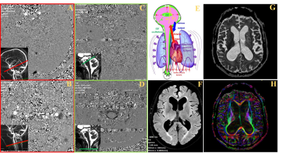 |
Impact of cerebrospinal and blood flow pulsatilities on periventricular white matter in patients with hydrocephalus
Fadoua Saadani-Makki1,2,3, Malek Makki3, Serge Metanbou4, Cyrille Capel 5, and Olivier Balédent1,2
1Department of Image Processing, University Hospital, Amiens, France, 2CHIMERE EA 7516, Research Team for Head & Neck, University of Picardie Jules Verne, Amiens, France, 3GIE Faire Face, CHU Amiens Picardie, Amiens, France, 4Department of Radiology, University Hospital, Amiens, France, 5Department of Neurosurgery, University Hospital, Amiens, France
The aim of this study was to assess the relationship between neuro-fluids dynamic and microstructure architecture of white matter fibers in hydrocephalus patients. Twenty-eight hydrocephalus patients underwent simultaneously diffusion tensor and phase contrast imaging. A statistical correlation between diffusion and flow parameters has shown a biological causal relationship between abnormal brain neuro-fluids dynamic and white matter alterations in hydrocephalus patients.
|
|
0649.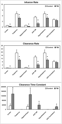 |
MRI Detection of Impairment of Glymphatic Function in Rat after Mild Traumatic Brain Injury
Lian Li1, Michael Chopp1,2, Guangliang Ding1, Esmaeil Davoodi-Bojd1, Qingjiang Li1, Yanlu Zhang1, Ye Xiong1, and Quan Jiang1
1Henry Ford Hospital, Detroit, MI, United States, 2Oakland University, Rochester, MI, United States Using dynamic MRI glymphatic measurement and our advanced mathematic model, the alterations of glymphatic function in the brain with mild TBI were investigated. Our data show that mild TBI leads to both impaired influx and efflux of contrast agent along the glymphatic pathway. The reduced efficiency of glymphatic function affects the multiple regions across the brain, which may decrease the clearance of waste metabolites and facilitate protein aggregation, contributing to subsequent cognitive deficits. The global change in brain clearance function, rather than the appearance of focal lesions, appears to provide a reliable measure indicating the injury of the brain. |
|
|
0650.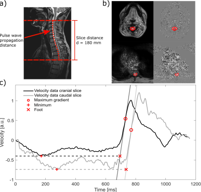 |
Cerebrospinal fluid pulse wave velocity measurements using multiband CINE phase-contrast MRI
Kristina Sonnabend1, Gerrit Brinker2, David Maintz1, Alexander Bunck1, and Kilian Weiss1,3
1University of Cologne, Faculty of Medicine and University Hospital Cologne, Institute for Diagnostic and Interventional Radiology, Cologne, Germany, 2University of Cologne, Faculty of Medicine and University Hospital Cologne, Department of General Neurosurgery, Center for Neurosurgery, Cologne, Germany, 3Philips GmbH, Hamburg, Germany
Intraspinal compliance is related to neurological diseases and can be measured by pulse wave velocity (PWV). A multiband CINE phase-contrast MRI sequence was developed to measure the intraspinal PWV between two simultaneously acquired slices along spine. The method was evaluated in-vitro, in healthy-subjects and in a normal pressure hydrocephalus patient. In-vitro results show good reproducibility and dependency on transmural pressure in agreement with theory. A higher PWV compared to healthy subjects is observed in the patient. A decline in PWV after shunt surgery is detected, making it a promising tool for investigation and treatment follow-up of neurological diseases.
|
0651.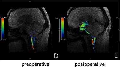 |
Evaluation of Cerebrospinal Fluid Dynamics in endoscopic third ventriculostomy for treating obstructive hydrocephalus with 4D Flow MRI
Liu Jia1, Cheng Xiaoqing 1, Lu Guang ming1, Dou Weiqiang2, and Shen Yong2
1Department of Medical Imaging,, Jinling Hospital , Medical School of Nanjing University, Nanjing, China, 2GE Healthcare, MR Research, Beijing, P.R. China, Nanjing, China We aim to investigate the clinial value of 4D flow MRI in assessing cerebrospinal fluid(CSF)dynamics. The optimal velocity encoding factor(VENC) and high test-retest reproducibility was firstly obtained in CSF measurements for healthy volunteers. Ensured by thses, 4D flow MRI has been further applied to evaluate the CSF dynamics for patients with obstructive hydrocephalus before and after endoscopic three ventriculostomy(ETV). Notable cerebrospinal fluid flow at the stoma has been found, indicating that a new cerebrospinal fluid circulation pathway was established. Our study thus demonstrated that 4D flow MRI is an effective tool to assess CSF dynamics quantitively. |
|
0652.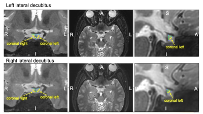 |
Analysis of Physiological Brain Shift and Optic Chiasm in the Closed Cranium due to Postural Position
Etsuko Kumamoto1, Shigeto Hayashi1,2, Ari Shinojima3, Koshi Yokota4, and Eiji Kohmura5
1Kobe University, Kobe, Japan, 2Department of Neurosurgery, Hyogo Emergency Medical Center, Kobe, Japan, 3Keio University, Tokyo, Japan, 4Japan Aerospace Exploration Agency, Tsukuba, Japan, 5school of medicine, Kobe University, Kobe, Japan
Methodological analysis related to physiological brain shift in the closed cranium is lacking. The spaceflight-associated neuro-ocular syndrome is attributed to the upward shift of the brain. We analyzed the relationship of brain shift and optic nerve shift by using MR volume data acquired in different body positions. The movement and rotation of each voxel, divided into 20 × 20 × 20 pixels3, were calculated using the block matching method. Experimental results show that the optic nerve transforms and deforms with the movement of the brain because of a change in body position.
|
 Watch the Video