
Scientific Session
Neurodegeneration 1
Session Topic: Neurodegeneration 1
Session Sub-Topic: Extrapyramidal Disease
Oral
Neuro
| Monday Parallel 2 Live Q&A | Monday, 10 August 2020, 14:30 - 15:15 UTC | Moderators: Maria Eugenia Caligiuri |
Session Number: O-03
 |
0201.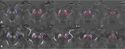 |
Introducing Substantia Nigra Iron Content and Neuromelanin Overlap to Distinguish Parkinson’s Patients from Healthy Controls
Naying He1, Kiarash Ghassaban2,3, Pei Huang4, Zenghui Cheng1, Yan Li1, Mojtaba Jokar5, Sean K. Sethi2,5, Weibo Chen6, Shengdi Chen4, Fuhua Yan1, and Ewart Mark Haacke1,2,5
Video Permission Withheld
1Radiology, Ruijin Hospital, Shanghai Jiaotong Univ. School of Medicine, Shanghai, China, Shanghai, China, 2Department of Radiology, Wayne State University, Detroit, MI, United States, 3Department of Biomedical Engineering, Wayne State University, Detroit, MI, United States, 4Department of Neurology, Ruijin Hospital, Shanghai Jiao Tong University School of Medicine, Shanghai, China, 5Magnetic Resonance Innovations, Inc., Bingham Farms, Bingham Farms, MI, United States, 6Philips Healthcare, Shanghai, China
A total of 40 Parkinson’s disease (PD) patients and 40 age- and sex-matched healthy controls (HC) were scanned using a single 3D gradient echo magnetization transfer sequence to measure neuromelanin and iron content, and the overlap between them. These measures showed reliable results indicative of powerful diagnostic biomarkers to differentiate PD patients from HCs. An increase in iron content was seen in the substantia nigra for the PD patients while the neuromelanin volume decreased. The best predictor, however, was found to be the combination of neuromelanin volume and its overlap with iron-containing substantia nigra which yielded an AUC of 98%.
|
0202.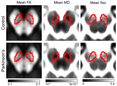 |
Reinterpreting Parkinson’s disease-related diffusion signal changes in substantia nigra
Jason Langley1, Daniel E Huddleston2, Evan Oculam3, Stewart Factor2, and Xiaoping Hu1,3
1Center for Advanced Neuroimaging, University of California Riverside, Riverside, CA, United States, 2Neurology, Emory University, Atlanta, GA, United States, 3Bioengineering, University of California Riverside, Riverside, CA, United States
Parkinson’s disease is a progressive, neurodegenerative disorder characterized by asymmetrical onset of motor symptoms such as bradykinesia, rigidity, and tremor. The principal pathology in Parkinson's disease is the loss of melanized dopamine neurons in the substantia nigra pars compacta (SNpc) with iron deposited alongside this neuronal loss. Loss of SNpc neurons should remove barriers for diffusion and increase diffusivity of water molecules in regions undergoing this loss. Studies examining Parkinsonian SNpc microstructural changes using a single tensor model have yielded conflicting results. Here, we investigate PD-related microstructural changes in multiple compartment and single tensor models.
|
|
|
0203.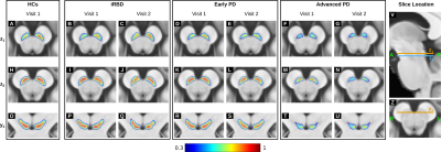 |
Investigating Spatiotemporal Changes in the Substantia Nigra of Patients with Prodromal and Clinical Parkinson's Disease
Emma Biondetti1,2,3, Rahul Gaurav1,2,3, Lydia Yahia-Cherif1,3, Graziella Mangone4, Nadya Pyatigorskaya1,2,3,5, Romain Valabrègue1,3, Claire Ewenczyk2,3, Matthew Hutchison6, Jean-Christophe Corvol3,4,7, Marie Vidailhet2,3,7, and Stéphane Lehéricy1,2,3,5
1Brain and Spine Institute - ICM, Centre for NeuroImaging Research - CENIR, Paris, France, 2Brain and Spine Institute - ICM, Team "Movement Investigations and Therapeutics", Paris, France, 3Brain and Spine Institute - ICM, INSERM U 1127, CNRS UMR 7225, Sorbonne University, Paris, France, 4National Institute of Health and Medical Research - INSERM, Clinical Investigation Centre, Pitié-Salpêtrière Hospital, Paris, France, 5Department of Neuroradiology, Pitié-Salpêtrière Hospital, Public Assistance - Paris Hospitals (AP-HP), Paris, France, 6Biogen Inc., Cambridge, MA, United States, 7Department of Neurology, Pitié-Salpêtrière Hospital, Public Assistance - Paris Hospitals (AP-HP), Paris, France
Parkinson's disease (PD) and idiopathic rapid eye movement sleep behaviour disorder (iRBD, a prodromal condition of Parkinsonism) are characterised by the progressive loss of neuromelanin-containing neurons in the substantia nigra (SN). Based on longitudinal neuromelanin-sensitive magnetic resonance imaging (NM-MRI) of healthy controls, patients with iRBD and patients with PD, and voxel-wise analysis of NM-MRI on a study-specific anatomical brain template, we showed the temporal evolution of SN atrophy in disease. We also found significant correlations between temporal changes in the NM-MRI signal-to-noise ratio and clinical scores of disease severity, reflecting the functional organisation (motor, cognition and behaviour/mood) of the SN.
|
|
0204.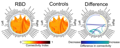 |
Brainstem structural connectivity changes in prodromal Parkinson’s disease by 7 Tesla HARDI
María Guadalupe García-Gomar1, Kavita Singh1, Matthew Stauder2, Laura D. Lewis1, Lawrence L. Wald1, Bruce R. Rosen1, Aleksandar Videnovic2, and Marta Bianciardi1
1Department of Radiology, Athinoula A. Martinos Center for Biomedical Imaging, Massachusetts General Hospital and Harvard Medical School, Boston, MA, United States, 2Department of Neurology, Massachusetts General Hospital and Harvard Medical School, Boston, MA, United States
REM-sleep-behavior-disorder (RBD) is a sleep disorder characterized by the absence of muscular atonia during REM sleep. RBD patients have a high risk of developing Parkinson’s disease (PD) within 10 years from RBD diagnosis. Thus, RBD allows the investigation of early/prodromal neurodegenerative-stages. Changes in brainstem-nuclei-connectivity are expected in RBD/prodromal-PD based on animal and ex-vivo human-studies. Yet, their investigation in living-humans is understudied. Through high-spatial-resolution 7 Tesla HARDI MRI and a recently-developed probabilistic-brainstem-nuclei-atlas, we built a brainstem-based structural-connectome in living RBD-patients and age-matched controls. Interestingly, in RBD-patients we detected structural-connectivity-changes within the brainstem in line with the pathophysiology of RBD in animal-models.
|
0205.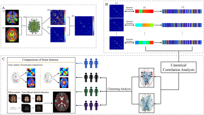 |
Clinical-related connectivity features define three biotypes of Parkinson’s disease
Tao Guo1, Xiaojun Guan1, Cheng Zhou1, Ting Gao2, Jingjing Wu1, Peiyu Huang1, Xiaojun Xu1, and Minming Zhang1
1Department of Radiology, Second Affiliated Hospital of Zhejiang University School of Medicine, Hangzhou, China, 2Department of Neurology, Second Affiliated Hospital of Zhejiang University School of Medicine, Hangzhou, China
We establish brain connectivity features that represented the disease signatures and identify Parkinson’s disease (PD) subtypes by data-driven approaches. Canonical correlation analysis (CCA) was performed to define the clinical related connectivity features, which were then used in hierarchical cluster analysis to identify the distinct biotypes of PD. Multimodal MRI including gray matter functional connectivity and white matter microstructure were further used to explore the neuropsychological significance of these biotypes. CCA revealed two significant clinical-related patterns in PD. Hierarchical cluster analysis identified three neurophysiological biotypes: mild, progressive depression-dominant and progressive motor-dominant. These three biotypes characterized by different neural substrate.
|
|
 |
0206.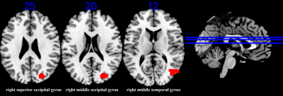 |
Association of ApoE gene polymorphism and caudate functional connectivity in mild cognitive impairment of Parkinson’s disease
Song'an Shang1, Weiqiang Dou2, Hongying Zhang3, Jing Ye3, and Jingtao Wu3
1Department of Radiology, Nanjing First Hospital, Nanjing Medical University, Nanjing, China, 2GE Healthcare, MR Research China, Beijing, China, 3Northern Jiangsu People’s Hospital, Yangzhou, China
In this study, we aimed to investigate the association of Apolipoprotein E (ApoE) gene polymorphism and caudate functional connectivity in mild cognitive impairment of Parkinson’s disease (PD-MCI), using resting-state functional magnetic resonance imaging (rs-fMRI) and genotyping. Our findings revealed that gene-brain-behavior associations involve alterations of caudate activity with posterior cortical, thereby provide potential imaging-based markers that contribute to the early diagnosis and monitoring of PD-MCI.
|
|
0207.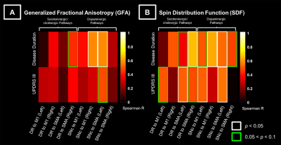 |
Dopaminergic premotor and motor pathways are dominant in the progression of motor disability in Parkinson’s disease
Yao Chia Shih1, Septian Hartono2,3, Amanda Choo2, Celeste Chen2, Isabel Chew1, Zheyu Xu2,3, Louis Tan2,3, ChingYu Cheng3,4, Eng King Tan2,3, and Ling Ling Chan1,3
1Department of Diagnostic Radiology, Singapore General Hospital, Singapore, Singapore, 2Department of Neurology, National Neuroscience Institute – SGH Campus, Singapore, Singapore, 3Duke-NUS Medical School, Singapore, Singapore, 4Department of Ophthalmology, Yong Loo Lin School of Medicine, National University of Singapore, Singapore, Singapore
White matter microstructural changes in relation to the neurotransmitter systems in Parkinson’s disease (PD) progression remains unclear. We used diffusion spectrum imaging and local connectome fingerprint analysis to investigate microstructural integrity of the the premotor and motor pathways in associations with various neurotransmitter systems in the brainstem, and disease duration and severity of motor-related symptoms. We found greater microstructural changes in the dopaminergic pathways in association with motor progression than for the other neurochemical pathways. Patients with longer disease duration or more severe motor dysfunctions showed increased anisotropic water diffusion in these pathways, suggesting a compensatory effect of axonal sprouting.
|
0208.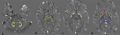 |
QUANTITATIVE SUSCEPTIBILITY MAPPING AS A DIAGNOSTIC TOOL TO DISTINGUISH TREMOR DOMINANT PD FROM ESSENTIAL TREMOR.
Shumyla Jabeen1, Jitender Saini1, Jaladhar Neelavalli2, Narayankrishna Rolla2, Shweta Prasad3, and Ravi Yadav4
1Neuroimaging and Interventional Radiology, National Institute of Mental Health and Neurosciences, Bangalore, India, 2Philips India, Bangalore, India, 3Clinical neurosciences, National Institute of Mental Health and Neurosciences, Bangalore, India, 4Neurology, National Institute of Mental Health and Neurosciences, bangalore, India
Tremor dominant PD and ET often pose a diagnostic difficulty in view of overlapping clinical features. We aimed at distinguishing the two using the novel technique of QSM to measure iron deposition in various gray matter nuclei including the substantia nigra pars compacta(SNPc). A statistically significant difference was seen in the QSM values of the SNPc, SNPr and caudate nucleus. ROC curve analysis showed a sensitivity and specificity of 90 and 87.5% respectively using a cut off value of 12 ppb for the SNPc. Thus, QSM is a potentially useful problem solving technique for distinguishing tremor dominant PD from ET.
|
|
0209.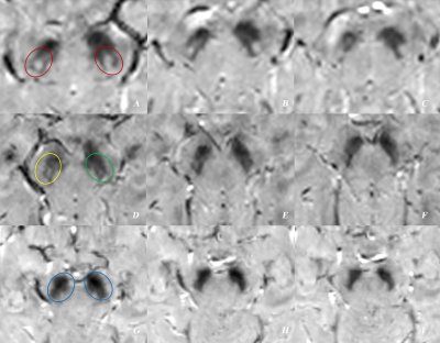 |
Total loss of “swallow tail sign”: a potential substitute of PET for detecting dopaminergic degeneration in early-stage Parkinson’s disease
Na Wang1, XueLing Liu2, and YuXin Li2
1Fudan university Huashan hospital, Shanghai, China, 2Huashan Hospital, Fudan University,, Shanghai, China
Whether swallow tail sign (STS) could serve as a substitute or complement for nuclear medical imaging remains unclear. In this study, we compared STS features on MRI with striatal uptake on positron emission tomography (PET) at per nuclei level, construct an evaluation scale based on bilateral STS changes at the patient level, and estimate the diagnostic performance of the scale in 39 early-stage PD and 28 healthy controls. STS alterations corresponded well with striatal uptake on PET in early-stage PD. Total loss of STS is a reliable sign of nigrostriatal dopaminergic degeneration and might be a potential substitute for PET.
|
|
0210.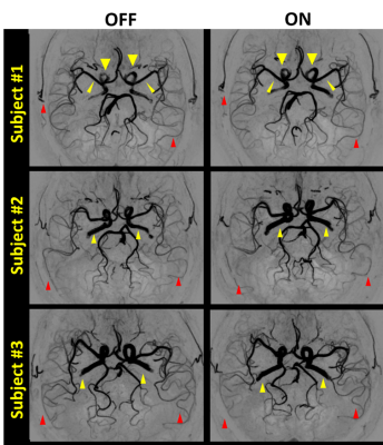 |
Effects of Levodopa Therapy on Cerebral Arteries and Brain Tissue Perfusion in Parkinson’s Disease Patients
Yuhui Xiong1,2, Lanxin Ji1, Le He1, Li Chen3, Xue Zhang1, Zhensen Chen3, Xuesong Li4, Huilin Zhao3, Manabu Shirakawa3, Chun Yuan3, Yu Ma5, and Hua Guo1
1Center for Biomedical Imaging Research, Department of Biomedical Engineering, School of Medicine, Tsinghua University, Beijing, China, 2Neusoft Medical Systems Co., Ltd., Shanghai, China, 3Vascular Imaging Laboratory, Department of Radiology, University of Washington, Seattle, WA, United States, 4School of Computer Science and Technology, Beijing Institute of Technology, Beijing, China, 5Tsinghua University Yuquan Hospital, Beijing, China
Parkinson’s Disease (PD) has shown to be associated with cerebrovascular abnormalities, but its non-dopaminergic pathological mechanism is less studied. This study investigated the regulatory effect of levodopa, the most-commonly used therapy for PD, on cerebral arteries and blood flow. 57 PD patients and 17 age-matched healthy controls were scanned for artery morphologic and cerebral perfusion imaging at baseline, then the patients were re-scanned 50 minutes after taking levodopa. Results indicated that levodopa elevated blood perfusion level of PD brains to normal levels and dilated proximal arteries. Plus, blood perfusion showed related to motor syndrome scale post-levodopa.
|
 Watch the Video