
Scientific Session
Acquisition & Processing in Neuro
Session Topic: Acquisition & Processing in Neuro
Session Sub-Topic: Neuroimaging Techniques: Acquisition & Processing 2
Oral
Neuro
| Tuesday Parallel 2 Live Q&A | Tuesday, 11 August 2020, 14:30 - 15:15 UTC | Moderators: Yunhong Shu |
Session Number: O-08
 |
0556.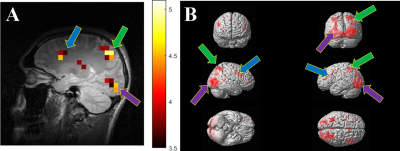 |
Hyperpolarized 129Xe functional brain mapping
Yurii Shepelytskyi1,2, Francis T Hane2,3, Vira Grynko1,2, Tao Li3, Ayman Hassan4, and Mitchell S Albert2,3,5
1Chemistry and Materials Science Program, Lakehead University, Thunder Bay, ON, Canada, 2Thunder Bay Regional Health Research Institute, Thunder Bay, ON, Canada, 3Chemistry, Lakehead University, Thunder Bay, ON, Canada, 4Thunder Bay Regional Health Science Centre, Thunder Bay, ON, Canada, 5Northern Ontario School of Medicine, Thunder Bay, ON, Canada
Functional magnetic resonance imaging (fMRI) localizes active regions of the brain during brain stimuli. In this work, we demonstrate hyperpolarized (HP) 129Xe fMRI in two classical fMRI experiments: a flashing visual stimulus and a fist-clenching motor stimulus. Using a chemical shift saturation recovery (CSSR) pulse sequence, our processed images localize brain activity to regions of the brain correlated to those identified using conventional Blood Oxygenation Level Dependent fMRI. The sensitivity of Xe fMRI was nearly two orders of magnitude greater than that of BOLD fMRI. In addition, 129Xe fMRI allows presenting stimuli with significantly smaller repetition frequencies.
|
0557.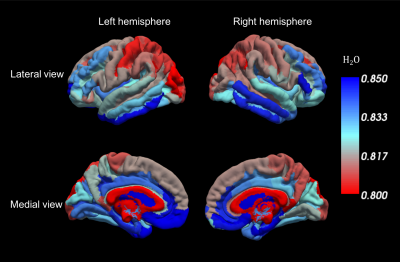 |
Water content for brain mapping at 7T: sub-mm resolution, sub-one percent precision
Ana-Maria Oros-Peusquens1, Ricardo Loução1, Monica Ferreira1, and N. Jon Shah1,2,3
1Research Centre Juelich, Juelich, Germany, 2Section JARA‑Brain, Jülich ‑Aachen Research Alliance (JARA), Aachen, Germany, 3Department of Neurology, RWTH Aachen University, Aachen, Germany
We present a method for high resolution, high precision measurements of water content in vivo, validated by comparison of the values obtained in the same brains at 3T and 7T. Applications relevant to brain structure and function are illustrated. The cortical distribution of water content simultaneously reflects its complement, the macromolecular content of tissue. Furthermore, a 3D “long TR” single-scan mapping method with 3deg excitation angle is proposed at 7T and delivers results consistent with the 2D method. Structural scans reflecting quantitative properties of tissue can thus be obtained in a short (7min or less) measurement time.
|
|
|
0558.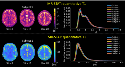 |
A five-minute multi-parametric high-resolution whole-brain MR-STAT exam: first results from a clinical trial
Stefano Mandija1,2, Federico D'Agata1,3, Hongyan Liu1,2, Oscar van der Heide1,2, Beyza Koktas2, Cornelis A.T. van den Berg1,2, Jeroen Hendrikse2, Anja van der Kolk2, and Alessandro Sbrizzi1,2
1Computational Imaging Group for MR diagnostic and therapy, Center for Image Sciences, University Medical Center Utrecht, Utrecht, Netherlands, 2Department of Radiology, University Medical Center Utrecht, Utrecht, Netherlands, 3Department of Neurosciences, University of Turin, Turin, Italy
MR-STAT is a recently developed technique which aims at reconstructing multi-parametric quantitative maps (T1, T2, PD, etc.) from a short cartesian acquisition. Previous research efforts have focused on the feasibility of the MR-STAT framework from a technical point of view. In this work, we present the implementation of a five-minute long high-resolution whole-brain MR-STAT protocol in a clinical trial and show the first results obtained from nine subjects. Synthetically generated contrast images as well as quantitative parametric maps show the robustness and the practical feasibility of the 5 minute long comprehensive MR-STAT protocol.
|
|
0559.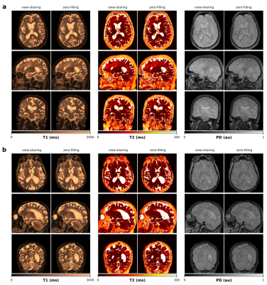 |
Accelerated 3D multiparametric MRI in glioma patients - Initial clinical experience
Carolin M Pirkl1,2, Laura Nuñez-Gonzalez3, Pedro A Gómez1, Sebastian Endt1,2, Rolf F Schulte2, Guido Buonincontri4,5, Marion Smits3, Bjoern H Menze1, Marion I Menzel2,6, and Juan A Hernandez-Tamames3
1Informatics, Technical University of Munich, Munich, Germany, 2GE Healthcare, Munich, Germany, 3Radiology & Nuclear Medicine, Erasmus MC, University Medical Center Rotterdam, Rotterdam, Netherlands, 4Fondazione Imago7, Pisa, Italy, 5IRCCS Fondazione Stella Maris, Pisa, Italy, 6Physics, Technical University of Munich, Munich, Germany
In brain tumor diagnosis, fully quantitative, multiparametric MRI offers great opportunities as it allows for comprehensive tissue and hence tumor characterization which is essential for treatment planning and monitoring the treatment response. With its highly accelerated acquisition, advanced rapid MR mapping techniques facilitate multiparametric imaging in clinically acceptable scan times, providing quantitative, reproducible and accurate diagnostic information that is less affected by system and interpretation biases. In this work, we present initial clinical results and demonstrate the feasibility of a novel 3D multiparametric quantitative transient-state imaging (QTI) acquisition scheme in glioma patients.
|
0560.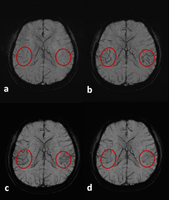 |
3D Flow Compensated Interleaved EPI with a Centric Reordering Scheme for Fast High-Resolution Susceptibility-Weighted Imaging
Wei Liu1 and Kun Zhou1
1Siemens Shenzhen Magnetic Resonance Ltd., Shenzhen, China In this study, we implemented a novel centric reordering scheme in a partial flow compensated 3D-iEPI to further reduce the flow effect and assessed its feasibility for a fast high-resolution SWI application. By properly dividing one interleave into two EPI shots sequentially acquired with opposite phase encoding gradient polarities and overlapping one line in the interleave center, we demonstrated that the partial flow compensated 3D-iEPI with such centric reordering scheme can significantly reduce the arterial contamination and obtain comparable contrast and image quality to 3D-GRE, whilst enjoying an approximate 2-fold reduction in acquisition time. |
|
 |
0561.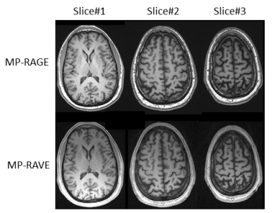 |
MP-RAVE: IR-Prepared T1-Weighted Radial Stack-of-Stars 3D GRE Imaging with Retrospective Motion Correction
Eddy Solomon1, Houchun H. Hu2, Kai Tobias Block1, Daniel K. Sodickson1, and Hersh Chandarana1
1Radiology, New York University School of Medicine, New York, NY, United States, 2Radiology, Nationwide Children's Hospital, Columbus, OH, United States
Inversion-recovery 3D T1 gradient echo sequences are commonly used in brain examinations for their excellent gray-/white-matter contrast. However, prominent motion artifacts can arise during lengthy Cartesian k-space sampling (typically 5-7 minutes) if the patient is not able to hold still, as is often the case for pediatric or elderly patients. Here, we present an alternative based on radial stack-of-stars imaging and show that comparable image contrast can be achieved, with lower sensitivity to head motion. Moreover, we demonstrate how the radial acquisition scheme can be utilized for additional retrospective motion correction to further improve robustness without increasing acquisition time.
|
|
0562.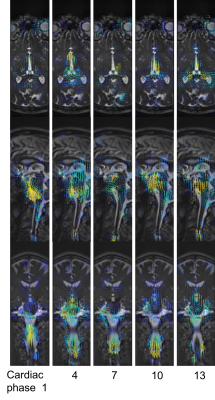 |
3D amplified MRI (aMRI) for visualizing pulsatile brain motion
Itamar Terem*1, Leo Dang*2, Allen Champagne3, Javid Abderezaei 4, Zainab Almadan 2, Anna-Maria Lydon 5, Mehmet Kurt4,6, Miriam Scadeng2,7,8, and Samantha J Holdsworth2,8
1Department of Electrical Engineering & Department of Structural Biology, Stanford University, Stanford, CA, United States, 2Department of Anatomy and Medical Imaging & Centre for Brain Research, University of Auckland, Auckland, New Zealand, 3Centre for Neuroscience Studies, Queen’s University, Kingston, ON, Canada, 4Department of Mechanical Engineering, Stevens Institute of Technology, Hoboken, NJ, United States, 5Centre for Advanced MRI, University of Auckland, Auckland, New Zealand, 6Translational and Molecular Imaging Institute, Icahn School of Medicine at Mount Sinai, New York, NY, United States, 7Department of Radiology, University of California, San Diego, CA, United States, 8Mātai Medical Research Institute, Gisborne-Tairāwhiti, New Zealand
Amplified Magnetic Resonance Imaging (aMRI) has been introduced as a new brain motion detection and visualization method. Originally employed to amplify pulsatile brain motion in 2D, aMRI has shown to be promising for differentiating abnormal from normal pulsatile brain motion in obstructive brain disorders. Here, we further improve aMRI with the introduction of a combined 3D aMRI acquisition and post-processing tool, with subsequent image processing with optical flow and strain mapping. The 3D aMRI tool is then tested on both multi-slice and volumetric data and its ability to capture 3D brain motion is analyzed.
|
|
0563.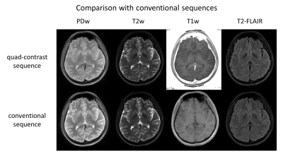 |
Quad-contrast imaging with quantitative relaxation maps for clinical neuro-evaluation
Sooyeon Ji1, Se-Hong Oh2, and Jongho Lee1
1Electrical and Computer Engineering, Seoul National University, Seoul, Korea, Republic of, 2Biomedical Engineering, Hankuk University of Foreign Studies, Yongin, Korea, Republic of
A 2D quad-contrast sequence is developed to generate four different contrast images commonly used in the clinical patient scan (PDw, T2w, T1w, and FLAIR) and two quantitative maps (T1- and T2- maps) in 4:14 of scan time. The proposed sequence provides comparable tissue contrasts to that of conventional sequences. In particular, native FLAIR contrast is acquired, which does not display hyperintense brain surface that arises from partial volume error during parameter mapping. Psuedo-contrast images with different TE and TI are also synthesized utilizing the quantitative maps, analogous to MAGiC.
|
0564.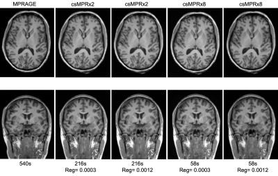 |
Optimizing rapid compressed-sensing MPRAGE acquisitions for repeat sampling of brain morphometry within individuals
Ross W. Mair1,2, Lindsay C. Hanford1,3, Emilie Mussard4,5,6, Tom Hilbert4,5,6, Tobias Kober4,5,6, and Randy L. Buckner1,2,3
1Center for Brain Science, Harvard University, Cambridge, MA, United States, 2Martinos Center for Biomedical Imaging, Massachusetts General Hospital, Charlestown, MA, United States, 3Department of Psychology, Harvard University, Cambridge, MA, United States, 4Advanced Clinical Imaging Technology, Siemens Healthcare, Lausanne, Switzerland, 5Department of Radiology, Lausanne University Hospital and University of Lausanne, Lausanne, Switzerland, 6LTS5, École Polytechnique Fédérale de Lausanne, Lausanne, Switzerland
Incoherent under-sampling and compressed-sensing reconstructions can reduce the scan time for a 1.0 mm MPRAGE down to as little as 60-90 seconds. Such time-savings permit the acquisition of multiple scans per session, allowing the variation of image metrics and brain morphometrics around their mean values to be quantified. We compared MPRAGE scans accelerated up to eight-fold with a fully-sampled MPRAGE; and using SNR, cortical thickness and gray matter volume, assessed the optimal regularization in the compressed-sensing reconstruction for each acceleration level to best match the values from the fully-sampled scan.
|
|
|
0565.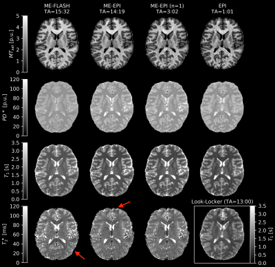 |
Fast Quantitative Multiparametric Mapping using 3D-EPI with Segmented CAIPIRINHA Sampling at 3T
Difei Wang1, Tony Stöcker1,2, and Rüdiger Stirnberg1
1German Center for Neurodegenerative Diseases (DZNE), Bonn, Germany, 2Department of Physics and Astronomy, University of Bonn, Bonn, Germany
By comparison to a gold standard multiparametric mapping (MPM) protocol at 3T, this study shows that multi-echo 3D-EPI with highly segmented CAIPIRINHA sampling can yield whole-head T1, PD*, MTsat and R2* maps of high quality at 1mm isotropic resolution in less than 3 minutes scan time. Even less than 1 minute of single-echo 3D-EPI is sufficient to yield accurate quantitative T1, PD* and MTsat maps. If necessary, SNR can be improved by including repeated EPI measurements. Optional motion- and distortion-correction across measurements may further improve results. Motion-robust MPM thus renders assessing quantitative parameter maps in clinical or population studies feasible.
|
 Watch the Video