
Scientific Session
Acquisition & Processing in Neuro
Session Topic: Acquisition & Processing in Neuro
Session Sub-Topic: Neuroimaging Techniques: Acquisition & Processing 1
Oral
Neuro
| Tuesday Parallel 2 Live Q&A | Tuesday, 11 August 2020, 14:30 - 15:15 UTC | Moderators: Jennifer McNab & Chantal Tax |
Session Number: O-11
 |
0547.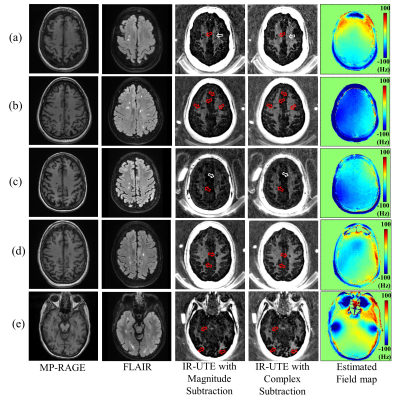 |
Improved Volumetric Myelin Imaging in Human Brain Utilizing Inversion Recovery Prepared Ultrashort Echo Time with Complex Echo Subtraction
Hyungseok Jang1, Zhao Wei1, Mei Wu1, Yajun Ma1, Eric Chang1,2, Jody Corey-Bloom1, and Jiang Du1
1University of California, San Diego, San Diego, CA, United States, 2VA San Diego Healthcare System, San Diego, CA, United States
Myelin accelerates neural signaling in the central and peripheral nervous systems. Ultrashort echo time (UTE)-based imaging techniques have been proposed for direct capture of magnetic resonance (MR) signal from myelin lipid protons with extremely short T2* (~0.3 ms). To suppress signal from long T2 water components and thereby improve myelin imaging, inversion recovery (IR)-based UTE techniques have been proposed. In this study, we explored the efficacy and feasibility of qualitative myelin imaging in vivo combining dual-echo IR-UTE with complex echo subtraction.
|
0548.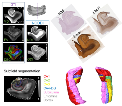 |
Post-mortem Diffusion MRI Analysis of Neuronal Pathways in the Human Hippocampus
Choong Heon Lee1, Jing Li2, Yulin Ge1, Timothy M Shepherd1, Youssef Zaim Wadghiri1, Jiangyang Zhang1, and David W Nauen3
Video Permission Withheld
1Radiology, New York University School of Medicine, New York, NY, United States, 2Peking Union Medical College Hospital, Beijing, China, 3Pathology, Johns Hopkins University School of Medicine, Baltimore, MD, United States
High-resolution diffusion MRI data of post-mortem adult human hippocampus specimens were acquired and compared to histology to identify major axonal pathways in the hippocampus. The complex microstructural organization in the hippocampus made it difficult to resolve axonal pathways based on conventional diffusion tensor data. In comparison, neurite density map using the NODDI toolbox revealed the locations of the perforant path, mossy fibers, and Schaffer collaterals confirmed by histology. We were able to reconstruct the fimbria/alveus and perforant pathways using tractography, and the results resembled in vivo results from the HCP dataset. Other pathways in the hippocampus remained difficult to delineate.
|
|
0549. |
Spatial matching of fiber orientation distribution functions (fODFs) & brain structure using fODF-based vs. tensor-based registration
Xiaoxiao Qi1, Yingjuan Wu1, Abdur Raquib Ridwan1, Shengwei Zhang1, Mohammad Rakeen Niaz1, and Konstantinos Arfanakis1,2
1Biomedical Engineering, Illinois Institute of Technology, Chicago, IL, United States, 2Rush Alzheimer’s Disease Center, Rush University Medical Center, Chicago, IL, United States
Group-wise spatial normalization of fiber orientation distribution functions (fODF) is an important step in fixel-based analysis. This work compared the accuracy in matching fODFs using fODF-based and tensor-based registration. It demonstrated superior fODF matching with fODF-based registration, as expected, even in conditions that are optimal for tensor but not fODF reconstruction. Nevertheless, it was shown that tensor-based registration has the ability to spatially match fODF features rather well, though less accurately than fODF-based registration. Finally, this work demonstrated that fODF-based transformations resulted in worse matching of structural information than tensor-based transformations.
|
|
0550.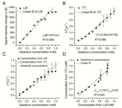 |
A real-time quantitative method of Gd-DTPA concentration in neuroimaging using T1 3D MP-RAGE sequence at 3.0T
Yumeng Cheng1,2,3, Hongbin Han1,2,3, Yajuan Gao2,3, Rui Wang2,3, Yu Song3, Xianjie Cai1,2,3, and Zeqing Tang1,2,3
1Institute of Medical Technology(IMT), Peking University Health Science Center(PKUHSC), Beijing, China, 2Department of Radiology, Peking University Third Hospital, Beijing, China, 3Key Laboratory of Magnetic Resonance Imaging Equipment and Technique, Beijing, China
We proposed a simple quantitative method based on the linear relationship between MR signal enhancement and Gd-DTPA concentration (C) by using T1 3D MP-RAGE for the real-time in vivo measurement of Gd-DTPA concentration in neuroimaging at 3.0 T. A good linear relationship between ΔSI and Gd-DTPA concentration existed over the concentration range of 0–1 mM (R2=0.985). Further, six human subjects with different brain tumors were enrolled for in vivo application of the novel method. All the results revealed that the quantitative method presented by our study is accurate, real-time and applicable.
|
|
0551.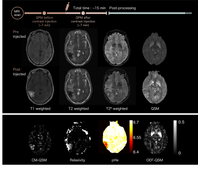 |
Calculation of Concentration of Contrast Media, Relaxivity, Extracellular pH and Oxygen Extraction Fraction for Brain Tumor Characterization
Yuki Matsumoto1, Masafumi Harada1, Yuki Kanazawa1, Takashi Abe1, Maki Otomo1, Yo Taniguchi2, Masaharu Ono3, and Yoshitaka Bito3
1Tokushima University, Tokushima, Japan, 2Research & Development Group, Hitachi, Ltd., Tokyo, Japan, 3Healthcare Business Unit, Hitachi, Ltd., Tokyo, Japan
Concentration of contrast agent (CM), relaxivity (r1), extracellular pH (pHe), and oxygen extraction fraction (OEF), maps were calculated for detecting changes in tissue environment of brain diseases. As a result, the pHe value on glioblastoma or brain metastasis region was significantly lower than that on radiation necrosis (see Fig.3; P < 0.001). The OEF value on glioblastoma region recorded significantly lower values than radiation necrosis and lung metastasis (P < 0.001) while there was no significant difference amongst glioblastoma, breast metastasis, and lung metastasis (P > 0.05).
|
|
|
0552.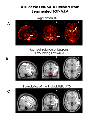 |
Downloadable Probabilistic Map of the Territorial Distribution of the Left – Middle Cerebral Artery Derived from TOF-MRA
Samantha Cote1, Jean-Francois Lepage2, and Kevin Whittingstall3
1Médecine Nucléaire et Radiobiologie, Université de Sherbrooke, Sherbrooke, QC, Canada, 2Pédiatrie, Université de Sherbrooke, Sherbrooke, QC, Canada, 3Radiologie Diagnostique, Université de Sherbrooke, Sherbrooke, QC, Canada
Standardized artery territorial distributions (ATD) are derived from variable post-mortem ATD yet assume a homogenous distribution. We developed a downloadable probabilistic territorial distribution of the left-MCA derived from Time-of-Flight Magnetic-Resonance-Angiography that can be used with other MRI modalities. We examined the probability of the arterial territory in Broca’s and Wernicke’s area and found it to be almost 3 times higher in Broca area than Wernicke’s area; however, both are traditionally believed to be supplied by the left-MCA. Combining the variability of arterial territories with functionally defined regions of interest can advance our knowledge of the consequences of cerebrovascular incidents.
|
 |
0553.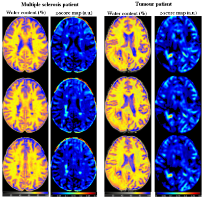 |
A novel MRI-based quantitative water content atlas of the human brain
N. Jon Shah1,2,3, Zaheer Abbas1, Dominik Ridder1, Markus Zimmermann1, and Ana-Maria Oros-Peusquens1
1Medical Imaging Physics, Institute of Neuroscience and Medicine 4, Jülich, Germany, 2Institute of Neuroscience and Medicine 11, INM 11, JARA, Jülich, Germany, 3Department of Neurology, Faculty of Medicine, Aachen, Germany
Measurement of quantitative, tissue-specific MR properties such as water content or relaxation times using quantitative-MRI at clinical field strength is a well-explored topic. However, none of the commonly used standard brain atlases, e.g., MNI or JHU, provide quantitative information. Utilising the framework of quantitative-MRI of the brain, this work reports on the development of the first quantitative in-vivo water content atlas based on twenty healthy volunteers datasets. Additionally, water content maps from patients with pathological changes in the brain were compared voxel-wise. These results suggest that quantitative-MRI in combination with water content atlas allows careful and quantitative interpretation of disease.
|
0554.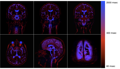 |
CSF Protein Cotent Estimation By T2 Component Analysis
Koichi Oshio1, Masao Yui2, Seiko Shimizu2, and Shinya Yamada3,4
1Department of Diagnostic Radiology, Keio University School of Medicine, Tokyo, Japan, 2Canon Medical Systems Corporation, Otawara-shi, Japan, 3Kugayama Hospital, Tokyo, Japan, 4Juntendo University, Tokyo, Japan
Although there is no lymphatic system in the CNS, there seems to be a mechanism to remove macro molecules from the brain. CSF and ISF are thought to be parts of this pathway, but the details are not known. In this study, MR signal of the extracellular water, including CSF, was decomposed into components with distinct T2’s, to estimate content of macromolecules in each compartment. Assuming that protein content is relatively high along the clearance pathway, it might be possible to have some insight about this pathway from the obtained T2 map.
|
|
0555.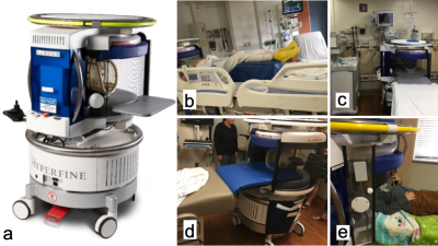 |
Use Environments and Clinical Feasibility of Portable Point-of-Care Bedside Brain MRI
E. Brian Welch1, Samantha By1, Gang Chen1, Hadrien Dyvorne1, Cedric Hugon1, Christopher McNulty1, Anne Nelson1, Rafael O'Halloran1, Michael Poole1, Laura Sacolick1, Nicholas Zwart1, Sean C.L. Deoni2, Joel M. Stein3,
Christopher Raio4, Kimon Bekelis5, Gerardo Chiricolo6, Kevin N. Sheth7, and Jonathan M. Rothberg1
1Hyperfine, Guilford, CT, United States, 2Advanced Baby Imaging Lab, Brown University School of Engineering, Providence, RI, United States, 3Department of Radiology, Hospital of the University of Pennsylvania, Philadelphia, PA, United States, 4Emergency Department, Good Samaritan Hospital Medical Center, West Islip, NY, United States, 5Department of Neurological Surgery, Good Samaritan Hospital Medical Center, West Islip, NY, United States, 6Department of Emergency Medicine, New York Presbyterian Brooklyn Methodist Hospital, Brooklyn, NY, United States, 7Department of Neurology, Yale University School of Medicine, New Haven, CT, United States
Using the world’s first truly portable point-of-care (POC) MRI scanner, it is possible to acquire the fundamental neuro MR imaging contrasts in settings such as the neuro intensive care unit, emergency department, outpatient clinic, and pediatric clinic. Results are presented of neuro MRI exams of children and adults (some with known pathology) using T1W, T2W, FLAIR, and DWI from a low-field portable MRI scanner that transports directly to the patient’s bedside.
|
 Watch the Video