
Scientific Session
Neurodegeneration 2
Session Topic: Neurodegeneration 2
Session Sub-Topic: Imaging & Spectroscopy of Traumatic Brain Injury
Oral
Neuro
| Wednesday Parallel 2 Live Q&A | Wednesday, 12 August 2020, 15:15 - 16:00 UTC | Moderators: Rebecca Feldman |
Session Number: O-12
 |
0919.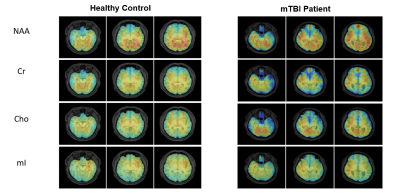 |
Altered neurometabolic changes in acute mild traumatic brain injury patients: a SPICE study
Tianyao Wang1, Jialin Hu2, Danni Wang2, Yujie Hu2, Jiahua Sun3, Jun Liu1, Yudu Li4,5, Rong Guo4,5, Yibo Zhao4,5, Ziyu Meng2,4, Zhipei Liang4,5, and Yao li2,6
1Radiology department, The Fifth People's Hospital of Shanghai, Shanghai, China, 2Institute for Medical Imaging Technology, School of Biomedical Engineering, Shanghai Jiao Tong University, Shanghai, China, 3Neurosurgery department, The Fifth People's Hospital of Shanghai, Shanghai, China, 4Beckman Institute for Advanced Science and Technology, University of Illinois at Urbana-Champaign, Urbana, IL, United States, 5Department of Electrical and Computer Engineering, University of Illinois at Urbana-Champaign, Urbana, IL, United States, 6Med-X Research Institute, Shanghai Jiao Tong University, Shanghai, China
Mild traumatic brain injury (mTBI) is the most prevalent form of brain injury but the underlying physiological mechanisms are still not fully understood. MRSI has long been recognized as a potentially powerful tool for detection of neurometabolic alterations induced by TBI but most existing studies are limited by low resolution. In this study, we used a 3D high-resolution MRSI technique, known as SPICE, to study neurometabolic alterations in acute mTBI patients. Our experimental results showed various metabolic changes in different areas of patients, which lay a foundation for further investigation to gain new insights into the pathophysiology underlying acute mTBI.
|
0920.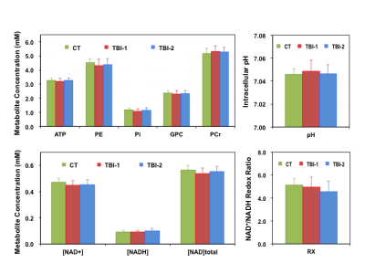 |
Quantitative 31P MRS Assessment of Neurometabolic Derangement in Pediatric Concussion
Xiao-Hong Zhu1, Byeong-Yeul Lee1, Katherine Ingram2, Wei Chen1, Robert Doss2,3, and Joseph Petronio2
Video Permission Withheld
1CMRR, Department of Radiology, University of Minnesota, Minneapolis, MN, United States, 2Children’s Minnesota Neuroscience Institute, St. Paul, MN, United States, 3Department of Neurology, University of Minnesota,, Minneapolis, MN, United States
Abnormal changes in brain metabolism and its role in pediatric concussion have not been well studied. We employed 31P MRS technique at 7T to assess the neurometabolic alteration in children with concussion. Phosphorous metabolites concentrations and other key physiological parameters were measured in patient and control cohorts. Metabolic differences between healthy and concussed brains were detected at two time points after the injury. We also found that mild head trauma reduced the age-dependences of high-energy phosphates and NAD contents in the developing brain, and it took much longer than clinically defined “recovery time” to fully restore such relationship.
|
|
 |
0921.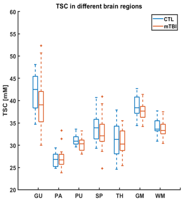 |
Quantitative 23Na MRI of mild traumatic brain injury: Initial findings
Teresa Gerhalter1, Rosemary Peralta1, Mickael Tordjman1, Julia Zabludovsky1, Seena Dehkharghani1, Alejandro Zarate2, Soo-Min Shin3, Ilya Aylyarov3, Tamara Bushnick2, Jonathan M. Silver4, Stephen P. Wall3, Brian S. Im2, Ryan Brown1,
Guillaume Madelin1, and Ivan I. Kirov1
1Center for Biomedical Imaging, Department of Radiology, New York University School of Medicine, New York, NY, United States, 2Department of Rehabilitation Medicine, New York University School of Medicine, New York, NY, United States, 3Ronald O. Perelman Department of Emergency Medicine, New York University School of Medicine, New York, NY, United States, 4Department of Psychiatry, New York University School of Medicine, New York, NY, United States
In this quantitative sodium MRI study, 24 mild traumatic brain injury (mTBI) and 9 controls were scanned at 3 T. Total sodium content (TSC) was calculated in five different subcortical regions, as well as in global grey and white matter. TSC in mTBI did not differ statistically from controls for the examined regions. Patients with findings on conventional 1H imaging (e.g. lesions, microhemorrhages) did not differ from patients without such findings in their TSC. More patients and controls are being recruited to strengthen the statistical power of these comparisons.
|
0922.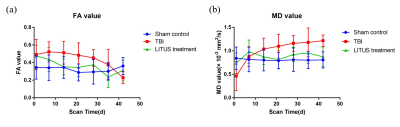 |
The usefulness of diffusion tensor imaging in evaluating the neuroprotective effect of LITUS to moderate traumatic brain injury with rat model
Zheng Tao1, Du Juan1, Yuan Yi2, Wu Shuo1, Wang Zhanqiu1, Liu Defeng1, Shi Qinglei3, Wang Xiaohan1, and Liu Lanxiang1
Video Permission Withheld
1MRI, Qinhuangdao Municipal No. 1 Hospital, Qinhuangdao, China, 2Institute of Electrical Engineering, Yanshan University, Qinhuangdao, China, 3Siemens Healthcare, MR Scientific Marketing, Beijing, China
In this study, we verified the feasibility of FA and MD values in evaluating the neuro therapeutic effect of LITUS with rat model. The neuro therapeutic effect of LITUS was attributed to promoting blood flow and the protein expression of BDNF.
|
|
0923.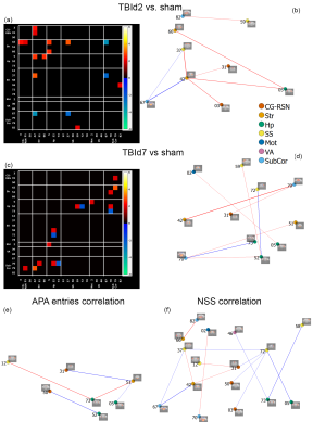 |
Repercussions of a single concussion in the mouse brain: insights from functional and structural Magnetic Resonance Imaging.
Xuan Vinh To1 and Fatima A. Nasrallah1,2
1The Queensland Brain Institute, The University of Queensland, Australia, Queensland, Australia, 2The Centre for Advanced Imaging, The University of Queensland, Australia, St Lucia, Australia
Resting-state functional connectivity in mouse model of concussion detected a process of functional adaptation at day 2 post-injury in compensation for white matter injuries: increased connectivity among the Default Mode and Hippocampal Networks and decreased or negative connectivity to the Midbrain. These adaptations maintained cognition and spatial learning but negatively affected the motor and balance functions. The functional adaptations were short-term: at day 7, increased cellularity were detected by Diffusion MRI in grey matter regions involved with day 2 functional adaptations.
|
|
0924.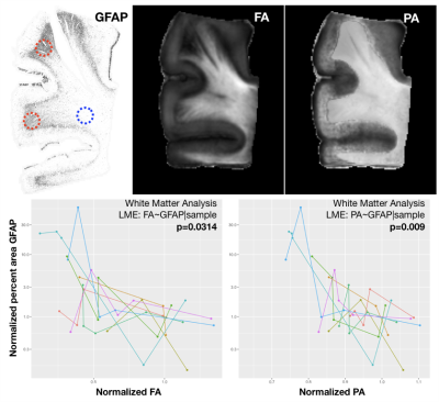 |
Correspondence of diffusion tensor and propagator metrics with quantitative histologic outcomes in chronic traumatic encephalopathy
Mihika Gangolli1, Elizabeth Hutchinson2, Ann McKee3, Joong Hee Kim1, Sinisa Pajevic1, and Peter Basser1
1National Institutes of Health, Bethesda, MD, United States, 2BME, University of Arizona, Tucson, AZ, United States, 3Boston University, Boston, MA, United States
Diffusion tensor and propagator metrics are compared directly in post-mortem cortex specimens from humans with chronic traumatic encephalopathy. Significant correlation was found between fractional anisotropy and non-Gaussianity with pTau staining in the sulcal depths. Additionally, GFAP staining of astrocytosis in the white matter was significantly correlated with Trace, FA, return-to-origin probability and propagator anisotropy. Cluster-based methods were also applied to explore the multivariate diffusion signature associated with CTE pathology. These findings suggest that diffusion metrics may be sensitive to CTE-related pathology.
|
|
| 0925. | Ultra-early versus early magnetic resonance imaging for mild traumatic brain injury: a CENTER-TBI Study
Sophie Richter1, Stefan Winzeck1, Evgenios Kornaropoulos 1, Marta Correia 2, Jan Verheyden3, Thijs Vande Vyvere 3, Guy Williams4, David Menon1, and Virginia Newcombe 1
1University Division of Anaesthesia, University of Cambridge, Cambridge, United Kingdom, 2MRC Cognition and Brain Sciences Unit, University of Cambridge, Cambridge, United Kingdom, 3Icometrix, Leuven, Belgium, 4Wolfson Brain Imaging Center, University of Cambridge, Cambridge, United Kingdom
Traumatic brain injury (TBI) is a major public health problem and is a leading cause of neurodisability. This study demonstrates the dynamic changes that occurs after mTBI as defined using conventional and advanced MRI including diffusion tensor imaging.
|
|
0926.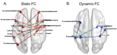 |
Abnormal static and dynamic functional connectivity in active professional fighters with repetitive head trauma: A resting-state fMRI study
Xiaowei Zhuang1, Virendra Mishra1, Zhengshi Yang1, Karthik Sreenivasan1, Sarah J Banks2, Lauren Bennett3, Bernick Charles1, and Dietmar Cordes1,4
1Lou Ruvo Center for Brain Health, Cleveland Clinic, Las Vegas, NV, United States, 2Department of Neuroscience, University of California, San Diego, La Jolla, CA, United States, 3Neuroscience Institute, Hoag Hospital, Irvine, CA, United States, 4University of Colorado, Boulder, Boulder, CO, United States
Both static and dynamic functional connectivity differences between cognitively impaired and non-impaired active professional fighters were first explored. Significant decreased static functional connections and trend-level increased dynamic functional connections among regions involved in memory and executive functions were found in cognitively impaired fighters, which adds brain functional reorganizations to previously observed structural damages in brain deficits related to repetitive head trauma. We further demonstrated that both static and dynamic functional connectivity were sensitive to cognitive declines in this fighter’s cohort, as both static and dynamic functional features can reliably predict cognitive impairment status in fighters.
|
|
|
0927.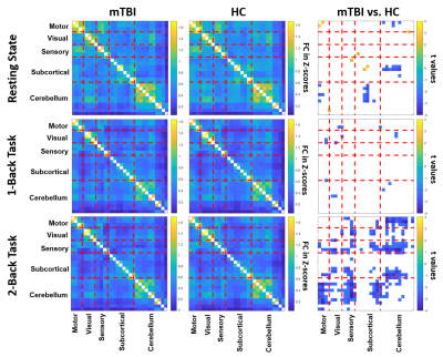 |
Association of Brain Functional Connectivity with Dizziness is Modulated by Executive Functions in Mild Traumatic Brain Injury
Jyun-Ru Chen1, Li-Chun Hsieh2,3,4, Cheng-Yu Chen2,3,4, and Chia-Feng Lu1
1Department of Biomedical Imaging and Radiological Sciences, National Yang Ming University, Taipei, Taiwan, 2Department of Medical Imaging, Taipei Medical University Hospital, Taipei, Taiwan, 3Department of Radiology, School of Medicine, College of Medicine, Taipei Medical University, Taipei, Taiwan, 4Translational Imaging Research Center, Taipei Medical University Hospital, Taipei, Taiwan
Dizziness is one of the frequent post-concussion symptoms, however neuroimaging evidence that supports symptom occurrence was less explored. This study started with the investigation of modulation effects from executive functions on functional connectivity (FC) between brain regions related to dizziness and balance followed by the correlation analysis to identify the imaging biomarker for elucidating the dizziness symptoms after mTBI.
|
 Watch the Video