
Scientific Session
Brain Tumors
Session Topic: Brain tumors
Session Sub-Topic: Brain Tumour: Metabolic & Biomarker Imaging
Oral
Neuro
| Tuesday Parallel 2 Live Q&A | Tuesday, 11 August 2020, 13:45 - 14:30 UTC | Moderators: Seung Hong Choi & Janine Lupo |
Session Number: O-13
 |
0398.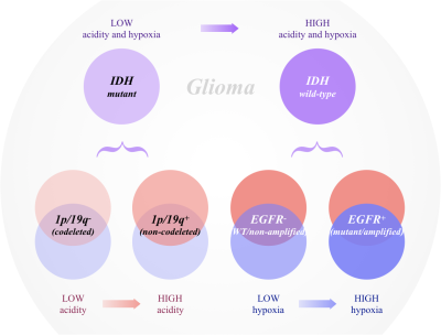 |
Metabolic characterization of human glioma subtypes using simultaneous pH- and oxygen-sensitive amine CEST-SAGE-EPI
Jingwen Yao1,2,3, Talia Oughourlian1,2,4, Timothy Cloughesy5,6, Phioanh L. Nghiemphu5,6, Albert Lai5,6, Linda M. Liau7, Richard G. Everson7, Whitney B. Pope2, Noriko Salamon2, David A. Nathanson8, and Benjamin M. Ellingson1,2,3,4,5
1Brain Tumor Imaging Laboratory (BTIL), Center of Computer Vision and Imaging Biomarker, David Geffen School of Medicine, UCLA, Los Angeles, CA, United States, 2Department of Radiological Sciences, David Geffen School of Medicine, UCLA, Los Angeles, CA, United States, 3Department of Bioengineering, Henry Samueli School of Engineering and Applied Science, UCLA, Los Angeles, CA, United States, 4Neuroscience Interdepartmental Program, David Geffen School of Medicine, UCLA, Los Angeles, CA, United States, 5UCLA Neuro-Oncology Program, David Geffen School of Medicine, UCLA, Los Angeles, CA, United States, 6Department of Neurology, David Geffen School of Medicine, UCLA, Los Angeles, CA, United States, 7Department of Neurosurgery, David Geffen School of Medicine, UCLA, Los Angeles, CA, United States, 8Department of Molecular and Medical Pharmacology, David Geffen School of Medicine, UCLA, Los Angeles, CA, United States
We simultaneously quantified acidity and hypoxia of human gliomas with IDH, 1p/19q, and EGFR genotypes using CEST-SAGE-EPI. Results suggest IDH mutant gliomas are significantly less acidic and hypoxic than IDH wild-type tumors. Within IDH mutants, 1p/19q codeletion is associated with lower tumor acidity, while IDH wild-type, EGFR amplified tumors were more hypoxic. Both MRI-derived acidity and hypoxia were correlated with patient survival, suggesting metabolic characteristics may be prognostic. CEST-SAGE-EPI may be useful for exploring metabolic changes that result from particular genetic alterations or useful as a biomarker for accelerating drug development in human brain tumors.
|
 |
0399.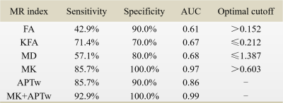 |
Discrimination between lower-grade glioma and glioblastoma with amide proton transfer and diffusion kurtosis imaging at 3 Tesla
Zongwei Xu1, Chao Ke2, Xiaofei Lv3, Jie Liu1, Long Qian4, Shijie Xu2, Xin Liu1, Hairong Zheng1, and Yin Wu1
1Paul C. Lauterbur Research Center for Biomedical Imaging, Shenzhen Institutes of Advanced Technology, Chinese Academy of Sciences, Shenzhen, China, 2Department of Neurosurgery, Sun Yat-Sen University Cancer Center, Guangzhou, China, 3Department of Medical Imaging, Sun Yat-Sen University Cancer Center, Guangzhou, China, 4GE Healthcare, Beijing, China
Preoperative assessment of histological tumor characteristics plays an essential role in evaluating prognosis and optimizing therapeutic strategies for glioma patients. This study aims to evaluate the feasibility of DKI and APT in differentiating lower-grade glioma (LGG) from glioblastoma at 3T. Twenty-four untreated patients were recruited and classified into LGG (grade II and III, N=10) and glioblastoma (grade VI, N=14). Results show comparable diagnostic performance of APTw and MK in differentiating the two groups with AUCs>0.85, superior to other DKI indices. Combining them further improves the discrimination accuracy, that may greatly facilitate prompt diagnosis and treatment decisions.
|
|
0400.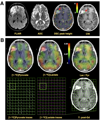 |
Initial experience: detection of aberrant HP-13C metabolism in patients with glioblastoma prior to resection
Adam Autry1, Jeremy Gordon1, Marisa LaFontaine1, Hsin-Yu Chen1, Javier Villanueva-Meyer1, Susan Chang2, Duan Xu1, Peder EZ Larson1, Daniel B Vigneron1, Jennifer Clarke2, Janine Lupo1, and Yan Li1
1Department of Radiology and Biomedical Imaging, University of California San Francisco, San Francisco, CA, United States, 2Department of Neurological Surgery, University of California San Francisco, San Francisco, CA, United States
Detection of [1-13C]pyruvate metabolism in GBM using hyperpolarized carbon-13 echo-planar imaging in a post-recurrence and pre-surgical setting
|
0401.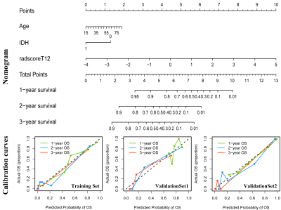 |
Radiomics profiling identifies the incremental value of MRI features to key molecular biomarkers for risk stratification of high-grade gliomas
Guoqiang Yang1, Shuaitong Zhang2, Xiaochun Wang1, Yan Tan1, Jingwei Wei2, Xiaoxu Chen3, Jie Tian2, and Hui Zhang1
1Department of Radiology, First Clinical Medical College, Shanxi Medical University, Taiyuan, Shanxi Province, China, 2Key Laboratory of Molecular Imaging, Institute of Automation, Chinese Academy of Sciences, Beijing, China, 3School of Economics and Management, Shanxi University, Taiyuan, Shanxi Province, China
To identify the incremental value of MRI features to the key molecular biomarkers for risk stratification of high-grade gliomas (HGGs). A comprehensive radiomics analysis integrated MRI features, clinical characteristics and genetic information was performed on 137 patients from TCGA/TCIA dataset and our institution. The combined model integrated radiomics signature with age and IDH genotype holds the best prognostic value. The radiomics signature has incremental prognostic value beyond the key molecular biomarkers, and could identify risk subgroups in various clinical and molecular subgroups. Our comprehensive radiomics analysis provided a potential tool to guide an individual diagnosis and treatment decisions for HGGs.
|
|
0402.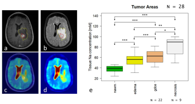 |
Sodium MRI at 7 Tesla as quantitative biomarker to assess tumor heterogeneity and histologic subtypes in glioma patients
Daniel Paech1, Sebastian Regnery2, Nicolas Behl3, Tanja Platt3, Nina Weinfurtner1, Mark Edward Ladd3, Jürgen Debus2, Sebastian Adeberg2, and Heinz-Peter Schlemmer1
1Radiology, German Cancer Research Center, Heidelberg, Germany, 2Radiooncology, University Hospital Heidelberg, Heidelberg, Germany, 3Medical Physics in Radiology, German Cancer Research Center, Heidelberg, Germany
23Na MRI provides information on physiologic and pathophysiologically altered tissue sodium concentrations in vivo. In this prospective trial, we investigated the potential of 23Na MRI at 7.0 Tesla to predict the tumor grade and genetic subtypes (such as isocitrate dehydrogenase (IDH) mutation and O6-methylguanine DNA methyltransferase (MGMT) promotor methylation) in a study cohort of 28 glioma patients. We show that that the quantitative 23Na signal correlates with tissue-specific tumor subcompartments and that the contrast may allow non-invasive assessment of the tumor grade and IDH mutation.
|
|
0403.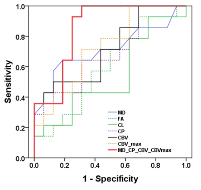 |
Differentiation of IDH Mutant from IDH wild-type High-grade Gliomas using Combined Analysis of Diffusion and Perfusion MRI
Santosh Kumar Yadav1, Sumei Wang2, Shadi Asadollahi2, MacLean Nasrallah3, Steven Brem4, Mohammad Haris1, Suyash Mohan2, and Sanjeev Chawla2
1Research, Sidra Medicine, Doha, Qatar, 2Department of Radiology, Perelman School of Medicine at the University of Pennsylvania, Philadelphia, PA, United States, 3Department of Pathology and Lab Medicine, Perelman School of Medicine at the University of Pennsylvania, Philadelphia, PA, United States, 4Department of Neurosurgery, Perelman School of Medicine at the University of Pennsylvania, Philadelphia, PA, United States
Accurate identification of isocitrate dehydrogenase (IDH) mutant high-grade glioma is clinically important. We investigated the combined utility of diffusion (DTI) and perfusion (DSC-PWI) MR imaging in distinguishing IDH mutant from IDH wild-type high-grade gliomas. Treatment naïve patients (n=30) with IDH-mutant (n=14) and IDH-wild-type (n=16) high-grade gliomas were recruited. A classification model comprising of mean diffusivity, coefficient of planar anisotropy and maximum relative cerebral blood volume differentiated two genotypes of gliomas with an accuracy of 85%, a sensitivity of 87.2%, and a specificity of 81.5%. Combined analysis of DTI and DSC-PWI may be helpful in distinguishing IDH profiles of high-grade gliomas.
|
|
0404.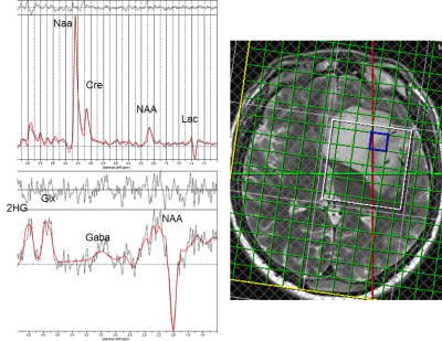 |
Glioma 2HG threshold setting based on normal appearing white matter increases the diagnostic value of 3D MEGA-LASER for IDH mutation detection
Marzena Wylezinska-Arridge1,2, Enrico De Vita2,3, Laura Mancini1,2, Ovidiu Andronesi4, Wolfgang Bogner5, Bernhard Strasser4, Tarek Yousry1,2, John Thornton1,2, and Sotirios Bisdas1,2
1Department of Neuroradiology, National Hospital for Neurology and Neurosurgery, London, United Kingdom, 2Institute of Neurology, University College London, London, United Kingdom, 3Department of Biomedical Engineering. School of Biomedical Engineering and Imaging Sciences, Kings College London, London, United Kingdom, 4Athinoula A. Martinos Center for Biomedical Imaging, Massachusetts General Hospital, Harvard Medical School, Boston, MA, United States, 5High Field MR Centre, Department of Biomedical Imaging and Image-guided Therapy, Medical University of Vienna, Vienna, Austria
It has been established that 2-hydroxyglutarate (2GH) MRS is important for non-invasive diagnosis of isocitrate dehydrogenase (IDH) status that holds prognostic value for the patient and is important for treatment planning. The purpose of this work was to investigate whether the threshold determination for identification of IDH mutation could be improved by using ‘control’ spectra from normal-appearing white matter, NAWM , in multi-voxel 3D MEGA-LASER acquisitions. The proposed approaches to threshold determination for 2HG detection in the tumour voxels provided increased sensitivity and specificity as compared with the cut-off thresholds based on CRLB% relative error.
|
|
0405.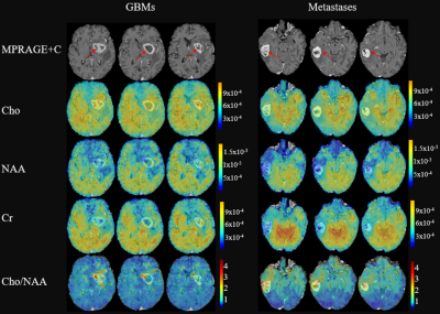 |
Differentiation between Glioblastomas and Cerebral Metastases using High-Resolution 3D MRSI
Pengcheng Yu1, Tianyao Wang2, Yujie Hu1, Yudu Li3,4, Rong Guo3,4, Yibo Zhao3,4, Ziyu Meng1,3, Hong Zhu5, Jun Liu6, Xin Yu7, Zhi-Pei Liang3,4, and Yao Li1
1Institute for Medical Imaging Technology, School of Biomedical Engineering, Shanghai Jiao Tong University, Shanghai, China, 2Radiology Department, The Fifth People's Hospital of Shanghai, Fudan University, Shanghai, China, 3Beckman Institute for Advanced Science and Technology, University of Illinois at Urbana-Champaign, Urbana, IL, United States, 4Department of Electrical and Computer Engineering, University of Illinois at Urbana-Champaign, Urbana, IL, United States, 5Department of Radiation Oncology, Minhang Branch of Cancer Hospital, Fudan University, Shanghai, China, 6Radiology department, The Fifth People's Hospital of Shanghai, Fudan University, Shanghai, China, 7Department of Biomedical Engineering, Case Western Reserve University, Cleveland, OH, United States Differentiation between glioblastomas (GBMs) and cerebral metastases based on MR structural images is often challenging due to the poor specificity. MRSI is a useful tool for mapping the metabolic fingerprints of tumors. In this study, we investigate the use of a high-resolution MRSI technique known as SPICE for differentiation between GBMs and cerebral metastases in 28 patients. Our results show the metabolic biomarkers are different between GBMs and metastases as well as in the enhancing ring and the core of neoplasms. SPICE potentially provides a non-invasive metabolic measure to differentiate GBMs from cerebral metastases with high resolution. |
|
0406.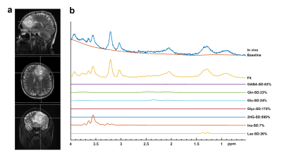 |
The Effect of Tumor Grade within IDH Wild-Type and IDH Mutant Gliomas Assessed by Proton Magnetic Resonance Spectroscopy at 3T
Esin Ozturk-Isik1,2, Banu Sacli Bilmez1, Ayca Ersen Danyeli2,3, Cengiz Yakicier4, Alpay Ozcan2,5,6, M. Necmettin Pamir2,5,7, Koray Ozduman2,5,7, and Alp Dincer2,5,8
1Institute of Biomedical Engineering, Bogaziçi University, Istanbul, Turkey, 2Brain Tumor Research Group, Acibadem Mehmet Ali Aydinlar University, Istanbul, Turkey, 3Department of Pathology, Acibadem Mehmet Ali Aydinlar University, Istanbul, Turkey, 4Department of Molecular Biology and Genetics, Acibadem Mehmet Ali Aydinlar University, Istanbul, Turkey, 5Center for Neuroradiological Applications and Research, Acibadem Mehmet Ali Aydinlar University, Istanbul, Turkey, 6Department of Medical Device Technologies, Acibadem Mehmet Ali Aydinlar University, Istanbul, Turkey, 7Department of Neurosurgery, Acibadem Mehmet Ali Aydinlar University, Istanbul, Turkey, 8Department of Radiology, Acibadem Mehmet Ali Aydinlar University, Istanbul, Turkey
Isocitrate dehydrogenase (IDH) mutation highly affects the overall survival of gliomas. In addition to the IDH mutation, the tumor histologic grade might play a role in patient prognosis. The aim of this study was to assess the metabolic variations between different tumor grades within IDH mutant (IDH-mut) and IDH wild-type (IDH-wt) gliomas using proton MR spectroscopy. Higher glycine in glioblastoma (GBM) within IDH-mut, and lower Cr and mIns in GBM within IDH-wt were the only statistically significant differences. Our study indicated similar metabolic profiles of different grades within IDH mutational subgroups, supporting IDH as a better predictor of clinical outcome.
|
|
0407.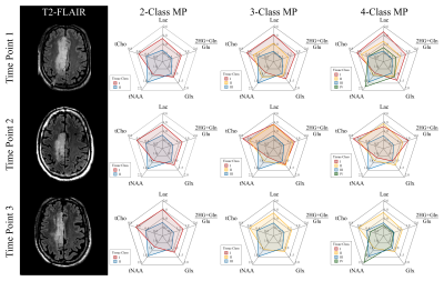 |
Long‑Term Monitoring of Brain Tumors Using MR Metabolic Phenotyping
Eduardo Coello1, Raymond Huang1, Molly F. Charney1, Wufan Zhao1, Huijun Liao1, Changho Choi2, and Alexander Lin1
1Radiology, Brigham and Women's Hospital, Boston, MA, United States, 2Radiology, University of Texas Southwestern Medical Center, Dallas, TX, United States
This work introduces the concept of MR metabolic phenotyping (MRMP), a method that combines the high-resolution anatomical context of MRI and the highly specific metabolic information of MR spectroscopic imaging via unsupervised learning. The value of this technique was shown for the long‑term follow up of IDH‑mutated low‑grade gliomas where high sensitivity to changes in the metabolic composition of the major tissue compartments was achieved.
|
 Watch the Video