
Scientific Session
Brain Tumors
Session Topic: Brain tumors
Session Sub-Topic: Brain Tumour: Quantitative MR Imaging
Oral
Neuro
| Tuesday Parallel 2 Live Q&A | Tuesday, 11 August 2020, 13:45 - 14:30 UTC | Moderators: Kader Oguz & Koji Sakai |
Session Number: O-14
0408.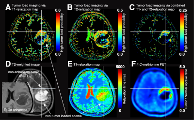 |
Stereotactic tissue sampling and T1-, T2-relaxometry compared with ADC for tissue cell density quantification in gliomas imaging
Manabu Kinoshita1,2, Masato Uchikoshi3, Souichiro Tateishi2, Shohei Miyazaki2, Mio Sakai2, Tomohiko Ozaki2, Katsunori Aasai2, Yuya Fujita2, Takahiro Matsuhashi2, Yonehiro Kanemura4, Eku Shimosegawa1, Jun Hatazawa1, Shin-ichi Nakatsuka2,
Haruhiko Kishima1, and Katsuyuki Nakanishi2
1Osaka University Graduate School of Medicine, Suita, Japan, 2Osaka International Cancer Institute, Osaka, Japan, 3Canon Medical Systems Corporation, Tochigi, Japan, 4National Hospital Organization Osaka National Hospital, Osaka, Japan
The authors attempted to elucidate the correlation of tumor cell density within the brain in glioma patients and T1-, T2-relaxometry and ADC. First, an exploratory study compared T1-, T2-relaxometry, and ADC with 11C-methionine PET followed by a validation study using intraoperative stereo-tactically obtained tissues. A range of T1 values indicative of high cell density was identified, which finding was confirmed by stereotactic tissue sampling. For T2 values and ADC, however, no statistically significant correlation was confirmed regarding tumor cell density. The proposed technique was further able to create predictive tumor cell density map by T1 supplemented by T2 values.
|
|
0409.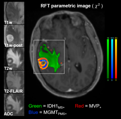 |
Statistical multiscale mapping of IDH1, MGMT, and microvascularity in human brain tumors from multiparametric MR and registered core biopsy
Jason Glenn Parker1, Emily E Diller2, Sha Cao3, Jeremy T Nelson4, Kristen Yeom5, Chang Ho1, and Robert Lober6
1Radiology & Imaging Sciences, Indiana University School of Medicine, Indianapolis, IN, United States, 2School of Health Sciences, Purdue University, West Lafayette, IN, United States, 3Biostatistics, Indiana University School of Medicine, Indianapolis, IN, United States, 4Military Health Institute, University of Texas Health San Antonio, San Antonio, TX, United States, 5Neuroradiology, Lucile Salter Packard Children’s Hospital and Stanford University Medical Center, Palo Alto, CA, United States, 6Neurosurgery, Dayton Children's Hospital, Dayton, OH, United States
We demonstrate statistical relationships between routine multiparametric imaging signatures and underlying cellular and molecular properties of brain tumors. We apply advanced statistical methods to correct for the family-wise error rate problem associated with whole-brain statistical parametric mapping, and show that the results have strong agreement with surgical biopsy. These results imply that cellular and molecular mapping of tumor heterogeneity from minimally-invasive images may be possible in the near future.
|
|
0410.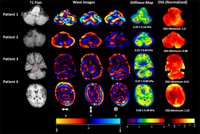 |
Preoperative assessment of stiffness and tumor-brain adhesion in schwannoma and meningioma patients and its comparison with surgical findings
Prateek Kalra1, Varun Varadarajan 2, Michael S Harris3, Ashonti Harper3, Omar Mohamed3, Oliver Adunka2, Daniel M. Prevedello4, and Arunark Kolipaka1
1Radiology, Ohio State University Wexner Medical Center, Columbus, OH, United States, 2Otolaryngology, Ohio State University Wexner Medical Center, Columbus, OH, United States, 3Ohio State University Wexner Medical Center, Columbus, OH, United States, 4Neurological Surgery, Ohio State University Wexner Medical Center, Columbus, OH, United States
Microsurgery in brain tumor patients aim to complete tumor resection without compromising neurological functionality. Inadequate preoperative knowledge of tumor may prolong surgical time and increase risk of postoperative complications which may depends upon tumor-brain adhesion and tumor stiffness. Previous studies have only looked into adhesion and stiffness separately using Magnetic Resonance Elastography (MRE). Previously, we proposed both adhesion and stiffness in vestibular schwannoma patients. Aim of this study is to also include meningioma cases in the analysis in order to broaden the tumor types and complexity. Preliminary results show good correlation between MRE-derived preoperative assessment of tumor and surgical findings.
|
|
 |
0411.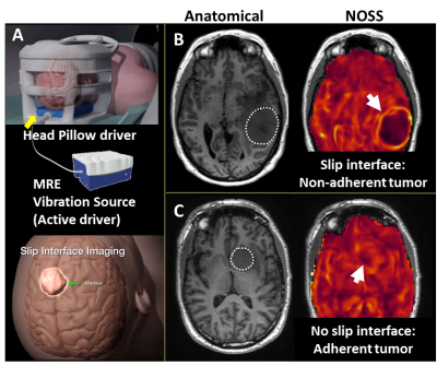 |
Quantification of meningioma-brain adhesion using MR-elastography based slip interface imaging
Ziying Yin1, Xin Lu1, Salomon Cohen Cohen2, Yi Sui1, Armando Manduca3, Jamie J Van Gompel2, Richard L. Ehman1, and John III Huston1
1Radiology, Mayo Clinic, Rochester, MN, United States, 2Neurosurgery, Mayo Clinic, Rochester, MN, United States, 3Physiology and Biomedical Engineering, Mayo Clinic, Rochester, MN, United States
Brain tumor adherence has been long recognized to impact surgical resection difficulty. Recently-developed slip interface imaging (SII) can preoperatively predict tumor-brain adhesion. In previous studies, subjectively-determined SII assessment of tumor adhesion has been shown to agree well with intraoperative findings. The purpose of this work was to develop an objective quantitative method for analyzing SII data for adherence, thereby minimizing inter- and intraobserver variability. We developed a radiomics-based metric (termed “adhesion degree”) based on SII to quantify the degree of tumor adhesion. In 46 meningiomas, the adhesion degree showed excellent accuracy in predicting completely adherent tumors (AUROC=0.96) from non-adherent tumors.
|
0412.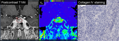 |
Pharmacokinetic analysis of DCE-MRI in pituitary adenoma: evaluation of tumor consistency and comparison with histological collagen content
Kiyohisa Kamimura1, Masanori Nakajo1, Tomohide Yoneyama1, Manisha Bohara1, Yoshihiko Fukukura1, Shingo Fujio2, Takashi Iwanaga3, Hiroshi Imai4, Marcel Dominik Nickel5, and Takashi Yoshiura1
1Radiology, Kagoshima University Graduate School of Medical and Dental Sciences, Kagoshima, Japan, 2Neurosurgery, Kagoshima University Graduate School of Medical and Dental Sciences, Kagoshima, Japan, 3Clinical Engineering Department Radiation Sectio, Kagoshima University Hospital, Kagoshima, Japan, 4Siemens Healthcare K.K., Tokyo, Japan, 5Siemens Healthcare, Erlangen, Germany
Preoperative information of tumor consistency is important in patients with pituitary adenoma. Our aim was to evaluate the possible role of high-temporal and spatial resolution dynamic contrast enhanced MR imaging (DCE-MRI) and quantitative pharmacokinetic analysis in differentiation of a hard adenoma from soft adenoma. The hard adenoma showed significantly higher extravascular extracellular space per unit volume of tissue (ve) than soft adenoma and the ve was significantly correlated to collagen IV content of pituitary adenomas. This quantitative pharmacokinetic parameter ve may be useful for differentiation of the hard adenoma from soft adenoma.
|
|
|
0413.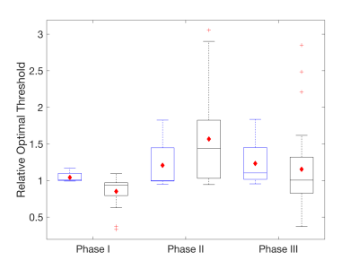 |
Evaluating the use of rCBV as a tumor grade classifier across NCI Quantitative Imaging Network sites: Part II of the DSC-MRI DRO Challenge
Laura C. Bell1, Natenael B. Semmineh1, Leland S. Hu2, Yuxiang Zhou2, Melissa Prah3, Kathleen M. Schmainda3, Jerrold L. Boxerman4, Hongyu An5, Cihat Eldeniz5, Richard Wahl5, Bradley Erickson6, Panagiotis Korfiatis6, Chengyue Wu7,
Thomas Yankeelov7, Anna Sorace7, Neal Rutledge7, Thomas Chenevert8, Dariya Malyarenko8, Yichu Liu9, Andrew Brenner9, Yi-Fen Yen10, Jayashree Kalpathy-Cramer10, Andrew Beers10, Mark Muzi11, Ananth J. Madhuranthakam12, Marco Pinho12,
Brian Johnson12, and C. Chad Quarles1
1Barrow Neurological Institute, Phoenix, AZ, United States, 2Mayo Clinic, Scottsdale, AZ, United States, 3Medical College of Wisconsin53226, Milwaukee, WI, United States, 4Rhode Island Hospital and Alpert Medical School of Brown University, Providence, RI, United States, 5Washington University in St. Louis, St. Louis, MO, United States, 6Mayo Clinic, Rochester, MN, United States, 7The University of Texas at Austin, Austin, TX, United States, 8University of Michigan, Ann Arbor, MI, United States, 9University of Texas Health Science Center at San Antonio, San Antonio, TX, United States, 10Massachusetts General Hospital, Boston, MA, United States, 11University of Washington, Seattle, WA, United States, 12The University of Texas Southwestern, Dallas, TX, United States
Using a dynamic susceptibility contrast (DSC) MRI DRO we previously characterized brain tumor relative cerebral blood volume (rCBV) reproducibility across 12 sites employing a range of imaging protocols and software platforms. Our goal in this study is to determine the impact of rCBV reproducibility for tumor grade classification. We found that varying software platforms produced a range of optimal thresholds, but the performance of these thresholds were similar. These results indicate that different software platforms are able to classify tumor grades, but the site-specific thresholds underscore the importance of standardizing acquisition and analysis protocols across sites and software benchmarking.
|
0414.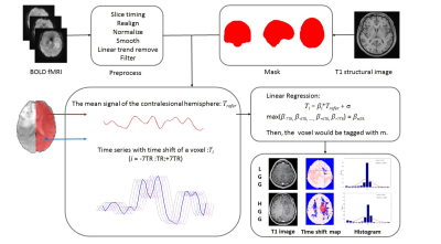 |
Altered Systemic Fluctuations of Blood Flow in Brains with Glioma: An Investigation with Temporal-Shift Resting-State fMRI
Siqi Cai1, Zhifeng Shi2, Shihui Zhou1,3, Chunxiang Jiang1,3, and Lijuan Zhang1
1Shenzhen Institutes of Advanced Technology, Chinese Academy of Sciences, Shenzhen, China, 2Huashan Hospital of Fudan University, Shanghai, China, 3University of Chinese Academy of Science, Beijing, China
The systemic oscillation of the blood flow in cerebrum and cerebellum may vary with gliomas of different malignancy, resulting in neurovascular uncoupling and abnormal perfusion. In this study, we investigated the implication of glioma on the oscillation of cerebral blood flow based on time-shifted rs-fMRI. HGGs induces more widely alterations in the spontaneous fluctuations of cerebral and cerebellar blood flow at the global scale. Vascular oscillation changes derived from rs-fMRI may provide a novel insight for the assessment of the functional plasticity and its clinical relevance in the interpretation of the psychological and psychiatric symptoms in subjects with glioma.
|
|
0415. |
Multimodal MRI to aid prediction of low-grade glioma growth characteristics
Franklyn Howe1, Timothy Jones2, Philip Rich2, Jordan Colman3, Guang Yang4, Felix Raschke5, Venus Liang1, Alex Denley1, and Thomas Barrick1
1Neurosciences Research Centre, St George's, University of London, London, United Kingdom, 2St George's University Hospitals NHS Foundation Trust, London, United Kingdom, 3Ashford and St Peter's Hospitals NHS Foundation Trust, Surrey, United Kingdom, 4National Lund and Heart Institute, Imperial College, London, United Kingdom, 5OncoRay—National Center for Radiation Research in Oncology, Dresden, Germany
1H MRS and DTI measures were assessed for their ability to predict the future growth and malignant transformation of low-grade gliomas. The tumour core NAA concentration and the mean diffusivity (MD) within the MRS voxel, combined with the FLAIR tumour volume, provided a good predictor of tumours with higher growth rates. A ROC analysis gave an AUC of 0.86 to predict tumours likely to undergo malignant progression, and AUC of 0.98 when including those undergoing early debulking. The combined NAA, MD and volumetric parameter provided a single time-point assessment of future growth characteristics.
|
|
0416.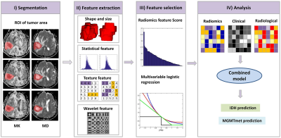 |
Whole-tumor radiomics analysis of DKI and DTI may improve the prediction of genotypes for astrocytomas: a preliminary study
Yan Tan1,2, Wei Mu3, Xiaochun Wang1,2, Guoqiang Yang1,2, Robert James Gillies3, and Hui Zhang1,2
1Department of Radiology, First Hospital of Shanxi Medical University, Taiyuan, China, 2College of Medical Imaging, Shanxi Medical University, Taiyuan, China, 3Department of Cancer Physiology, H. Lee Moffitt Cancer Center and Research Institute, Tampa, FL, United States
Assessment of glioma genotypes by quantifying MR diffusion imaging heterogeneity of whole tumour may serve as a powerful tool to instruct therapeutic decision-making. This study evaluated the role and incremental value of whole-tumor radiomics analysis based on DKI and DTI images in determining the IDH and MGMTmet genotypes of astrocytomas. A radiomics models based on whole-tumor MK and MD maps showed good diagnostic efficiency in predicting IDH and MGMTmet genotypes. Furthermore, the combined model constructed by radiomics score, edema degree and age further improved the performance of predicting IDH, while the combined model did not benefit for MGMTmet prediction.
|
|
0417.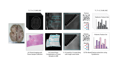 |
Information-based assessment of the radiomic-histomic relationship in brain cancer patients
Samuel Bobholz1, Allison Lowman2, Alexander Barrington3, Michael Brehler2, Sean McGarry1, Jennifer Connelly4, Elizabeth Cochran5, Anjishnu Banerjee6, and Peter LaViolette2,3
1Biophysics, Medical College of Wisconsin, Wauwatosa, WI, United States, 2Radiology, Medical College of Wisconsin, Wauwatosa, WI, United States, 3Biomedical Engineering, Medical College of Wisconsin, Wauwatosa, WI, United States, 4Neurology, Medical College of Wisconsin, Wauwatosa, WI, United States, 5Pathology, Medical College of Wisconsin, Wauwatosa, WI, United States, 6Biostatistics, Medical College of Wisconsin, Wauwatosa, WI, United States
This study sought to provide a biological basis of radiomics-based analyses by assessing the relationship between MR features and analogous histomic features of the underlying tissue using coregistered histology samples taken at autopsy from brain cancer patients. Several radiomic features demonstrated substantial mutual information with their histomic analogs, with first order features showing the strongest associations. These histomic-preserving features were shown to be stable across potential confounds such as differences in scanner vender and acquisition field strength. These findings suggest that MR radiomic features reflect information about the texture of the underlying tissue.
|
 Watch the Video