
Scientific Session
Neurovascular Imaging
Session Topic: Neurovascular imaging
Session Sub-Topic: Beyond the Lumen: Vessel Wall Imaging in Cerebrovascular Diseases
Oral
Neuro
| Thursday Parallel 2 Live Q&A | Thursday, 13 August 2020, 14:20 - 15:05 UTC | Moderators: Jae Song & Lena Vaclavu |
Session Number: O-16
1061.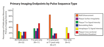 |
Imaging Endpoints by Pulse Sequence Type for Intracranial Atherosclerosis using Vessel Wall MR Imaging
Jae W Song1, Athanasios Pavlou1, Jiayu Xiao2, Steven R Messe3, Scott E Kasner3, Zhaoyang Fan2, and Laurie A Loevner1
1Radiology, University of Pennsylvania, Philadelphia, PA, United States, 2Radiology, Cedars Sinai Medical Center, Los Angeles, CA, United States, 3Neurology, University of Pennsylvania, Philadelphia, PA, United States
Intracranial atherosclerosis is a common cause of ischemic stroke. Variability in protocol/pulse sequence design of intracranial vessel wall MR imaging (VWI) has led to different imaging endpoints to detect and characterize atherosclerosis. We systematically reviewed the literature to identify VWI investigations studying atherosclerosis to identify commonly reported imaging endpoints. The most common imaging endpoints using T1-weighting included wall enhancement, thickening, plaque quadrant in cross-section, and stenosis; on T2-weighting, intraplaque T2 signal intensity and wall thickening were common endpoints. Establishing diagnostically accurate imaging endpoints to validate as atherosclerosis biomarkers are critical to understand where efforts for technique optimization should be directed.
|
|
 |
1062.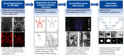 |
Automated Morphology Analysis of Intracranial and Extracranial Vessel Wall Using Convolutional Neural Network
Liwen Wan1, Na Zhang1, Lei Zhang1, Shi Su1, Cheng Wang1, Baochang Zhang1, Hao Peng1, Haoxiang Li1, Dong Liang1, Xin Liu1, and Hairong Zheng1
1Shenzhen Institutes of Advanced Technology, Chinese Academy of Sciences, Shenzhen, China
Intracranial and extracranial atherosclerotic disease are major causes of ischemic stroke. Manual analyses of intracranial and extracranial artery vessel wall are time consuming and experience dependent. The purpose of this study was to develop an automated method to analyze 3D intra- and extracranial arterial vessel wall images, including vessel centerline tracking, vessel straightened reformation, vessel wall segmentation based on CNN, and morphological quantification. In conclusion, the proposed method facilitates the large-scale quantitative analysis of vessel wall, and is promising in promoting the clinical applications of MR vessel wall imaging.
|
1063.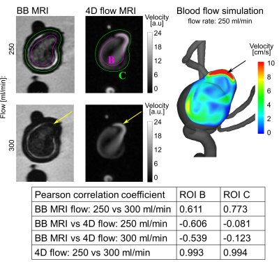 |
Investigation of black blood MRI signal enhancement in a patient-specific aneurysm model.
Mariya Stanislavovna Pravdivtseva1, Carson Hoffman2, Leonardo A. Rivera-Rivera2, Rafael Medero2, Lindsay Bodart2, Alejandro Roldan-Alzate2, Michael A. Speidel2, Charles M. Strother2, Kevin M. Johnson2, Oliver Wieben2, Olav Jansen3, Naomi Larsen3, Philipp Berg4,
Eva Peschke1, and Jan-Bernd Hövener1
1Neuroradiology and Radiology, Section Biomedical Imaging, Molecular Imaging North Competence Center (MOIN CC), Department of Radiology and Neuroradiology, University Medical Center Schleswig-Holstein (UKSH), Kiel University, Kiel, Germany, 2Department of Medical Physics, University of Wisconsin School of Medicine and Public Health, Madison, Wisconsin, USA, Madison, WI, United States, 3Neuroradiology and Radiology, Department of Radiology and Neuroradiology, University Medical Center Schleswig-Holstein (UKSH), Kiel, Germany, 4Research Campus STIMULATE, University of Magdeburg, Magdeburg, Germany, Magderburg, Germany
Intracranial aneurysm is a life-threatening disease. Vessel wall enhancement may be used as a marker to identify an aneurysm with a high risk of rupture. Accumulation of contrast agent in the vessel wall and slow or turbulent flow can contribute to the formation of vessel wall enhancement. In the current study enhanced signal on black blood MRI was observed in printed model of an intracranial aneurysm with and without Gd administration. The found signal was associated with the slow flow in the aneurysm. Additionally, the impact of spatial resolution, flow rate, MSDE preparation and contrast concentration was considered.
|
|
 |
1064.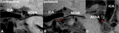 |
Atherosclerotic Plaques on Perforating Arteries Can be Detected by Vessel Wall Imaging at 7T in Patients with Single Subcortical Infarction
Qingle Kong1,2,3, Haiqiang Qin4, Ning Wei5, Jing An6, Yan Zhuo1,2,3, and Zihao Zhang1,2,3
1State Key Laboratory of Brain and Cognitive Science, Beijing MR Center for Brain Research, Institute of Biophysics, Chinese Academy of Sciences, Beijing, China, 2University of Chinese Academy of Sciences, Beijing, China, 3CAS Center for Excellence in Brain Science and Intelligence Technology, Beijing, China, 4Department of neurology, Beijing Tiantan Hospital, Capital Medical University, Beijing, China, 5China National Clinical Research Center for Neurological Diseases, Beijing Tiantan Hospital, Capital Medical University, Beijing, China, 6Siemens Shenzhen Magnetic Resonance Ltd., Shenzhen, China
Branch atheromatous disease (BAD) refers to small, deep brain infarcts that are predominantly caused by the occlusion of perforating arteries, which may lead to single subcortical infarction (SSI). However, there is no in-vivo radiological evidence of plaques in the perforating arteries due to their small caliber. In this study, we used high-resolution black-blood imaging at 7T to display the vessel wall of the anterior choroidal artery (AChA), and analyzed atherosclerotic plaques of AChA in patients with isolated infarcts on the posterior limb of internal capsule. The delineation of AChA plaques provides direct imaging evidence for the etiological diagnosis of BAD.
|
|
1065.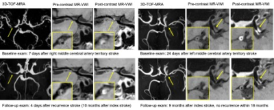 |
Serial MR Vessel Wall Imaging Reveals Medical Treatment Response of Symptomatic Intracranial Atherosclerotic Plaque
Jiayu Xiao1, Shlee Song1, Konrad Schlick1, Shuang Xia2, Tao Jiang3, Tong Han4, Robert Jackson1, Oana Dumitrascu1, Marcel Maya1, Patrick Lyden1, Debiao Li1,5, Qi Yang6, and Zhaoyang Fan1,5
1Cedars-Sinai Medical Center, Los Angeles, CA, United States, 2Tianjin First Central Hospital, Tianjin, China, 3Beijing Chaoyang Hospital, Beijing, China, 4Tianjin Huanhu Hospital, Tianjin, China, 5University of California, Los Angeles, Los Angeles, CA, United States, 6Beijing Xuanwu Hospital, Beijing, China
Ischemic stroke is a leading cause of disability and death, also has a high recurrence rate. Serial magnetic resonance vessel wall imaging (MR-VWI) was used to quantify the morphological changes of intracranial atherosclerotic plaque during the medical treatment of ischemic stroke patients. Changes of quantitative plaque features were compared between patients with different clinical outcomes. Our study showed an increasing trend in most progression patients. Maximum wall thickness, pre-contrast plaque-wall contrast ratio
|
1066.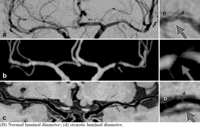 |
Accuracy of 3D High-Resolution Vessel Wall Imaging in Evaluating Internal Carotid and Intracranial Arterial Stenotic Lesions
Yan Gong1, Chen Cao2, Yu Guo3, Song Liu2, Zhu Jinxia4, Shuang Xia3, Xiudi Lu3, Ying Zou3, and Wen Shen3
1Tianjin Medical University NanKai Hospital, Tianjin, China, 2Department of Radiology, Tianjin Huanhu Hospital, Tianjin, China, 3Department of Radiology, Tianjin First Central Hospital, Tianjin, China, 4siemens-healthineers, Tianjin, China
This study compared high-resolution vessel wall imaging (HR-VWI) and time-of-flight magnetic resonance angiography (TOF-MRA) for evaluation of stenosis using digital subtraction angiography (DSA) as the criterion standard. Compared with TOF-MRA, HR-VWI produced results that more closely agreed with DSA, showed better reproducibility and accuracy with smaller variance, and provided additional information on vessel wall pathology. HR-VWI may therefore be useful as an adjunct to DSA to diagnose stenosis and evaluate changes in intracranial vessel walls.
|
|
|
1067.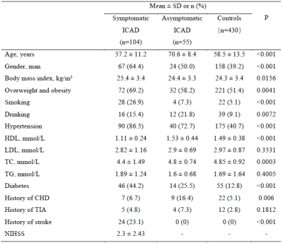 |
Association of conventional vascular risk factors with asymptomatic and symptomatic intracranial atherosclerosis
Yongjun Han1,2, Runhua Zhang3, DanDan Yang1,2, Hualu Han2, Huiyu Qiao2, Dongye Li4, Shuo Chen2, Gaifen Liu3, and Xihai Zhao2
1Center for Brain Disorders Research, Capital Medical University and Beijing Institute of Brain Disorders, Beijing, China, 2Center for Biomedical Imaging Research, Department of Biomedical Engineering, Tsinghua University School of Medicine, Beijing, China, 3Department of Neurology, Beijing Tiantan Hospital, Capital Medical University; China National Clinical Research Center for Neurological Diseases, Beijing, China, 4Department of radiology, Sun Yat-sen Memorial hospital, Sun Yat-sen University, Beijing, China
This study investigated the association of vascular risk factors with asymptomatic and symptomatic ICAD using MR vascular wall imaging. Compared with controls, there was a positive association between hypertension and asymptomatic ICAD; and a positive association of hypertension, LDL, and diabetes and an inverse association of HDL with symptomatic ICAD (all p<0.05). Compared to asymptomatic ICAD, there was an inverse association between hyperlipidemia and symptomatic ICAD (p<0.001). We found that hypertension was a risk factor of asymptomatic ICAD and hypertension, diabetes and higher LDL were risk factors for symptomatic ICAD, whereas HDL was inversely associated with symptomatic ICAD.
|
 Watch the Video