
Scientific Session
Cardiovascular Techniques
Session Topic: Cardiovascular Techniques
Session Sub-Topic: Myocardial Perfusion & Function
Oral
Cardiovascular
| Thursday Parallel 3 Live Q&A | Thursday, 13 August 2020, 15:50 - 16:35 UTC | Moderators: Edward DiBella & Pedro Ferreira |
Session Number: O-18
1309.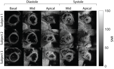 |
Multi-slice arterial spin labelled myocardial perfusion imaging with single shot EPI
Ahsan Javed1 and Krishna S Nayak1
1Electrical and Computer Engineering, University of Southern California, Los Angeles, CA, United States
Arterial spin labelled cardiac magnetic resonance (ASL-CMR) imaging is a non-contrast myocardial perfusion (MP) imaging technique which can detect clinically relevant changes in MP under vasodilatory stress. Existing ASL-CMR techniques have limited spatial coverage because they cannot acquire multiple slices during the limited duration of pharmacologically induced peak stress (~3-4 min). In this work, we demonstrate the feasibility of a using carefully designed single shot echo planar imaging sequence for multi slice ASL-CMR at 3T.
|
|
1310. |
Interaction of Aortic Flow and Myocardial Motion in Patients with Repaired Tetralogy of Fallot
Xiao-Qing Zhang1, Meng-Chu Chang1, Ming-Ting Wu2, Ken-Pen Weng3,4, and Hsu-Hsia Peng1
1Department of Biomedical Engineering and Environmental Sciences, National Tsing Hua University, Hsinchu, Taiwan, 2Department of Radiology, Kaohsiung Veterans General Hospital, Kaohsiung, Taiwan, 3Department of Pediatrics, Kaohsiung Veterans General Hospital, Kaohsiung, Taiwan, 4Department of Pediatrics, National Yang-Ming University, Taipei, Taiwan
We aimed to investigate the abnormal aortic flow and its adverse interaction with regional myocardial motion in repaired tetralogy of Fallot (rTOF) patients. The rTOF patients were divided into rTOF1and rTOF2 groups according to their indexed right ventricular end-systolic volume (RVESVi). The rTOF2 group demonstrated increased aortic retrograde fraction and there was a correlation exhibited between retrograde fraction and systolic myocardial motion. In conclusion, the assessments of abnormal artic flow and altered myocardial motion were helpful in elucidating the possibly adverse interaction between the characteristics of the aorta and myocardium in rTOF patients with different degrees of RV dilatation.
|
|
| WITHDRAWN | ||
 |
1312. |
Fully Self-gated Free-breathing 3D Cartesian Cardiac CINE with Isotropic Whole-heart Coverage in Less Than 2 Minutes
Thomas Küstner1, Aurelien Bustin1, Olivier Jaubert1, Radhouene Neji1,2, Claudia Prieto1, and René M Botnar1
1Biomedical Engineering Department, School of Biomedical Engineering and Imaging Sciences, King's College London, London, United Kingdom, 2MR Research Collaborations, Siemens Healthcare Limited, Frimley, United Kingdom
Free-breathing continuous acquisitions, so called free-running, enable 3D whole-heart coverage for motion-resolved functional cardiac MRI. In prior work approaches based on 3D radial imaging were proposed with scan times of ~10-15min which also require computationally demanding reconstructions. In this work, we propose a 3D Cartesian free-running water-selective sequence that provides isotropic 3D whole-heart CINE imaging in <2min. Data is acquired with a variable-density spiral-like 3D Cartesian out-inward sampling and sequence-adaptive tiny-golden and golden angle increment. Respiratory motion-corrected and cardiac motion-resolved CINE images are obtained from a multi-bin-PROST reconstruction which exploits spatial-temporal redundancies. High agreement to conventional 2D CINE was observed.
|
|
1313.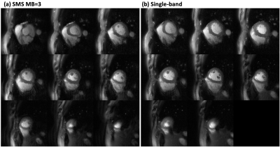 |
High resolution spiral simultaneous multi‐slice first‐pass perfusion imaging with whole-heart coverage at 1.5 T and 3 T
Junyu Wang1, Yang Yang2,3, Ruixi Zhou1, Changyu Sun1, Mathews Jacob4, Daniel S. Weller5, Frederick H. Epstein1,6, and Michael Salerno1,3,6
1Biomedical Engineering, University of Virginia, Charlottesville, VA, United States, 2Biomedical Engineering and Imaging Institute and Department of Radiology, Icahn School of Medicine at Mount Sinai, New York, NY, United States, 3Medicine, University of Virginia, Charlottesville, VA, United States, 4Electrical and Computer Engineering, University of Iowa, Iowa City, IA, United States, 5Electrical and Computer Engineering, University of Virginia, Charlottesville, VA, United States, 6Radiology, University of Virginia, Charlottesville, VA, United States
First-pass contrast-enhanced myocardial perfusion imaging is a useful noninvasive tool to evaluate patients with known or suspected coronary artery disease, but current techniques are still limited in spatial-temporal resolution and ventricular coverage. We designed a spiral pulse sequence with simultaneous multi-slice (SMS) acquisition and utilized the SMS-L1-SPIRiT reconstruction technique to achieve ultra-high resolution (1.5 mm at 1.5 T and 1.25 mm at 3 T) perfusion imaging with whole-heart coverage. The proposed spiral SMS perfusion acquisition strategy was tested on heathy volunteers and clinical patients. High image quality was demonstrated with an SMS factor of 3 at both 1.5T and 3T.
|
 |
1314.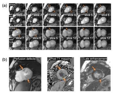 |
Whole-heart, ungated, free-breathing myocardial perfusion MRI by using CRIMP
Ye Tian1, Jason Mendes1, Brent Wilson2, Alexander Ross2, Edward DiBella1, and Ganesh Adluru1
1Radiology, University of Utah, Salt Lake City, UT, United States, 2Cardiology, University of Utah, Salt Lake City, UT, United States
We propose Continuous Radial Interleaved simultaneous Multi-slice acquisitions at sPoiled steady-state (CRIMP) for whole-heart, ungated, free-breathing myocardial perfusion assessment. The simultaneous multi-slice (SMS) sequence captures multiple cardiac phases in all image slices simultaneously and keeps the inner slices at steady-state. We use a patch-based motion-compensated locally low-rank method to reconstruct the images. Quantitative perfusion analysis was also performed with an arterial input function estimated from a separate low-dose injection.
|
|
1315.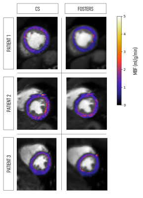 |
High-Resolution Free-Breathing Quantitative Myocardial Perfusion MRI Using Multi-Echo Dixon
Joao Tourais1,2, Torben Schneider3, Cian Scannell4, Russell Franks4, Javier Sanchez-Gonzalez5, Mariya Doneva6, Christophe Schuelke6, Jakob Meineke6, Jochen Keupp6, Jouke Smink1, Marcel Breeuwer1,2, Amedeo Chiribiri4, Markus Henningsson7,
and Teresa Correia4
1MR R&D – Clinical Science, Philips Healthcare, Best, Netherlands, 2Department of Biomedical Engineering, Eindhoven University of Technology, Eindhoven, Netherlands, 3Philips Healthcare, Guildford, Surrey, United Kingdom, 4School of Biomedical Engineering and Imaging Sciences, King’s College London, London, United Kingdom, 5Philips Healthcare Iberia, Madrid, Spain, 6Philips Research Europe, Hamburg, Germany, 7Department of Medical and Health Sciences, Linkoping University, Linkoping, Sweden
First-pass perfusion cardiac MR (FP-CMR) allows the detection of myocardial ischemia. Also, quantitative methods enable a reliable and operator-independent assessment of myocardial perfusion. However, conventional FP-CMR has limited spatial resolution and should be performed under breath-hold. Therefore, diagnostic accuracy is compromised by respiratory induced motion artifacts and false-positive defects due to dark-rim artifacts. We propose, a k-t accelerated dual-saturation FP-CMR multi-echo Dixon sequence to increase the spatial resolution, estimate respiratory motion from fat images and measure T2*-related signal loss from the multi-echo images. Thus, perfusion quantification is improved by minimizing dark-rim artifacts, correcting for respiratory motion and T2*.
|
1316.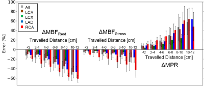 |
Analysis of Location-dependent Errors of Myocardial Blood Flow (MBF) Estimates using Computational Fluid Dynamics (CFD)-Simulations
Tim A. Jedamzik1, Johannes Martens1, Sabine Panzer1, Maria Siebes2, Jeroen P. H. M. van den Wijngaard2,3, and Laura M. Schreiber1
1Chair of Cellular and Molecular Imaging, Comprehensive Heart Failure Center (CHFC), University Hospital Würzburg, Würzburg, Germany, 2Dept. of Biomedial Engineering & Physics - Translational Physiology, Amsterdam UMC, University of Amsterdam, Amsterdam Cardiovascular Sciences, Amsterdam, Netherlands, 3Dept. of Clinical Chemistry and Hematology, Diakenessenhuis, Utrecht, Netherlands
To analyze systematic errors and regional variability of the myocardial blood flow (∆MBF) and myocardial perfusion reserve (∆MPR) estimates in dynamic contrast-enhanced perfusion MRI, computational fluid dynamic (CFD)-simulations were performed in a realistic 3D coronary vasculature model of an ex-vivo porcine heart. Simulations were performed down to the pre-arteriolar level for the myocardial segments. The simulations show a strong spatial variance in the resulting ∆MBF and ∆MPR values of up to 60%. The errors are increasing with distance from the model inlet as well as with lower flow velocities. Errors are more pronounced in the right coronary artery.
|
|
|
1317.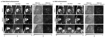 |
Rapid Dealiasing of Undersampled, Radial First-Pass Cardiac Perfusion MR Images using 3D Residual U-Net
Lexiaozi Fan1,2, Daming Shen1,2, Hassan Haji-Valizadeh3, Nivedita K Naresh4, James C. Carr1, Benjamin H. Freed5, Daniel C. Lee5, and Daniel Kim1,2
1Department of Radiology, Northwestern University Feinberg School of Medicine, Chicago, IL, United States, 2Department of Biomedical Engineering, Northwestern University, Evanston, IL, United States, 3Department of Medicine (Cardiovascular Division), Beth Israel Deaconess Medical Center & Harvard Medical School, Boston, MA, United States, 4Department of Radiology, University of Colorado Denver, Denver, CO, United States, 5Division of Cardiology, Internal Medicine, Northwestern University Feinberg School of Medicine, Chicago, IL, United States
Compressed sensing (CS) is capable of accelerating cardiac perfusion MRI for achieving high spatial resolution (1.6 mm x 1.6 mm x 8 mm) and extensive spatial coverage (6+ slices per heartbeat), but the lengthy image reconstruction time (~8 min per slice with 64 frames using GPU) hinders its clinical translation. In this study, we sought to, for the first time, rapidly reconstruct accelerated cardiac perfusion data using a 3D residual U-net for clinical translation.
|
1318.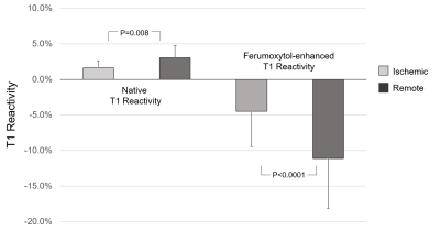 |
Ferumoxytol contrast increases the normalized relative difference in T1 reactivity between remote and ischemic myocardium
Caroline M. Colbert1,2, Anna Le3, Jiaxin Shao1, Jesse Currier3, Peng Hu1, and Kim-Lien Nguyen1,2,3
1Department of Radiological Sciences, David Geffen School of Medicine at UCLA, Los Angeles, CA, United States, 2Physics and Biology in Medicine Graduate Program, David Geffen School of Medicine at UCLA, Los Angeles, CA, United States, 3Division of Cardiology, David Geffen School of Medicine at UCLA, Los Angeles, CA, United States
T1 reactivity can be used as a marker for myocardial perfusion reserve in the setting of ischemia or hypoperfusion. We hypothesize that ferumoxytol, as a pure intravascular agent with high r1 relaxivity, sensitizes T1 reactivity for assessment of myocardial perfusion. We selectively induced acute myocardial hypoperfusion in twelve healthy male Yorkshire swine. We then performed native and ferumoxytol-enhanced adenosine stress testing with the MOLLI sequence at 3.0T. Ferumoxytol increased absolute T1 reactivity in remote regions by 4.62-fold. The normalized difference in T1 was 4.5-fold greater in FE images compared to native T1.
|
 Watch the Video