
Scientific Session
Cardiovascular Techniques
Session Topic: Cardiovascular Techniques
Session Sub-Topic: MRA & Atherosclerosis Imaging
Oral
Cardiovascular
| Thursday Parallel 3 Live Q&A | Thursday, 13 August 2020, 15:50 - 16:35 UTC | Moderators: Xihai Zhao |
Session Number: O-20
1319.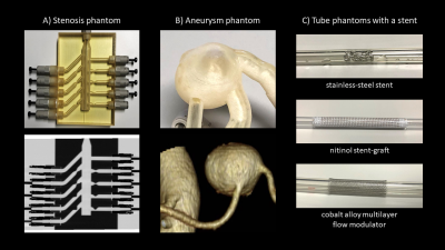 |
Ultrashort TE Time-Spatial Labeling Inversion Pulse MRA for Simulated Visceral Arterial Diseases Indicated for Endovascular Interventions.
Ryuichi Mori1, Hideki Ota2, Simon TUPIN3, Tomoyoshi Kimura1, Hironobu Sasaki1, Tatsuo Nagasaka1, Takashi Nishina4, Sho Tanaka4, Yoshiaki Morita2, Yoshimori kassai4, and Kei Takase2
1Department of Radiology, Tohoku University Hospital, Sendai, Japan, 2Diagnostic Radiology, Tohoku University Hospital, Sendai, Japan, 3Institute of Fluid Science, Tohoku University, Sendai, Japan, 4Canon Medical Systems corp., Tochigi, Japan
Visceral arterial diseases should be evaluated before and after endovascular interventions. We compared ultrashort TE (UTE) and steady-state free precession (SSFP) time-SLIP MRAs regarding their signal decay in pulsatile flow phantoms reflecting stenosis, aneurysm, and metallic stents. In all phantom models, UTE time-SLIP MRA provided superior visualization of target lumens to SSFP time-SLIP MRA. UTE time-SLIP MRA demonstrated minimal signal decay except for in-stent lumen of a stainless-steel stent. Our results indicated robustness of UTE time-SLIP MRA for intra-voxel spin dephasing caused by accelerated flow at the stenosis, turbulent flow in the aneurysm and susceptibility effects from metallic devices.
|
|
1320.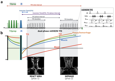 |
REACT-MD: simultaneous non-contrast-enhanced subclavian MRA and fat suppressed direct thrombus imaging (MPRAGE) with a large field-of-view
Masami Yoneyama1, Shuo Zhang2, Yasuhiro Goto3, Michinobu Nagao4, Kayoko Abe4, Osamu Togao5, Isao Shiina3, Kazuo Kodaira3, Yutaka Hamatani3, and Marc Van Cauteren6
1Philips Japan, Tokyo, Japan, 2Philips Healthcare, Hamburg, Germany, 3Department of Radiological Services,, Tokyo Women’s Medical University, Tokyo, Japan, 4Department of Diagnostic Imaging and Nuclear Medicine, Tokyo Women’s Medical University, Tokyo, Japan, 5Department of Clinical Radiology, Graduate School of Medical Sciences, Kyushu University, Fukuoka, Japan, 6Philips Healthcare, Best, Netherlands
Direct visualization of the plaques and vessel wall lesions in addition to luminal changes is of clinical importance for management of patients with atherosclerotic disease. In this work, the recently proposed REACT (Relaxation-Enhanced Angiography without Contrast and Triggering) technique was further developed and particularly optimized with Multiple Delays (REACT-MD) to simultaneously provide non-contrast-enhanced MR angiogram and MPRAGE (magnetization prepared rapid gradient echo) type images with uniform background tissue suppression over a large field of view. Initial results in patients showed great promise in detection of luminal changes and plaques for assessment of systemic atherosclerosis in one single scan.
|
|
 |
1321.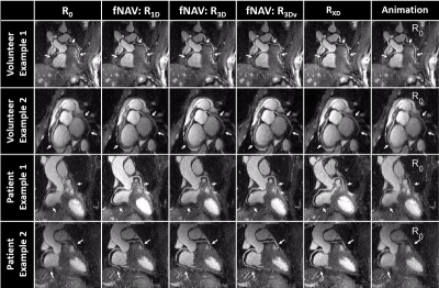 |
Motion Compensated Coronary MRA using Focused Navigation (fNAV)
Christopher W. Roy1, John Heerfordt1,2, Davide Piccini1,2, Juerg Schwitter3, and Matthias Stuber1,4
1Radiology, Lausanne University Hospital (CHUV) and University of Lausanne (UNIL), Lausanne, Switzerland, 2Advanced Clinical Imaging Technology (ACIT), Siemens Healthcare AG, Lausanne, Switzerland, 3Division of Cardiology and CMR-Center, Lausanne University Hospital (CHUV), Lausanne, Switzerland, 4Center for Biomedical Imaging (CIBM), Lausanne, Switzerland
Robust visualization of the coronary vessels is challenging due to cardiac and respiratory motion. Several strategies exist that resolve respiratory motion (XD-GRASP) or compensate for it using N-dimensional image-based navigators to correct for N dimensions of motion. We present a novel self-navigation method wherein the minimization of an image metric is used to estimate 3D non-rigid respiratory motion from a 1D navigator signal (focused navigation: fNAV). We validate fNAV for free-breathing cardiac triggered whole-heart CMRA in a realistic numerical phantom, demonstrate its use in cohorts of healthy volunteers and patients, and quantitatively compare fNAV reconstructions to XD-GRASP.
|
|
1322.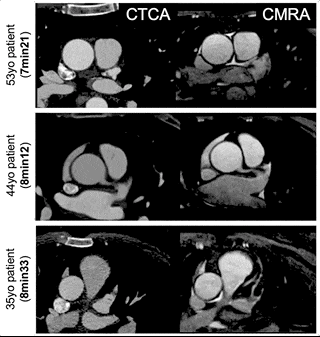 |
Impact of sub-millimeter isotropic resolution on the visualization of coronary arteries in patients undergoing accelerated whole-heart CMRA
Aurelien Bustin1, Reza Hajhosseiny1, Imran Rashid1, Gastao Cruz1, Ronak Rajani1, Tevfik Ismail1, René Botnar1, and Claudia Prieto1
1Biomedical Engineering Department, School of Biomedical Engineering and Imaging Sciences, King's College London, London, United Kingdom
The recent integration of undersampled acquisitions with image-navigators and non-rigid motion-correction have enabled free-breathing 3D whole-heart coronary MR angiography (CMRA) with sub-millimeter isotropic resolution in clinically feasible scan times in healthy subjects. The high acceleration factor and spatial resolution however must be balanced with the need for a robust and high-quality image reconstruction. We sought to assess whether this highly accelerated sub-millimeter isotropic resolution contrast-free CMRA framework could reliably improve the visualization of coronary arteries in comparison to lower resolution (and lower acceleration factor) CMRA in patients with suspected coronary artery disease who underwent CT coronary angiography.
|
1323.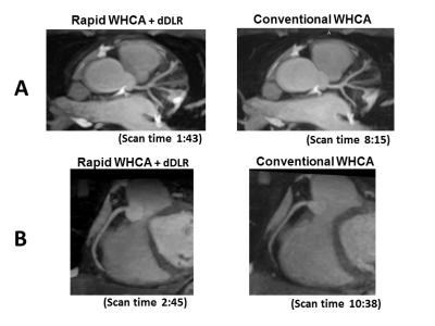 |
Rapid Whole Heart Coronary MRA with 100% respiratory gating efficiency: Fast 3D Wheel data sampling with denoising deep learning reconstruction
Yoshiaki Morita1, Hideki Ota1, Atsuro Masuda1, Takashi Nishina2, Sho Tanaka2, Yuichi Yamashita2, Yoshimori Kassai2, and Kei Takase1
1Department of Radiology, Tohoku University Hospital, Sendai, Miyagi, Japan, 2Canon Medical Systems Corporation, Otawara, Tochigi, Japan
Our proposed Whole Heart Coronary MR Angiography with 100% respiratory gating efficiency using Fast 3D Wheel data acquisition implementing denoising deep learning reconstruction allowed the rapid data acquisition consistently within 3 minutes in spite of irregular breath pattern while maintaining the image quality and contrast ratio of conventional scan. This technique will improve the ease-of-use of coronary artery imaging for practical use.
|
|
|
1324.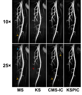 |
Highly Accelerated Subtractive NCE-MRA using Advanced k-space Subtraction and Magnitude Subtraction Reconstruction Methods
Hao Li1, Martin John Graves2, Nadeem Shaida2, Akash Prashar2, David John Lomas1, and Andrew Nicholas Priest2
1Department of Radiology, University of Cambridge, Cambridge, United Kingdom, 2Department of Radiology, Addenbrooke’s Hospital, Cambridge, United Kingdom
We implemented two advanced reconstruction methods for highly accelerated subtractive NCE-MRA, which can exploit the sparsity of subtracted angiograms. One method is based on k-space subtraction of complex raw data with phase and intensity correction (KSPIC). Another method is to reconstruct bright- and dark-blood data with an additional magnitude subtraction term in the cost function to exploit the sparsity. The performance of the two methods was evaluated in both retrospective and prospective accelerated datasets using quantitative metrics and qualitative scoring. Compared with conventional methods, they both showed improved image reconstruction quality, while KSPIC had the best performance.
|
 |
1325.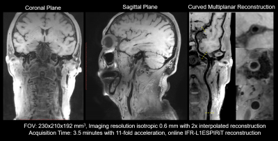 |
Highly accelerated vessel wall imaging using CAIPIRINHA accelerated SPACE and IFR-CS
Sen Jia1,2, Zhilang Qiu1,2, Lei Zhang2, Xin Liu2, Hairong Zheng2, and Dong Liang2
1University of Chinese Academy of Sciences, Shenzhen, China, 2Shenzhen Institutes of Advanced Technology, Chinese Academy of Sciences, Shenzhen, China
Joint intracranial and carotid 3D vessel wall imaging (VWI) with an isotropic spatial resolution between 0.5-0.6 mm is promising in diagnosing arterial wall diseases but leads to clinically impractical scan time. CAIPIRINHA 3x3 undersampling with elliptical scanning is used to expedite the VWI by 11-fold without less sampling the high-frequency region of k-space. Iterative L1-ESPIRiT algorithm is employed for reconstruction with GRAPPA result as the initialization. The scheme of Iterative Feature Refinement is embedded into L1-ESPIRiT iteration to avoid potential over-smoothing issue. Finally, the 3D 11x VWI scan at an isotropic resolution of 0.6 mm takes only 3.5 minutes.
|
|
1326.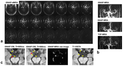 |
iSNAP sequence for simultaneous measurement of whole brain dynamic MRA, MRA, intracranial vessel wall and T1W structural brain MRI
Zhensen Chen1, Zechen Zhou2, Niranjan Balu1, Haikun Qi3, Baocheng Chu1, Thomas S Hatsukami4, and Chun Yuan1
1Vascular Imaging Lab and BioMolecular Imaging Center, Radiology, University of Washington, Seattle, WA, United States, 2Philips Research North America, Cambridge, MA, United States, 3Biomedical Engineering, King's College London, London, United Kingdom, 4Surgery, University of Washington, Seattle, WA, United States
In this study, a whole brain sequence named iSNAP, with 0.8 mm isotropic voxel size and 6 min 30 sec acquisition time, was developed to simultaneously obtain four different image contrasts (dynamic MRA [dMRA] for blood flow monitoring, MRA for luminal stenosis measurement, black blood image for vessel wall [VW] measurement and T1W for brain structural imaging). Preliminary testing results on a healthy volunteer and two patients with cerebrovascular diseases demonstrated the feasibility and potential of the sequence.
|
1327.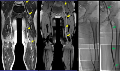 |
Outcome of Catheter-direct Thrombolysis for Deep Vein Thrombosis using T1-weighted Magnetic Resonance Black-blood Thrombus Imaging
Chen Huang1, Guoxi Xie2, Xueping He1, Yufeng Ye1, Wei Deng1, Jianke Liang1, Zhuonan He1, Xin Liu3, Debiao Li4, Zhaoyang Fan4, and Hanwei Chen1
1Guangzhou panyu central hospital, Guangzhou, China, 2Guangzhou medical university, Guangzhou, China, 3Shenzhen Institutes of Advanced Technology, Shenzhen, China, 4Cedars-Sinai Medical Center, Los Angeles, CA, United States
The present study aims to evaluate the outcome of thrombolysis for acute DVT using the thrombus signal characteristics generated by a T1-weighted MR black-blood thrombus imaging (BTI) technique.The patients were divided into iso- or hyper-intense thrombus groups according to the BTI images and the additional CDT were performed .The thrombolysis ratio of patients with iso-intense signals was significantly higher than hyper-intense ones .However, the patients with iso-intense thrombus had a lower incidence rate of PTS at 6 and 12 months.So,the thrombus signal characteristics on BTI images maybe used to predict the outcome of DVT treated with the lytic therapy.
|
|
1328.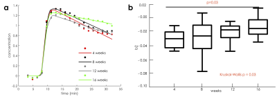 |
Self-gated dynamic contrast enhanced magnetic resonance imaging of the aortic root in atherosclerotic mice: a natural progression study
Claudia Calcagno1, John David2, Abdallah Motaal2, Thijs Beldman2, Alexandra Corbin1, Arnav Kak1, Sarayu Ramachandran1, Alison Pruzan1, Raphael Soler1, Christopher Faries1, Zahi A. Fayad1, Willem Mulder1, and Gustav Strijkers2
1Icahn School of Medicine at Mount Sinai, New York, NY, United States, 2University of Amsterdam, Amsterdam, Netherlands
Enhanced endothelial permeability is an important hallmark of atherosclerotic plaques at high-risk for causing severe cardiovascular events. Here we present a the application of a novel, self-gated DCE-MRI acquisition combined with compressed sensing reconstruction to quantify endothelial permeability in the mouse aortic root. In a longitudinal natural disease progression study in ApoE-/- mice, we find that plaque contrast agent washout computed from this acquisition changes significantly over time, with washout being slower in older mice.
|
 Watch the Video