
Scientific Session
Cardiovascular Techniques
| Thursday Parallel 3 Live Q&A | Thursday, 13 August 2020, 15:50 - 16:35 UTC | Moderators: Liang Zhong |
 |
1329.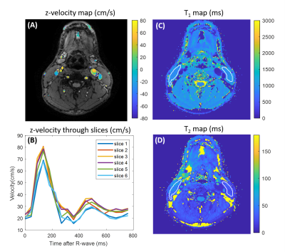 |
Time-resolved 3D Flow-MRF for relaxation and velocity quantification in the carotid arteries
Lisa Leroi1, Sebastian Flassbeck2,3, and Sebastian Schmitter1,2
1Physikalisch-Technische Bundesanstalt Berlin (PTB), Braunschweig and Berlin, Germany, 2Medical Physics in Radiology, German Cancer Research Center (DKFZ), Heidelberg, Germany, 3Faculty of Physics and Astronomy, Heidelberg University, Heidelberg, Germany
The simultaneous quantification of blood velocity and tissue relaxation times could be a valuable tool for clinicians, especially in the carotid arteries, where atheroma plaques could occur. This can be achieved using the recently presented Flow-MRF technique that relies on the acquisition of randomly distributed gradients m1 momentum using a FISP MRF-sequence, with varying flip angle and fixed TR. In this work, Flow-MRF is extended to a time-resolved 3D acquisition and successfully applied in-vivo to the carotid bifurcation at 3T. T1, T2 and 3D time-resolved flow maps are recovered in a 3D slab.
|
|
1330.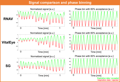 |
Retrospective camera-based respiratory binning for 4D flow MRI – A comparison with liver navigator and self-gating
Lukas M. Gottwald1, Joao Tourais2,3, Eva S. Peper1, Jouke Smink2, Bram F. Coolen4, Gustav J. Strijkers4, Pim van Ooij1, and Aart J. Nederveen1
1Radiology and Nuclear Medicine, Amsterdam UMC, Amsterdam, Netherlands, 2MR R&D – Clinical Science, Philips Healthcare, Best, Netherlands, 3Department of Biomedical Engineering, University of Technology, Eindhoven, Netherlands, 4Biomedical Engineering and Physics, Amsterdam UMC, Amsterdam, Netherlands This study aimed to compare the performance of the novel camera-based respiratory navigation sensor (VitalEye) in retrospective respiratory binned Cartesian 4D flow MRI to conventional liver navigator and self-gating. Analyzed were the cross-correlation of the respiratory signals, peak flow rate error compared to 2D flow and the image quality in terms of edge sharpness of the liver/diaphragm border and signal-to-noise ratio. The novel camera-based respiratory navigation sensor VitalEye performed as good as conventional liver navigator and self-gating. Respiratory signal, flow rate error, and image quality showed no significant difference, but VitalEye has the advantage of a 10-times higher sampling frequency. |
 |
1331.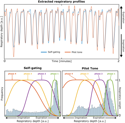 |
Extracting the respiratory signal from Pilot Tone for highly accelerated, respiratory-resolved whole-heart 4D flow imaging
Aaron Pruitt1, Peter Speier2, Chong Chen1, Yingmin Liu3, Ning Jin4, Orlando Simonetti5, and Rizwan Ahmad1
1Biomedical Engineering, The Ohio State University, Columbus, OH, United States, 2Siemens Healthcare GmbH, Erlangen, Germany, 3Dorothy M. Davis Heart and Lung Research Institute, The Ohio State University, Columbus, OH, United States, 4Siemens Medical Solutions USA, Inc., Columbus, OH, United States, 5Cardiovascular Medicine and Radiology, The Ohio State University, Columbus, OH, United States Pilot Tone has recently been proposed as a novel approach towards physiological signal monitoring. Unlike self-gating, often relied upon by free-running, respiratory-resolved imaging sequences, Pilot Tone is generalizable to a multitude of imaging techniques without requiring additional pulse sequence modification or specialized k-space sampling. In this work, we combine Pilot Tone with our previously described highly accelerated and fully self-gated whole-heart 4D flow framework to reconstruct respiratory-resolved 4D flow images in three healthy subjects. We compare Pilot Tone and self-gating derived respiratory binning and demonstrate good agreement in aortic and pulmonary artery flow quantification between the two methods. |
|
1332.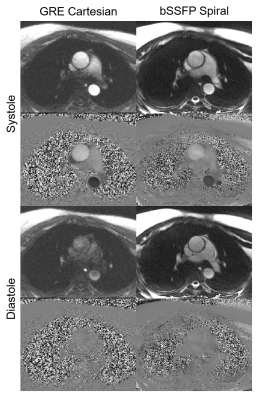 |
Spiral bSSFP Phase-contrast Flow at 0.55T
Rajiv Ramasawmy1, Daniel Herzka1, Robert Lederman1, and Adrienne Campbell-Washburn1
1National Heart, Lung & Blood Institute, National Institutes of Health, Bethesda, MD, United States
A balanced SSFP (bSSFP) phase-contrast using a spiral readout was implemented for quantitative flow measurements at 0.55T. bSSFP flow is challenging at 1.5T and 3T due to off-resonance. However, at 0.55T, this sequence exploits the improved field inhomogeneity for a long readout (TR = 7.2ms) bSSFP spiral acquisition. This sequence provided improved signal-to-noise ratio (SNR) normalized by voxel, especially during diastole (Cartesian gradient echo SNR/voxel = 3.6, spiral bSSFP SNR/voxel = 9.4), to produce quality flow measurements at 0.55T.
|
| 1333. | Longitudinal study of 4D flow MRI derived aortic hemodynamics in bicuspid aortic valve patients with repaired coarctation.
Gilles Soulat1, Michael Scott1, Ashitha Pathrose1, Kelly Jarvis1, Haben Berhane2, Bradley Allen1, Ryan Avery1, Cynthia Rigsby2, and Michael Markl3
1Department of Radiology, Feinberg School of Medicine, Northwestern University, Chicago, IL, United States, 2Department of Medical Imaging, Ann & Robert H. Lurie Children’s Hospital of Chicago,, Chicago, IL, United States, 3Department of Radiology, Feinberg School of Medicine, and Department of Biomedical Engineering, McCormick School of Engineering; Northwestern University, Chicago, IL, United States
Bicuspid aortic valve (BAV) patients with history of aortic coarctation are considered higher risk for aortic complications. We evaluated 4D flow aortic metrics in 15 BAV adults with coarctation repair (mean age 35y) retrospectively reviewed at baseline and follow-up (3.98y [2.10 to 4.96y ]). Areas of higher wall shear stress were mainly located in the arch, and 4D flow metrics remained stable at follow-up. Aortic growth was slow, with a significant increase in the anterior arch (0.25mm/y) and diaphragmatic aorta (0.27mm/y). At baseline, peak velocity at the coarctation repair site was inversely correlated to mid arch and diaphragmatic aortic growth.
|
|
1334.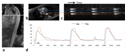 |
Evaluation of Vascular Reactivity of Maternal Cardiovascular Adaptations During Pregnancy with Quantitative MRI
Michael C Langham1, Felix W Wehrli1, Alessandra S Caporale1, and Nadav Schwartz2
1Radiology, University of Pennsylvania, Philadelphia, PA, United States, 2Maternal Fetal Medicine, University of Pennsylvania, Philadelphia, PA, United States
Significant maternal cardiovascular adaptations take place during pregnancy. One such alteration is a decrease in peripheral vascular resistance to accommodate an increase in cardiac output. In this pilot study, we aimed to evaluate changes in vascular reactivity during normal pregnancy by quantifying MRI surrogate markers of endothelial function. A novel quantitative MRI protocol was performed on 14 healthy pregnant women to evaluate peripheral micro- and macrovascular reactivity and central arterial stiffness. Preliminary results indicate attenuated peripheral vascular reactivity consistent with previous studies of brachial artery reactivity using ultrasound in pregnant women.
|
|
1335.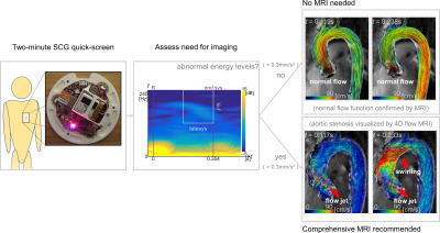 |
Screening Efficacy of Wearable Seismocardiography to Recommend MRI for Stratification of Aortic Valve Diseases
Ethan M I Johnson1, J. Alex Heller2, Flori Garcia Vicente2, Mozziyar Etemadi1,2, Alex Barker3, and Michael Markl1,4
1Biomedical Engineering, Northwestern University, Evanston, IL, United States, 2Anesthesiology, Northwestern University, Chicago, IL, United States, 3Radiology and Biomedical Engineering, University of Colorado, Anschutz Medical Campus, Aurora, CO, United States, 4Radiology, Northwestern, Chicago, IL, United States
Aortic valve diseases (AVD) require regular monitoring of aortic size and blood speeds. MRI can quantify morphology and flow parameters, such as velocity and wall shear stress, with high accuracy and reproducibility, especially as compared to echocardiography. However, the value proposition may be low for performing repeated MRI if there has been no change in disease state. A quick, inexpensive and easy to use test that identifies potential need for MR imaging could significantly raise the cost-effectiveness of using MRI for monitoring AVD. Here we show high potential screening efficacy of using seismocardiography to select AVD patients needing MRI examination.
|
|
1336.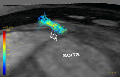 |
Towards Coronary Flow Reserve Assessment with 4D Flow MRI
Carmen PS Blanken1, Eva S Peper1, Lukas M Gottwald1, Bram F Coolen2, Gustav J Strijkers2, R Nils Planken1, Aart J Nederveen1, and Pim van Ooij1
1Radiology and Nuclear Medicine, Amsterdam UMC, Amsterdam, Netherlands, 2Biomedical Engineering, Amsterdam UMC, Amsterdam, Netherlands
Coronary flow reserve (CFR) is a clinical test that interrogates the function of the entire coronary vasculature, indicating the presence of coronary stenoses, microvascular disease or both in patients with ischemic heart disease. We used 15 times accelerated 4D flow MRI with compressed sensing reconstruction at an isotropic spatial resolution of 1.0 mm to measure diastolic flow in the left coronary artery of six healthy subjects. Mean diastolic flow was 1.15±0.18 ml/s with a mean scan-rescan difference of 0.06 ml/s. 4D flow MRI-based diastolic flow quantification in the LCA is feasible and could enable non-invasive CFR measurement.
|
|
1337.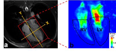 |
Myocardial and Intraventricular Kinetic Energy in Patients with Repaired Tetralogy of Fallot
Shi-Ying Ke1, Meng-Chu Chang1, Ming-Ting Wu2, Ken-Pen Weng3,4, and Hsu-Hsia Peng1
1Department of Biomedical Engineering and Environmental Sciences, National TsingHua University, Hsinchu, Taiwan, 2Department of Radiology, Kaohsiung Veterans General Hospital, Kaohsiung, Taiwan, 3Department of Pediatrics, Kaohsiung Veterans General Hospital, Kaohsiung, Taiwan, 4Department of Pediatrics, National Yang-Ming University, Taipei, Taiwan
We aimed to investigate the interaction between myocardial kinetic energy (KEmyo) and intraventricular KE (KEven) in left- and right-ventricle (LV, RV) for repaired tetralogy of Fallot (rTOF) patients. The rTOF group displayed higher systolic RV KEven, earlier LV myocardial diastolic time-to-peak (TTPmyo), earlier RV TTPmyo in both systole and diastole, earlier LV TTPven in both systole and diastole, and earlier RV TTPven in systole. In conclusion, from an insight of energy conversion, rTOF patients demonstrated undermined interaction between LV KEmyo and KEven in an early stage. The dilated RV potentially have impacts on the RV KEven in
rTOF patients.
|
|
|
1338.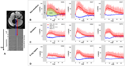 |
Quantitative MRI and serum biomarkers detect acute and chronic vascular effects of e-cigarette use
Alessandra Caporale1, Shampa Chatterjee2, Michael C Langham1, Wensheng Guo3, Frank Leone4, Andrew Strasser5, and Felix W Wehrli1
1Radiology, Laboratory for Structural, Physiologic and Functional Imaging, Perelman School of Medicine, University of Pennsylvania, Philadelphia, PA, United States, 2Physiology, Institute for Environmental Medicine, Perelman School of Medicine, Philadelphia, PA, United States, 3Biostatistics and Epidemiology, Perelman School of Medicine, Philadelphia, PA, United States, 4University of Pennsylvania Medical Center, Pulmonary, Allergy & Critical Care Division, Philadelphia, PA, United States, 5Psychiatry, Center for Interdisciplinary Research on Nicotine Addiction, Philadelphia, PA, United States
The vascular effects of e-cigarette use were investigated in young adults (19-35 years). Blood draws and 3T-MRI data were collected from seven e-cigarette users, seven smokers, thirty nonsmokers, the latter replicating the measurements after one nicotine-free e-cigarette vaping session. MRI-protocol measured peripheral vascular reactivity in response to cuff-induced ischemia, quantifying femoral artery luminal flow mediated dilation (FMDL), blood flow velocity, venous saturation (SvO2). FMDL decreased by 33% acutely after vaping, consistent with 20% NOx reduction and elevated inflammation (C-reactive protein increased by 95%). Reactive hyperemia was blunted as a chronic effect of both smoking and vaping, paired with anomalous biomarkers.
|
 Watch the Video