
Scientific Session
Novel Clinical Applications of CMR
Session Topic: Novel clinical applications of CMR
Session Sub-Topic: CMR to Study Mechanisms & Etiology of Cardiovascular Disease
Oral
Cardiovascular
| Thursday Parallel 3 Live Q&A | Thursday, 13 August 2020, 15:05 - 15:50 UTC | Moderators: Marcus Carlsson & Nivedita Naresh |
Session Number: O-22
 |
1194.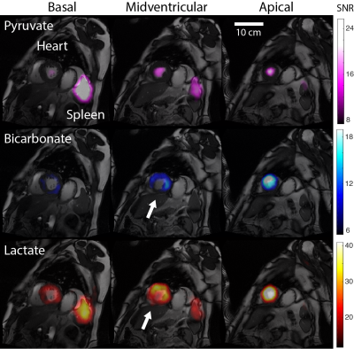 |
Human 13C Hybrid-Shot Spiral (HYSS) Imaging of Pathological Cardiac Metabolism Following Myocardial Infarction
Justin YC Lau1,2, Andrew Apps1, Jack JJJ Miller1,2,3, Andrew Tyler2, Liam AJ Young1, Andrew JM Lewis1, Gareth Barnes4, Claire Trumper1, Stefan Neubauer1, Oliver J Rider1, and Damian J Tyler1,2
1Oxford Centre for Clinical Magnetic Resonance Research, Division of Cardiovascular Medicine, Radcliffe Department of Medicine, University of Oxford, Oxford, United Kingdom, 2Department of Physiology, Anatomy & Genetics, University of Oxford, Oxford, United Kingdom, 3Department of Physics, University of Oxford, Oxford, United Kingdom, 4Royal Brompton & Harefield NHS Foundation Trust, Harefield, United Kingdom
This study reports the clinical translation of a hybrid-shot spiral sequence (HYSS) for imaging the diseased heart and spleen. Two subjects with myocardial infarction from coronary artery disease were imaged following administration of hyperpolarized [1-13C]pyruvate. High lactate and bicarbonate signal were seen in the region adjacent to the infarcted zone, with no signal within the infarct. A large splenic lactate signal was observed in a subject with an active systemic inflammatory response post myocardial infarction.
|
|
1195.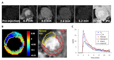 |
Tracer kinetic modeling of nitroxide-enhanced MRI to quantify oxidative stress in mouse models of heart disease
Soham Shah1, Yu Wang1, Christopher Waters1, Lanlin Chen1, Brent French1, and Frederick Epstein1
1University of Virginia, Charlottesville, VA, United States
Oxidative stress plays a significant role in the pathogenesis of heart disease. Nitroxide free radicals have been used as redox-sensitive MRI contrast agents where oxidative stress is correlated to the nitroxide-enhanced signal decay rate. We developed a two-compartment exchange and reduction model (2CXRM) to quantify both myocardial nitroxide exchange and reduction and hypothesized that dynamic nitroxide-enhanced MRI can comprehensively assess nitroxide kinetics in mouse models of angiotensin II infusion (ANGII) and myocardial infarction (MI). The 2CXRM detected elevated reduction rates in ANGII and post-MI mice indicative of oxidative stress and reduced nitroxide delivery, consistent with microvascular damage, in post-MI mice.
|
 |
1196.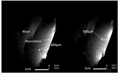 |
Initial technical developments of local RF coil for sub-millimeter cardiovascular MRI.
Marylène Delcey1,2,3,4, Isabelle Saniour5, Pierre Bour1,2,4, Fanny Vaillant1,2,4, Emma Abell1,2,4, Wadie Benhassen3, Marie Poirier-Quinot5, and Bruno Quesson1,2,4
1IHU Liryc, Electrophysiology and Heart Modeling Institute, Fondation Bordeaux Université, Pessac, France, 2Univ. Bordeaux, Centre de recherche Cardio-Thoracique de Bordeaux, U1045, Bordeaux, France, 3Siemens Healthcare SAS, Saint-Denis, France, 4INSERM, Centre de recherche Cardio-Thoracique de Bordeaux, U1045, Bordeaux, France, 5IR4M, UMR8081, Université Paris-Sud/CNRS, Université Paris-Saclay, Orsay, France
In the context of cardiovascular diseases, precise determination of the extent and locations of the arrhythmogenic substrate could significantly improve diagnosis and treatment for both atrial and ventricular electrical diseases. However, the current spatial resolution and signal-to-noise ratio (SNR) in clinical scanners remain insufficient to provide relevant information of small structures like atrial wall or sub-millimeter fatty infiltration in ventricle. To address this limitation in SNR, two receiver coils were implemented at 1.5T for high resolution cardiac imaging, with different active decoupling techniques (safety aspects). Images at 200 µm in-plane spatial resolution were successfully obtained on a beating heart.
|
|
1197. |
Chronic Myocardial Infarcts with Iron Deposits Exhibit Lower Rest Perfusion and Elevated Nitric Oxide Synthase Activity
Eric Johnson1,2, Anand Nair2, Ivan Cokic1,2, Hsin-Yung Yang2, Andreas Kumar3, and Rohan Dharmakumar1,2
1UCLA, Los Angeles, CA, United States, 2Cedars Sinai, Los Angeles, CA, United States, 3Northern Ontario School of Medicine, Thunder Bay, ON, Canada
Hemorrhagic myocardial infarction (hMI) patients are predisposed to adverse outcomes in the chronic stage of MI, yet physiological underpinnings contributing to this observation are not well understood. We hypothesized that hMI areas containing iron deposits would negatively impact endothelial function and tested our hypothesis by evaluating perfusion defects in patients and dogs with a history of hMI; with histological staining for iron, endothelial cells and nitric oxide synthase (NOS) in excised myocardial sections of dogs with chronic MI. Hemorrhagic subjects had significantly reduced perfusion and markedly elevated NOS activity.
|
|
1198.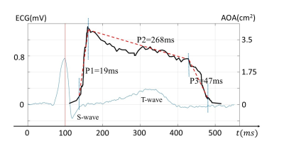 |
Capture the Opening and Closing of Human Aortic Valve Using MRI with Sub-Millisecond Temporal Resolution
Zheng Zhong1,2, Kaibao Sun2, Guangyu Dan1,2, Muge Karaman1,2, and Xiaohong Joe Zhou1,2,3,4
1Bioengineering, University of Illinois at Chicago, Chicago, IL, United States, 2CMRR, University of Illinois at Chicago, Chicago, IL, United States, 3Radiology, University of Illinois at Chicago, Chicago, IL, United States, 4Neurosurgery, University of Illinois at Chicago, Chicago, IL, United States
Stenosis and regurgitation are two common valvular diseases currently diagnosed using echocardiography. Cardiac MR has potential to diagnose these two diseases, however, faces the challenge of inadequate temporal resolution for capturing the rapid opening or closing of aortic valve. Using a variation of a recently proposed technique, coined Sub-millisecond Periodic Event Encoded Dynamic Imaging or SPEEDI (formerly called SMILE), we demonstrated that this process can be visualized using MRI with sub-millisecond temporal resolution. This new capability has improved the accuracy and reliability in studying the dynamics of aortic valve, opening new opportunities to detect stenosis and regurgitation using MRI.
|
|
1199.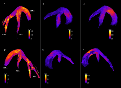 |
Evaluation of potential hemodynamic biomarkers in experimental PAH using center-out stack-of-stars 4D phase contrast velocity mapping
Ali Nahardani1,2, Simon Leistikow2,3, Katja Grün4, Martin Krämer1, Karl Heinz Herrmann1, Andrea Schrepper5, Reinhard Bauer6, Christian Jung7, Alexander Berndt8, P. Christian Schulze4, Lars Linsen3, Jürgen R. Reichenbach1, Marcus Franz4,
and Verena Hoerr1,2,9
1Institute of Diagnostic and Interventional Radiology, Medical Physics Group, University Hospital Jena, Jena, Germany, 2Institute of Medical Microbiology, University Hospital Jena, Jena, Germany, 3Institute of Computer Science, Department of Mathematics and Computer Science, Westfälische Wilhelms-Universität Münster, Muenster, Germany, 4Department of Internal Medicine I, Division of Cardiology, Angiology, Pneumology, and Intensive Medical Care, University Hospital Jena, Jena, Germany, 5Department of Cardiothoracic Surgery, University Hospital Jena, Jena, Germany, 6Institute of Molecular Cell Biology, Center of Molecular Biomedicine, University Hospital Jena, Jena, Germany, 7Department of Internal Medicine, Division of Cardiology, University Hospital Düsseldorf, Düsseldorf, Germany, 8Institute of Legal Medicine, Section of Pathology, University Hospital Jena, Jena, Germany, 9Department of Clinical Radiology, University Hospital Muenster, Muenster, Germany
Potential hemodynamic biomarkers of pulmonary arterial hypertension (PAH) and consecutive right ventricular remodeling were investigated by 4D flow center-out stack-of-stars velocity mapping in a rat model of monocrotaline induced PAH in comparison to healthy controls and a treatment group taking Macitentan. The averaged-mean values of blood flow velocities of pulmonary tract were substantially decreased in the diseased animal group compared to the control and under-treatment group. Diseased animals further showed a pronounced pressure gradient drop between the pulmonary artery bronchial branches and pulmonary veins. The effect of vascular resistance was additionally noted in the velocity-time curve of the pulmonary arteries.
|
1200.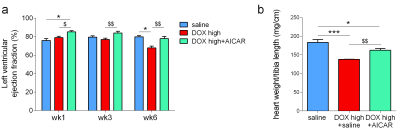 |
AICAR prevents heart failure in a rat model of doxorubicin-induced cardiotoxicity
Kerstin N Timm1, Vicky Ball1, Benjamin Thackray1, Michael P Murphy2, Lisa C Heather1, and Damian J Tyler1
1Department of Physiology, Anatomy and Genetics, University of Oxford, Oxford, United Kingdom, 2MRC Mitochondrial Biology Unit, University of Cambridge, Cambridge, United Kingdom
Doxorubicin (DOX) is a commonly used chemotherapeutic agent for the treatment of many cancers. However, DOX has serious cardiotoxic side effects culminating in congestive heart failure. We have previously shown in a clinically-relevant rat model of DOX-induced heart failure (DOX-HF), that this is due to loss and dysfunction of mitochondria. We show here that 5-aminoimidazole-4-carboxamide ribonucleotide (AICAR), an activator of AMPK, can prevent heart failure in DOX-treated rats. This cardioprotective effect appears to be, at least in part, achieved through improved fatty acid oxidation in cardiac mitochondria which can be indirectly assessed with hyperpolarized [2-13C]pyruvate MRS.
|
|
1201.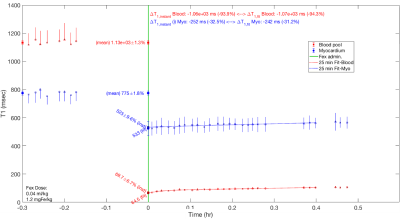 |
Estimating Blood Volume with Ferumoxytol at 0.55 T
Daniel A. Herzka1, Rajiv A. Ramasawmny1, Toby Rogers1, Kendall O'Brien1, Delaney McGuirt1, Adrienne Campbell-Washburn1, and Robert J. Lederman1
Video Permission Withheld
1National Heart Lung and Blood Institute, National Institutes of Health, Bethesda, MD, United States
Off-label use of ferumoxytol as an intravascular contrast agent for cardiovascular imaging is increasing. Measurements of circulating blood volume using dilution techniques has been previously demonstrated with ferumoxytol at 1.5 T. The relaxivity of ferumoxytol at low field (0.55 T) is increased, making it an attractive approach potentially requiring reduced dosages. Here we successfully demonstrate the feasibility of measurement of total circulating blood volume at lower field strength in swine.
|
|
|
1202.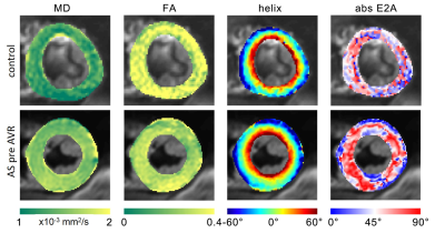 |
Microstructural cardiac remodelling in aortic stenosis and its reversibility following valve replacement – a CMR diffusion tensor imaging study
Alexander Gotschy1,2,3, Constantin von Deuster1, Lucas Weber4, Mareike Gastl5, Martin O. Schmiady6, Robbert J. H. van Gorkum1, Jochen von Spiczak1,4, Robert Manka2,4, Sebastian Kozerke1, and Christian T. Stoeck1
Video Permission Withheld
1Institute for Biomedical Engineering, University and ETH Zurich, Zurich, Switzerland, 2Department of Cardiology, University Hospital Zurich, Zurich, Switzerland, 3Great Ormond Street Hospital, London, United Kingdom, 4Institute of Diagnostic and Interventional Radiology, University Hospital Zurich, Zurich, Switzerland, 5Division of Cardiology, Pulmonology and Vascular Medicine, University Hospital Duesseldorf, Duesseldorf, Germany, 6Department of Cardiac Surgery, University Hospital Zurich, Zurich, Switzerland
CMR diffusion tensor imaging (CMR DTI) allows for the assessment of cardiac microstructure in diseased hearts. We investigated changes of myocardial diffusion properties and myocyte orientation in patients with aortic stenosis (AS) before and after valve replacement (AVR) using DTI and T1-mapping. Mean diffusivity (MD), fractional anisotropy (FA), E2A sheet angle and the transmural helix angle (HA)-slope were altered in AS patients, while native T1 was not significantly different. After AVR, the HA-slope was the only parameter with reversible changes, whereas MD, FA and E2A remained abnormal. This study indicates that AS-induced alterations of myocardial microstructure partly persist following AVR.
|
1203.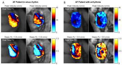 |
Cardiac Rhythm Impacts Left Atrial Hemodynamics Measured with 4D Flow and Real Time PC MRI in Controls and Patients with Atrial Fibrillation
Amanda L DiCarlo1, Hassan Haji-Valizadeh2, Suvai Gunasekaran1, Patrick McCarthy3, Rod Passman4, Philip Greenland4, Daniel C Lee1, Daniel Kim1, and Michael Markl1,5
1Radiology, Northwestern University, Chicago, IL, United States, 2Department of Medicine (Cardiovascular Division), Beth Israel Deaconess Medical Center & Harvard Medical School, Boston, MA, United States, 3Cardiac Surgery, Northwestern University, Chicago, IL, United States, 4Preventive Medicine, Northwestern University, Chicago, IL, United States, 5Biomedical Engineering, Northwestern University, Chicago, IL, United States
Stroke prevention is a major therapeutic goal in atrial fibrillation (AF) management. Flow quantification using MRI can provide information about left atrium hemodynamics implicated in stroke risk. This study evaluates the impact of cardiac arrhythmia on velocity and stasis, reflective of slow flow, measurements using both 4D-flow and real time phase contrast techniques in a cohort of healthy controls and AF patients in sinus rhythm and arrhythmia. Both real time phase contrast and 4D-flow showed a similar increase in left atrium stasis between controls and patients and between patients with low and high heart rate variability, but real time phase contrast was more sensitive to differences.
|
 Watch the Video