
Scientific Session
Hepatobiliary & Pancreas
Session Topic: Hepatobiliary & Pancreas
Session Sub-Topic: Liver: Methods & Applications
Oral
Body
| Monday Parallel 3 Live Q&A | Monday, 10 August 2020, 15:15 - 16:00 UTC | Moderators: Shintaro Ichikawa |
Session Number: O-25
0305.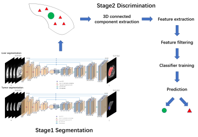 |
Automatic Liver Tumor Detection using Deep learning based segmentation and Radiomics guided Candidate Filtering
Rencheng Zheng1, Qidong Wang2, Ziying Feng3, Chengyan Wang3, and He Wang1,3
1Institute of Science and Technology for Brain-Inspired Intelligence, Fudan University, Shanghai, China, 2Radiology department, The first affiliated hospital, College of medicine, Zhejiang University, Hangzhou, China, 3Human Phenome Institute, Fudan University, Shanghai, China
The objective of this study is to perform automatic multi DCE phases liver tumor detection using deep learning based segmentation and radiomics guided candidate filtering. The proposed model consists mainly of two stages, primary segmentation based on a U-net architecture neural network in stage1, and suspected tumor discrimination mechanism using multi DCE phases radiomics features including shape features, texture features, time dimension features and location information in stage 2. The proposed two-stage model exhibits superior performance in HCC tumor segmentation with a mean Dice score of 0.7928 in test set.
|
|
0306.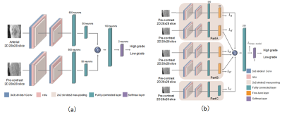 |
Improving the performance of non-enhanced MR for predicting the grade of hepatocellular carcinoma by transfer learning
Wu Zhou1, Wanwei Jian1, and Guangyi Wang2
1School of Medical Information Engineering, Guangzhou University of Chinese Medicine, Guangzhou, China, 2Department of Radiology, Guangdong General Hospital, Guangzhou, China
Contrast agent has several limitations in clinical practice, and the diagnostic performance of non-enhanced MR for lesion characterization should be thoroughly exploited. Inspired by the work of cross-modal learning framework,we propose a deeply supervised cross-modal transfer learning method to remarkably improve the malignancy characterization of HCC in non-enhanced MR, in which the cross-modal relationship between the non-enhanced modal and contrast-enhanced modal is explicitly learned and subsequently transferred to another CNN model for improving the characterization performance of non-enhanced MR. The visualization method Grad-CAM is also applied to verify the effectiveness of the proposed cross-modal transfer learning model.
|
|
0307.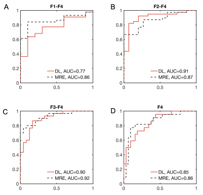 |
Fully Automated Prediction of Liver Fibrosis using Deep Learning Analysis of Gadoxetic acid-enhanced MRI
Stefanie Hectors1,2,3, Paul Kennedy1,2, Kuang-Han Huang1,4, Hayit Greenspan5, Scott Friedman6, and Bachir Taouli1,2
1BioMedical Engineering and Imaging Institute, Icahn School of Medicine at Mount Sinai, New York, NY, United States, 2Department of Diagnostic, Molecular and Interventional Radiology, Icahn School of Medicine at Mount Sinai, New York, NY, United States, 3Department of Radiology, Weill Cornell Medicine, New York, NY, United States, 4Prealize Health, Palo Alto, CA, United States, 5Medical Imaging Processing Lab, Tel Aviv University, Tel Aviv, Israel, 6Division of Liver Diseases, Icahn School of Medicine at Mount Sinai, New York, NY, United States
In this study we developed a fully automated deep learning algorithm based on gadoxetic acid-enhanced MRI for noninvasive prediction of liver fibrosis. We found good-to-excellent performance of the algorithm in an independent test set (AUC 0.77 – 0.91), which was equivalent to the diagnostic performance of MR elastography (AUC 0.86 – 0.92, p-values between methods >0.134). The developed algorithm may potentially allow for noninvasive liver fibrosis assessment, without the need for invasive biopsies.
|
|
0308.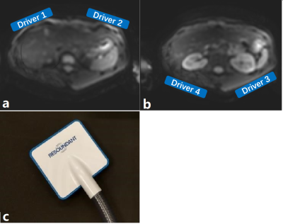 |
Simultaneous stiffness measurement of the upper abdomen with multiple acoustic actuators MRE: feasibility and repeatability
Jie Chen1,2, Jun Chen1, Jeremiah Heilman1, Kevin Glaser1, Richard Ehman1, and Meng Yin1
Video Permission Withheld
1Mayo Clinic, Rochester, MN, United States, 2West China Hospital, Sichuan University, Chengdu, China
Simultaneous stiffness assessment of multiple organs in patients with systemic diseases may be beneficial. Here we used four passive MRE drivers to assess the feasibility and repeatability of simultaneous stiffness measurements of the liver, spleen and kidneys using 60-Hz vibrations. No significant bias was observed in the stiffness measurements of the liver and kidneys with 1 versus multiple drivers. Splenic stiffness was lower with four drivers. With four drivers, it is possible to obtain simultaneous stiffness measurements of the liver, spleen and kidneys with repeatability coefficients similar to those using a single driver while also overcoming attenuation in the liver.
|
|
 |
0309.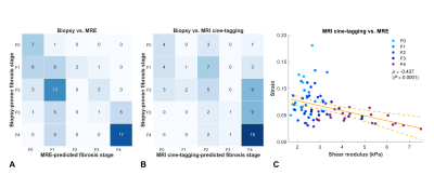 |
Prospective Comparison of MR Elastography with MRI Cine-Tagging of Cardiac-Induced Motion for Noninvasive Staging of Liver Fibrosis
Thierry Lefebvre1,2,3, Léonie Petitclerc1,2,4, Giada Sebastiani5, Jeanne-Marie Giard2,6, Marie-Pierre Sylvestre2,7, Bich N. Nguyen8, Guillaume Gilbert1,9, Guy Cloutier1,10,11, and An Tang1,2,10
1Department of Radiology, Radio-Oncology and Nuclear Medicine, Université de Montréal, Montreal, QC, Canada, 2Centre de recherche du Centre hospitalier de l'Université de Montréal (CRCHUM), Montréal, QC, Canada, 3Medical Physics Unit, McGill University, Montréal, QC, Canada, 4C.J. Gorter Center for High Field MRI, Department of Radiology, Leiden University Medical Center (LUMC), Leiden, Netherlands, 5Department of Medicine, Division of Gastroenterology and Hepatology, McGill University Health Centre (MUHC), Montréal, QC, Canada, 6Department of Medicine, Division of Hepatology and Liver Transplantation, Université de Montréal, Montréal, QC, Canada, 7Department of Social and Preventive Medicine, École de santé publique de l’Université de Montréal (ESPUM), Montréal, QC, Canada, 8Service of Pathology, Centre hospitalier de l'Université de Montréal (CHUM), Montréal, QC, Canada, 9MR Clinical Science, Philips Healthcare Canada, Markham, ON, Canada, 10Institute of Biomedical Engineering, Université de Montréal, Montréal, QC, Canada, 11Laboratory of Biorheology and Medical Ultrasonics (LBUM), Centre de recherche du Centre hospitalier de l'Université de Montréal (CRCHUM), Montréal, QC, Canada
MR elastography for staging liver fibrosis assesses the right liver lobe and requires external hardware. MRI cine-tagging evaluates cardiac-induced strain and shows promise for assessing fibrosis in the left lobe without additional hardware. Shear modulus measured by MRE provided higher AUCs than that of strain measured on tagged images for distinguishing stages F0 vs. ≥F1 (0.87 vs. 0.81, P=0.083) and ≤F3 vs. F4 (0.91 vs. 0.87, P=0.043). Hence, MRE provided a diagnostic accuracy similar or higher than that of MRI cine-tagging for staging of liver fibrosis. Strain could be evaluated on screening abdominal MRI to assess the left liver.
|
0310.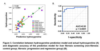 |
Noninvasive assessment of water content and collagen extent of liver tissue with multiparametric magnetic resonance elastography (MRE)
Jingbiao Chen1,2, Jin Wang3, Jiahui Li1, Jie Chen1, Xin Lu1, Hiroaki Yashiro4, Jenifer Siegelman4, Christopher T Winkelmann4, Richard L Ehman1, Vijay H Shah2, and Meng Yin1
1Radiology, Mayo Clinic, Rochester, MN, United States, 2Gastroenterology and Hepatology, Mayo Clinic, Rochester, MN, United States, 3the Third Affiliated Hospital of Sun Yat-Sen University, Guangzhou, China, 4Research and Development, Takeda Pharmaceuticals International Co., Cambridge, MA, United States
Liver biopsy remains the gold standard for staging liver fibrosis. However, its invasive nature makes it unacceptable for long-term disease dynamic monitoring. In addition, current histopathological scoring systems for staging liver fibrosis are not quantitative. Also, the inflammatory response of increased interstitial fluid volume is precursory and occult with histological analysis. It is critical to address the overlooked fluid-associated inflammatory response and semi-quantitative fibrosis grading. Here, we use a novel technique, magnetic resonance elastography, combined with ALT, as a noninvasive quantitative method to quantify and monitor hepatic water and fibrosis content in a mouse model with varying disease progression/regression.
|
|
|
0311.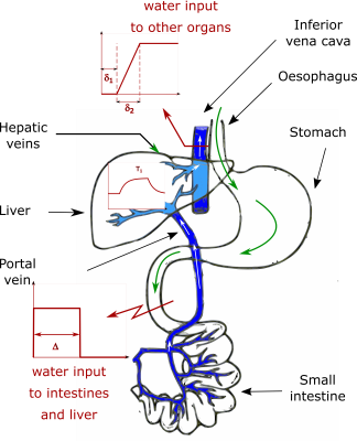 |
The thirsty liver: dynamic T1 mapping after fluid intake in healthy volunteers
Ferenc E. Mozes1, Emmanuel A. Selvaraj1, Michael Pavlides1,2, Matthew D. Robson1,3, and Elizabeth M. Tunnicliffe1
1OCMR, RDM Cardiovascular Medicine, University of Oxford, Oxford, United Kingdom, 2Translational Gastroenterology Unit, University of Oxford, Oxford, United Kingdom, 3Perspectum Diagnostics Ltd., Oxford, United Kingdom
Non-alcoholic fatty liver disease is on the rise and liver biopsies used to diagnose it need to be replaced by non-invasive methods such as T1 mapping. Guidance for MRI scans allow for free water consumption before scans, increasing measurement variability. We therefore aimed to assess the effect of hydration on liver T1 by acquiring serial shMOLLI T1 maps after participants drank 1 litre of isotonic water. As water passed through the stomach and small intestines it first reached the portal circulation and later the systemic circulation, causing an increase in liver T1 followed by an increase in spinal muscle T1.
|
 |
0312.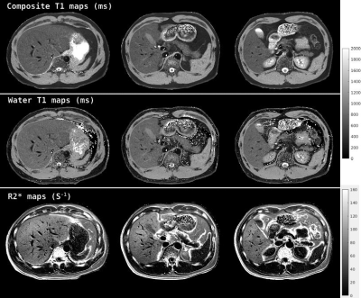 |
Rapid multi-slice fat and water separated T1 and composite R2* mapping using a dual-echo radial inversion recovery SPGR pulse sequence
Zhitao Li1, John Pauly2, and Shreyas Vasanawala1
1Department of Radiology, Stanford University, Stanford, CA, United States, 2Electrical Engineering, Stanford University, Stanford, CA, United States
A radial dual-echo IR-SPGR technique combined with a principal component based iterative reconstruction algorithm are demonstrated for fat and water separated rapid high-resolution abdominal multi-parameter mapping. This method can yield high-quality fat-signal-fraction map, B0 field map, composite T1 map, water component T1 map as well as a R2* map from a scan as fast as 3seconds/slice. The in-plane resolution of the resulting parameter maps is 1.25mm and the through-plane resolution is 5.00mm. With a selective inversion pulse, multiple slices can be acquired within a breath-hold.
|
|
0313.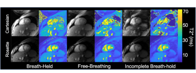 |
Ungated, Motion Robust, Simultaneous Cardiac and Liver T2* Quantification via Rosette k-Space Sampling
Adam Michael Bush1, Christopher Michael Sandino2, Shreya Ramachandran3, Nicholas Dwork4, Frank Ong1, Marcus Alley1, and Shreyas Vasanawala1
1Radiology, Stanford University, Palo Alto, CA, United States, 2Electrical Engineering, Stanford University, Palo Alto, CA, United States, 3Electrical Engineering, University of California Berkeley, Berkeley, CA, United States, 4Radiology, University of California San Francisco, San Francisco, CA, United States
In this work we introduce a novel method for T2* determination using rosette k-space sampling and locally low-rank reconstruction. This approach produced comparable T2* quantitation with higher spatial resolution, fewer motion artifacts and lessened variability without the use of gating. This approach offers a child-friendly, rapid, free-breathing, comprehensive assessment of liver and cardiac iron. |
|
0314.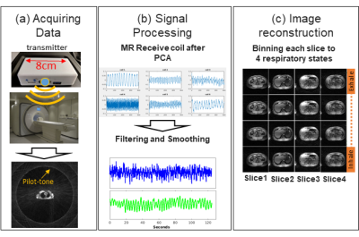 |
Free-Breathing Radial Imaging using a Pilot-Tone RF Transmitter for Detection of Respiratory Motion
Eddy Solomon1, Thomas Vahle2, Jan Paska1, Kai Tobias Block1, Daniel K. Sodickson1, Fernando Boada1, and Hersh Chandarana1
1Radiology, New York University School of Medicine, New York, NY, United States, 2Siemens Healthcare GmbH, Erlangen, Germany
The sensitivity of MRI sequences to motion impairs their reliability and diagnostic utility for examining the chest and abdomen. Established motion-compensation techniques are not accurate enough, come at the cost of patient comfort, and are limited by the MR imaging parameters. Here, we demonstrate a novel approach that detects respiratory signal from the amplitude modulation of a transmitted RF reference signal, termed ‘pilot-tone’ (PT). We show how the use of this simple RF transmitter, with its small dimensions, high sampling rate, and low interference with the MR acquisition, can produce motion corrected-images under free-breathing conditions.
|
 Watch the Video