
Scientific Session
Hepatobiliary & Pancreas
Session Topic: Hepatobiliary & Pancreas
Session Sub-Topic: Diffuse Liver & Metabolism
Oral
Body
| Monday Parallel 3 Live Q&A | Monday, 10 August 2020, 15:15 - 16:00 UTC | Moderators: Frederick Kelcz |
Session Number: O-28
0315.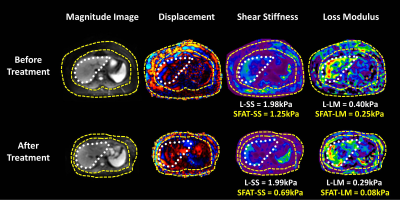 |
Assessment of obesity-induced metabolic disorders in adipose tissue by multi-parametric MR Elastography (MRE)
Jiahui Li1, Marzanna Obrzut1, Xin Lu1, Alina Allen2, Sudhakar K. Venkatesh1, Taofic Mounajjed3, Jun Chen1, Kevin J. Glaser1, Armando Manduca1, Vijay Shah2, Richard L. Ehman1, and Meng Yin1
1Radiology, Mayo Clinic, Rochester, MN, United States, 2Gastroenterology, Mayo Clinic, Rochester, MN, United States, 3Mayo Clinic, Rochester, MN, United States
We performed multiparametric 3D MR Elastography (MRE) in 37 obese patients who underwent bariatric surgeries. MRI/MRE, anthropometrics, and liver biopsy were obtained within three months of bariatric surgery and one year later. 12/37 (32%) patients have biopsy-proven non-alcoholic fatty liver disease (NAFLD) at the time of surgery. The MRE-assessed shear stiffness (SS) and loss modulus (LM) of subcutaneous adipose tissue decreased significantly after the surgery, as well as the liver tissue. MRE-assessed SS and LM have potential in distinguishing the obesity-induced metabolic disorder in the adipose tissues. The mechanical change may correlate with the therapeutic response in these obese patients.
|
|
 |
0316.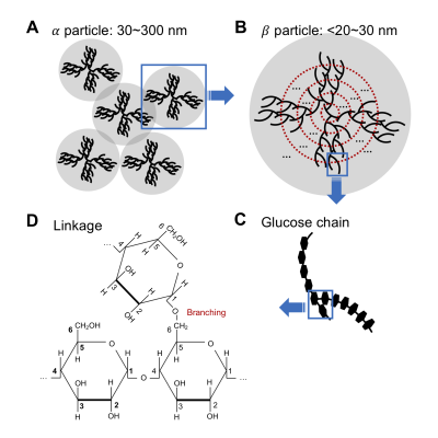 |
Mapping glycogen concentration in vivo based on the nuclear Overhauser enhancement (NOE) with water (glycoNOE)
Yang Zhou1,2, Peter C.M. van Zijl1,2, Xiang Xu1,2, Jiadi Xu1,2, Yuguo Li1,2, and Nirbhay N. Yadav1,2
1The Russell H. Morgan Department of Radiology, The Johns Hopkins University School of Medicine, Baltimore, MD, United States, 2F.M. Kirby Research Center for Functional, Brain Imaging, Kennedy Krieger Institute, Baltimore, MD, United States
We recently reported a method for the enhanced detection of glycogen using the nuclear Overhauser enhancement (NOE) between glycogen and water (glycoNOE). Here we show that the glycoNOE signal is linearly dependent on glycogen concentration both in vitro and in mouse liver in vivo. The glycoNOE signal is affected by glycogen particle size, but not pH or temperature. glycoNOE MRI can non-invasively quantify liver glycogen levels in vivo and thus has the potential to assess disease where glycogen metabolism is altered.
|
|
0317.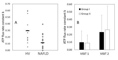 |
Mapping Metabolic Inflexibility in NAFLD: Comparison With Healthy Volunteers and Following L-Carnitine Intervention Using Advanced MRS.
Stephen Bawden1,2, Prarthana Thiagarajan1, Elizabeth Simpson1, Olivier Mougin2, Paul Greenhaff3, Penny Gowland2, and Guruprasad P Aithal1
1NIHR Nottingham Biomedical Research Centre, University of Nottingham, Nottingham University Hospitals NHS Trust and the University of Nottingham, Nottingham, United Kingdom, 2Sir Peter Mansfield Imaging Centre, University of Nottingham, Nottingham, United Kingdom, 3School of Life Sciences, University of Nottingham, Nottingham, United Kingdom
Advanced MRI techniques such as 1H MRS at 7T and saturation transfer 31P MRS offer unique capabilities to map metabolic conditions and complement current physiological methodologies. The aim of this study was to build a metabolic profile for NAFLD v healthy volunteers by measuring physiological metabolic markers alongside 1H MRS measurement of liver lipid, intra- (IMCL) and extra- (EMCL) myocellular lipid fraction, and dynamic 31P MRS measurements of ATP flux rates, and to investigate the effect of a 24 week L-carnitine intervention. Interim analysis shows metabolic inflexibility in NAFLD patients compared with HV and a potential benefit of L-carnitine supplementation
|
0318.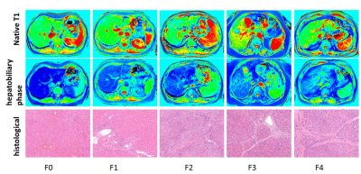 |
B1 inhomogeneity-corrected T1 mapping in quantitative evaluation of liver fibrosis using Gd-BOPTA enhanced MR imaging
Xinya Zhao1, Xianshun Yuan1, Xiang Feng2, Mengxiao Liu3, Xiangtao Lin1, and Ximing Wang1
1Department of Radiology, Shandong Provincial Hospital, Jinan, China, 2MR Scientific Marketing, Siemens Healthcare, Beijing, China, 3MR Scientific Marketing, Siemens Healthcare, Shanghai, China
The purpose of this study was to determine whether B1 inhomogeneity-corrected volumetric T1 mapping of Gd-BOPTA enhanced liver MR imaging are able to evaluate the degree of liver cirrhosis and to investigate their relationship with the histological grading. Our study found that B1 inhomogeneity-corrected T1 mapping using Gd-BOPTA enhanced MR imaging could be used in the quantitative evaluation of liver fibrosis.
|
|
0319.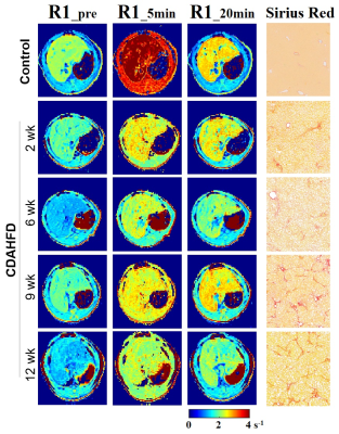 |
Gd-EOB-DTPA-enhanced MRI in Nonalcoholic Steatohepatitis (NASH): Liver Fibrosis or Liver Function?
Iris Y. Zhou1, Chuantao Tu2, Veronica Clavijo Jordan1, Nicholas J. Rotile1, Mozhdeh Sojoodi3, Bryan C. Fuchs3, Kenneth K. Tanabe3, and Peter Caravan1
1Athinoula A. Martinos Center for Biomedical Imaging, Institute for Innovation in Imaging (i3), Department of Radiology, Massachusetts General Hospital and Harvard Medical School, Charlestown, MA, United States, 2Department of Gastroenterology and Hepatology, Zhongshan Hospital, Fudan University, Shanghai, China, 3Division of Surgical Oncology, Massachusetts General Hospital and Harvard Medical School, Boston, MA, United States
Nonalcoholic steatohepatitis (NASH) promotes fibrotic remodeling of the liver parenchyma, which may lead to cirrhosis, liver failure, or hepatocellular carcinoma. Gd-EOB-DTPA is a hepatobiliary T1 MRI contrast agent, receiving increasing attention as a tool for detecting and staging liver fibrosis. Here, using a choline-deficient high-fat diet (CDAHFD) for different durations we modeled NASH disease progression in rats and performed Gd-EOB-DTPA-enhanced MRI at different disease stages, correlating imaging histological measures of fibrosis as well as liver function tests. Gd-EOB-DTPA-enhanced MRI correlated well with liver function tests but not with liver fibrosis.
|
|
0320.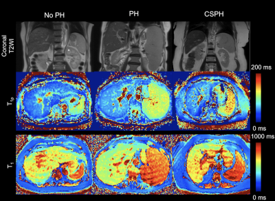 |
Splenic T1ρ as a noninvasive biomarker for portal hypertension
Stefanie Hectors1,2,3, Octavia Bane1,2, Daniel Stocker1,2, Paul Kennedy1,2, Thomas Schiano4, Swan Thung5, Aaron Fischman2, and Bachir Taouli1,2
1BioMedical Engineering and Imaging Institute, Icahn School of Medicine at Mount Sinai, New York, NY, United States, 2Department of Diagnostic, Molecular and Interventional Radiology, Icahn School of Medicine at Mount Sinai, New York, NY, United States, 3Department of Radiology, Weill Cornell Medicine, New York, NY, United States, 4Recanati/Miller Transplantation Institute, Icahn School of Medicine at Mount Sinai, New York, NY, United States, 5Department of Pathology, Icahn School of Medicine at Mount Sinai, New York, NY, United States
In this study we explored the used of liver and spleen T1 and T1ρ parameters for noninvasive assessment of portal hypertension (PH) and compared the performance of the MRI relaxation parameters with that of radiological assessment. Spleen T1ρ showed a strong significant positive correlation with quantitative portal pressure measurements (r=0.613, P=0.001), while the other relaxation parameters did not. Spleen T1ρ also outperformed other relaxation parameters and radiological assessment for prediction of (clinically significant) PH (AUC 0.778 – 0.817). Our results indicate that spleen T1ρ may be a suitable noninvasive biomarker for prediction of PH.
|
|
0321.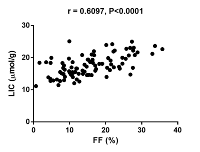 |
Proton-density fat fraction-derived R2* liver iron concentration – an exploratory study of Revita-2 phase II trial data
Manil D Chouhan1, Naomi Sakai1, Francisco Torrealdea2, Kelly White3, Juan Carlos Lopez Talavera3, Alan Bainbridge2, and Stuart A Taylor1
1UCL Centre for Medical Imaging, University College London, London, United Kingdom, 2Department of Medical Physics, University College London Hospitals NHS Trust, London, United Kingdom, 3Fractyl Laboratories Inc., Lexington, MA, United States
R2* derived liver iron concentration (LIC) measurements from proton density fat fraction (PDFF) data obtained in patients with normal LIC levels may be useful. Here we present data from the Revita-2 trial demonstrating strong significant positive correlations between baseline liver fat fraction (FF) and LIC. Significant and stronger correlations between relative % change in liver FF and LIC in the treatment arms of the trial raise the possibility of treatment-related mechanistic effects on hepatic iron metabolism.
|
|
0322.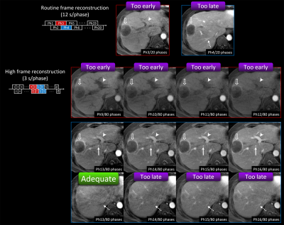 |
Utility of Stack-of-stars Acquisition for Arterial Phase Imaging without Breath-holding on Dynamic MRI of the Liver
Shintaro Ichikawa1, Daiki Tamada1, Tetsuya Wakayama2, Sagar Mandava3, Ty A Cashen4, Hiroshi Onishi1, and Utaroh Motosugi1
1Department of Radiology, University of Yamanashi, Chuo, Japan, 2MR Collaboration and Development, GE Healthcare, Hino, Japan, 3MR Collaboration and Development, GE Healthcare, Tucson, AZ, United States, 4MR Collaboration and Development, GE Healthcare, Madison, WI, United States
We compared arterial phase (AP) images using conventional (Cartesian) breath-hold liver acquisition with volume acceleration (LAVA) and stack-of-stars acquisition without breath-holding (LAVA-Star) on hepatic dynamic MRI. In Cartesian breath-hold LAVA group, 8.7% of patients showed inadequate scan timing of AP, while only 1 patient (4.0%) in LAVA-Star group (12 s/phase) showed inadequate scan timing. One advantage of LAVA-Star was that the adequate scan timing of AP can be obtained by using additional high frame rate reconstruction (3 s/phase) in the patient with inadequate scan timing in routine reconstruction. LAVA-Star was useful to obtain adequate scan timing in all patients.
|
|
|
0323.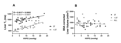 |
Quantitative MRI to assess portal hypertension in cirrhosis patients
Chris R Bradley1,2, Rob A Scott2, Eleanor F Cox1,2, Naaventhan Palaniyappan2, I Neil Guha2, Guruprasad P Aithal2, and Susan T Francis1,2
1Sir Peter Mansfield Imaging Centre, University of Nottingham, Nottingham, United Kingdom, 2NIHR Nottingham Biomedical Research Centre, Nottingham University Hospitals NHS Trust and University of Nottingham, Nottingham, United Kingdom
Hepatic venous pressure gradient (HVPG) is the gold standard method for the assessment of portal pressure, but highly invasive. We scanned patients with portal hypertension at both 1.5T and 3T to assess MRI parameters related to portal pressure as defined by HVPG. Iron-corrected liver T1 highly correlated over the full range of HVPG (3T p<0.0002, 1.5T p<0.0001), spleen T1 and superior mesenteric artery velocity correlated up to HVPG of 15 mmHg (spleen T1: 3T p<0.0003, 1.5T p<0.0006; SMA velocity: p<<0.00001), after which at HVPG >15 mmHg no correlation was observed.
|
0324.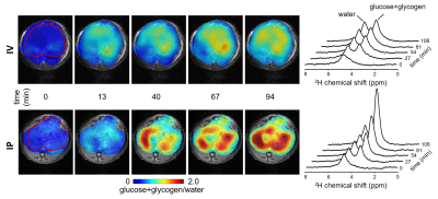 |
Visibility of deuterium-labeled liver glycogen in vivo.
Henk M. De Feyter1, Monique A. Thomas1, Kevin L. Behar2, and Robin A. de Graaf1
1Dept. of Radiology and Biomedical Imaging, Yale University, New Haven, CT, United States, 2Dept. of Psychiatry, Yale University, New Haven, CT, United States
Deuterium metabolic imaging (DMI) is a novel technique for mapping metabolism in vivo, that combines 2H MRSI with administration of a 2H-labeled substrate. DMI combined with [6,6'-2H2]-glucose has the potential to detect glycogen synthesis in the liver. However, the similar 2H chemical shifts of glucose and glycogen make unambiguous detection and separation difficult in vivo. Here we investigate the NMR-detectability of glycogen using high resolution 2H NMR of 2H-labeled glycogen isolated from mouse liver, and show that 2H-labeled glycogen is not detectable with DMI under in vivo conditions.
|
 Watch the Video