
Scientific Session
Muscle, Cartilage, Knee Stabilizers
Session Topic: Muscle, Cartilage, Knee stabilizers
Session Sub-Topic: Muscle
Oral
Musculoskeletal
| Monday Parallel 4 Live Q&A | Monday, 10 August 2020, 15:15 - 16:00 UTC | Moderators: Chiara Giraudo |
Session Number: O-34
 |
0339.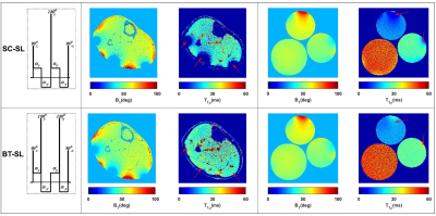 |
T1, T2, and T1ρ Relaxation Mapping of the Lower Leg Muscle in Diabetic Neuropathy Patients with MR-Fingerprinting (MRF): Exercise Intervention
Azadeh Sharafi1, Smita Rao2, Martijn A. Cloos1, Ryan Brown1, and Ravinder R. Regatte1
1Radiology, NYU Langone Health, New York, NY, United States, 2Physical Therapy, New York University, New York, NY, United States
Diabetic peripheral neuropathy (DPN) is characterized by metabolic and microvascular impairment (1) that damage peripheral nerves (2) and cause ischemic conditions and muscle degeneration in the lower extremities (3). Researchers have investigated the possibility of reversing DPN symptoms through exercise therapy (4). Such studies will benefit from quantitative biomarkers to evaluate therapeutic strategies targeting muscle function. In this work, we developed a magnetic resonance fingerprinting (MRF) technique that is insensitive to B1 imperfections for simultaneous T1, T2, and T1ρ relaxation mapping of skeletal muscle in DPN patients in response to exercise intervention at 3T.
|
0340.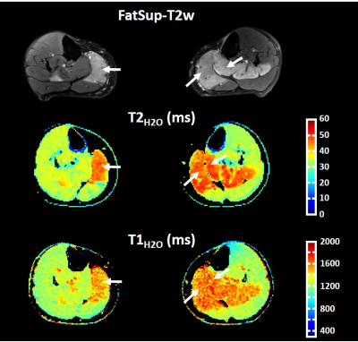 |
Water T1: a quantitative biomarker of disease activity in neuromuscular disorders
Benjamin Marty1,2, Harmen Reyngoudt1,2, Jean-Marc Boisserie1,2, Pierre-Yves Baudin3, and Pierre G. Carlier1,2
1NMR Laboratory, Neuromuscular Investigation Center, Institute of Myology, Paris, France, 2NMR Laboratory, CEA/DRF/IBFJ/MIRCen, Paris, France, 3Consultants for Research in Imaging and Spectroscopy, Tournai, Belgium
Recently, MR fingerprinting with water and fat separation was proposed to quantify water T1 (T1H2O) in the muscles of patients with neuromuscular disorders. In this study, we investigated the sensitivity of T1H2O as a quantitative biomarker of disease activity, by comparing it with fat suppressed T2-weighted (FatSup-T2w) imaging and quantitative water T2 (T2H2O) mapping, in a dataset of 61 subjects with different NMDs. We observed a significant increase of T1H2O values in muscles with FatSup-T2w signal hyperintensities. We also investigated different hypothesis explaining the moderate correlation (ρ = 0.54) observed between T1H2O and T2H2O in the muscles of these patients.
|
|
 |
0341.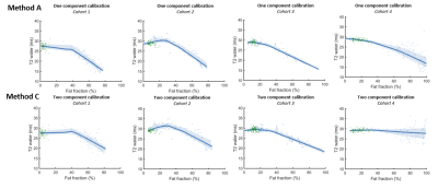 |
T2 mapping in healthy and diseased muscle using optimized extended phase graph algorithms in four clinical cohorts
Kevin Keene1,2, Jan-Willem Beenakker1,3, Melissa Hooijmans4, Karin Naarding2,5, Erik Niks2, Louise Otto6, Ludo van der Pol6, Martijn Tannemaat2, Hermien Kan1,5, and Martijn Froeling7
1Department of Radiology, C.J. Gorter center for high field MRI, Leiden University Medical Center, Leiden, Netherlands, 2Department of Neurology, Leiden University Medical Center, Leiden, Netherlands, 3Department of Ophthalmology, Leiden University Medical Center, Leiden, Netherlands, 4Amsterdam University Medical Center, Amsterdam, Netherlands, 5Duchenne Center Netherlands, Utrecht, Netherlands, 6Department of Neurology, UMC Utrecht Brain Center, University Medical Center Utrecht, Utrecht, Netherlands, 7Department of Radiology, University Medical Center Utrecht, Utrecht, Netherlands
Multi-echo spin-echo transverse relaxometry mapping using multi-component models is used to study disease activity in neuromuscular disease. A recent model using extended phase graphs (EPG) was introduced to obtain separate T2 values for water and fat, accounting for B1 and stimulated echoes. We improved this model and showed the importance of including flip angle slice profiles with a chemical shift displacement in the slice direction and correct calibration methods for the T2 of the fat component. We showed its performance in four clinical cohorts, and showed a gradual decline in T2water with increasing fat fractions.
|
|
0342.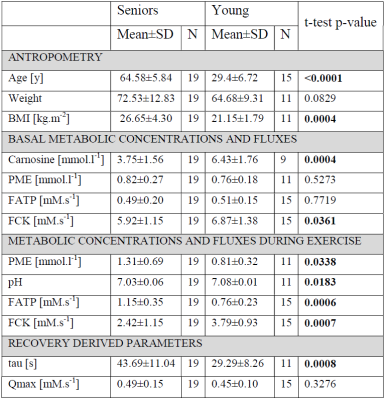 |
Multinuclear MRS at 7T uncovers exercise driven differences in skeletal muscle energy metabolism between young and seniors
Patrik Krumpolec1,2, Radka Klepochová1, Ivica Just Kukurová1, Marjeta Tušek Jelenc1, Jozef Ukropec2, Ivan Frollo3, Christopher Rodgers4,5, Barbara Ukropcová2,6, Siegfried Trattnig1,7, Martin Krššák1,7,8, and Ladislav Valkovič1,3,4
1High-field MR Center, Department of Biomedical Imaging and Image-guided Therapy, Medical University of Vienna, Vienna, Austria, 2Institute of Experimental Endocrinology, Biomedical Research Center of Slovak Academy of Sciences, Bratislava, Slovakia, 3Department of Imaging Methods, Institute of Measurements Science, Slovak Academy of Sciences, Bratislava, Slovakia, 4Oxford Centre for Clinical Magnetic Resonance Research, BHF Centre of Research Excellence, University of Oxford, Oxford, United Kingdom, 5Wolfson Brain Imaging Centre, Department of Clinical Neurosciences, University of Cambridge, Cambridge, United Kingdom, 6Institute of Pathophysiology, Faculty of Medicine, Commenius University, Bratislava, Slovakia, 7Christian Doppler Laboratory for Clinical Molecular MR Imaging, Vienna, Austria, 8Division of Endocrinology and Metabolism, Department of Internal MedicineIII, Medical University of Vienna, Vienna, Austria
The aim of this study was to investigate effect on the demand driven ATP production and carnosine content in the aging muscle. We utilized dynamic and saturation transfer 31P- and 1H-MRS. The dynamic experiment included acquisition of baseline data during two minutes of rest, six minutes of plantar flexion exercise (3.5 minutes long FAST measurement was performed), and six minutes of recovery. We report excessive Pi-to-ATP flux and increase of PME concentration during exercise as well as lower muscle carnosine concentration leading to lower pH after exercise in seniors, which could be linked to deprived metabolic flexibility in this population.
|
|
0343.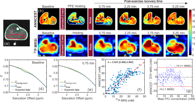 |
High-resolution phosphocreatine mapping using artificial neural network-based CEST MRI at 3T: A validation study
Lin Chen1,2, Michael Schär1,3, Kannie W.Y. Chan1,2,4, Jianpan Huang4, Zhiliang Wei1,2, Hanzhang Lu1,2, Qin Qin1,2, Robert G. Weiss1,3, Peter C.M. van Zijl1,2, and Jiadi Xu1,2
1Department of Radiology and Radiological Science, The Johns Hopkins University School of Medicine, Baltimore, MD, United States, 2F.M. Kirby Research Center for Functional Brain Imaging, Kennedy Krieger Research Institute, Baltimore, MD, United States, 3Division of Cardiology Department of Medicine, Johns Hopkins University School of Medicine, Baltimore, MD, United States, 4Department of Biomedical Engineering, City University of Hong Kong, Hong Kong, China
Phosphocreatine (PCr) plays a vital role in neuron and myocyte energy homeostasis, and measurement of PCr provides a unique way to achieve insight into cellular energetics. Our previous study demonstrated that high-resolution PCr mapping of human skeletal muscle can be obtained on standard 3T clinical MRI scanner using artificial neural network-based chemical exchange saturation transfer (ANNCEST). Here, for further validation, we applied ANNCEST to measure PCr changes in exercised skeletal muscle and compared the measures with those from 31P magnetic resonance spectroscopy. The feasibility of estimating spatially resolved PCr recovery rate constants using ANNCEST was also demonstrated.
|
|
0344. |
Quantitative MRI of skeletal muscle in a cross-sectional cohort of spinal muscular atrophy type 2 and type 3
Louise A.M. Otto1, Ludo W.L. van der Pol1, Lara Schlaffke 2, Camiel A. Wijngaarde1, Marloes Stam1, Renske I. Wadman1, Inge Cuppen3, Ruben P.A. van Eijk1,4, Fay-Lynn Asselman1, Bart Bartels5, Danny van der Woude5, Jeroen Hendrikse6, and Martijn Froeling6
1Neurology, UMC Utrecht Brain Center, University Medical Center, Utrecht, Utrecht, Netherlands, 2Neurology, BG-University Hospital Bergmannsheil, Ruhr-University Bochum, Bochum, Germany, 3Neurology and Child Neurology, UMC Utrecht Brain Center, University Medical Center, Utrecht, Utrecht, Netherlands, 4Biostatistics & Research Support, Julius Center for Health Sciences and Primary Care, University Medical Center Utrecht, Utrecht, Netherlands, 5Child Development and Exercise Center, UMC Utrecht Brain Center, University Medical Center, Utrecht, Utrecht, Netherlands, 6Radiology, UMC Utrecht Brain Center, University Medical Center, Utrecht, Utrecht, Netherlands
qMRI of skeletal muscle has shown promising results in other neuromuscular diseases, but multi-parametric imaging has not been executed in Spinal Muscular Atrophy. We investigated a cohort of 31 patients and 20 controls with protocol consisting of DIXON, T2 mapping and DTI on a 3T MR scanner. All parameters differed significantly between patients and controls. DTI elucidates distinct properties of the muscle, suggesting atrophy by a lowered MD and increased FA. DTI shows correlation with muscle strength and motor function. This suggests the potential of diffusion tensor imaging of muscle in monitoring disease progression in SMA.
|
0345. |
The effect of intramuscular fat on the large strain mechanical properties of skeletal muscle as measured by anisotropic MRE
Max Kaplan1,2, Alice Hatt1, Bezahd Babaei1,3, Lauriane Jugé1,2, and Lynne Bilston1,2
1Neuroscience Research Australia, Randwick, Australia, 2University of New South Wales, Kensington, Australia, 3University of Melbourne, Parkville, Australia
Intramuscular fat (IMF) increases with BMI and age, but it is unknown how it affects skeletal muscle viscoelastic properties, despite the key role skeletal muscle mechanical properties play in our capacity to move. We studied the effects of IMF on the anisotropic mechanical properties under large deformation of the calf muscles in healthy and obese participants, using an advanced approach incorporating diffusion tensor imaging data into magnetic resonance elastography reconstructions. Results show that intramuscular fat had no significant effect on muscle shear moduli, but stretching or shortening muscle altered the parallel and/or perpendicular stiffness and viscosity of some muscles.
|
|
|
0346. |
Non Invasive Imaging of Human Motor Units
Matthew Birkbeck1,2,3, Linda Heskamp1, Ian Schofield1, Andrew Blamire1, and Roger Whittaker1
1Newcastle University, Newcastle upon Tyne, United Kingdom, 2Newcastle Biomedical Research Centre, Newcastle upon Tyne, United Kingdom, 3Regional Medical Physics, Newcastle upon Tyne, United Kingdom
Motor units are fundamental components in the process of contraction of skeletal muscle. Motor unit morphology changes in response to pathologies including motor neurone disease and sarcopenia. Currently the clinical method to investigate motor unit morphology and activity is invasive needle electromyography. Here we present a novel diffusion weighted imaging technique, motor unit MRI (MUMRI). MUMRI has been used to investigate the morphology of single human motor units, producing quantitative data on cross sectional area and dimensions of human motor units. This data agrees with current literature. MUMRI has detected statistically significant changes in the morphology of motor units.
|
|
0347.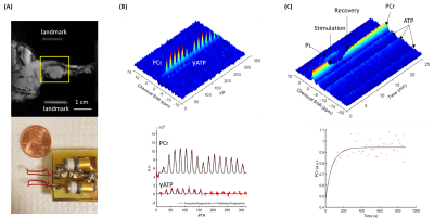 |
Repeated Muscle Contraction increases Creatine Kinase Reaction Rate and Shortens Phosphocreatine Recovery in Mouse Skeletal Muscle
Kihwan Kim1, Yuning Gu1, Gahee Kim1, Mei Wong1, Bryan Clifford 2,3, Sherry Huang1, Zhi-Pei Liang2,3, and Xin Yu1,4
1Department of Biomedical Engineering, Case Western Reserve University, Cleveland, OH, United States, 2Electrical and Computer Engineering, University of Illinois at Urbana-Champaign, Urbana, IL, United States, 3Beckman Institute for Advanced Science and Technology, University of Illinois at Urbana-Champaign, Urbana, IL, United States, 4Case Center for Imaging Research, Case Western Reserve University, Cleveland, OH, United States
This study examined the effects of muscle contraction, induced by electrical stimulation, on creatine kinase (CK) reaction rate and the rate of phosphocreatine (PCr) recovery after its transient depletion in mouse skeletal muscle using phosphorous-31 (31P) magnetic resonance fingerprinting and dynamic 31P magnetic resonance spectroscopy. Our results showed that electrical stimulation induced a significant increase in CK reaction rate by ~14%, as well as an increased in PCr recovery rate by 26%, suggesting a positive preconditioning effect induced by electrical stimulation.
|
|
0348.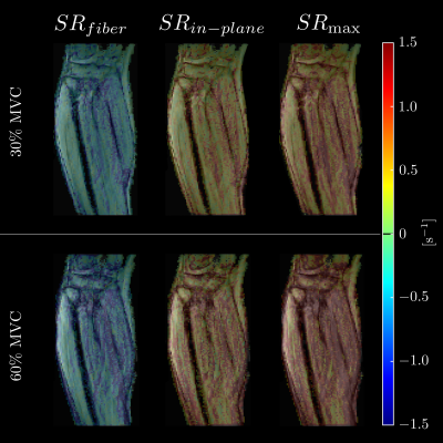 |
Principal Axis and Fiber Aligned 3D Strain / Strain Rate Mapping with Compressed Sensing Velocity Encoded Phase Contrast MRI to study Aging Muscle
Vadim Malis1, Usha Sinha2, and Shantanu Sinha3
1Physics, UC San Diego, La Jolla, CA, United States, 2Physics, San Diego State University, San Diego, CA, United States, 3Radiology, UC San Diego, La Jolla, CA, United States
Strain and Strain rate tensors can be computed from velocity encoded phase contrast imaging. The study of the variation of deformation indices with force output (% Maximum Voluntary Contraction (MVC)) can provide information on the aging muscle. However, such studies have been limited by the long acquisition time precluding its use at high MVCs and in older subjects. We developed a compressed sensing VE-PC technique to enable acquisitions across a range of MVCs and applicable to older subjects as well. Significant differences in the deformation indices were seen between 11 young / 8 senior subjects as well between different %MVCs.
|
 Watch the Video