
Scientific Session
Muscle, Cartilage, Knee stabilizers
Session Topic: Muscle, Cartilage, Knee stabilizers
Session Sub-Topic: Cartilage
Oral
Musculoskeletal
| Monday Parallel 4 Live Q&A | Monday, 10 August 2020, 15:15 - 16:00 UTC | Moderators: Ashley Williams |
Session Number: O-35
0349.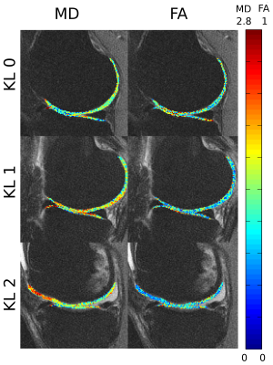 |
DTI of articular cartilage as a biomarker for OA diagnosis, staging and progression in a population with early stages of the disease
Elisa Ramos Gavila1, Alejandra Duarte1, Jenny Bencardino2, Jonathan Samuel2, Svetlana Krasnokutsky2, and Jose Raya Garcia del Olmo2
1Radiology, NYU Langone Health Hospital, New York, NY, United States, 2NYU Langone Health Hospital, New York, NY, United States
Early detection of knee osteoarthritis can be achieved by identifying early compositional changes of degenerative articular cartilage. The purpose of this case-control longitudinal study is to validate DTI as a biomarker for OA diagnosis, staging and progression in early stages of the disease. 60 patients with incipient OA (KL1) underwent 3 visits (baseline, 1.5 year and 3 years follow up). Clinical evaluation, Xray and MRI was performed. Positive correlation was demonstrated with morphological changes (KL and WORMS score). In addition, DTI showed changes in the follow up at 1.5 years that were not apparent in clinical MRI.
|
|
 |
0350.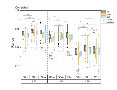 |
Gray Level Co-occurrence Matrix Based 3D Texture Analysis of Knee Articular Cartilage using 3D DESS Images
Ari Väärälä1,2, Arttu Peuna3, Egor Panfilov1,2, Victor Casula1,2, Marianne Haapea2,4, Eveliina Lammentausta1,2,4, and Miika T Nieminen1,2,4
1Research Unit of Medical Imaging, Physics and Technology, University of Oulu, Oulu, Finland, 2Medical Research Center, University of Oulu and Oulu University Hospital, Oulu, Finland, 3Medical Imaging, Central Finland Health Care District, Jyväskylä, Finland, 4Department of Diagnostic Radiology, Oulu University Hospital, Oulu, Finland
In the present study, a gray level co-occurrence matrix-based 3D texture analysis of knee 3D DESS images was used to investigate longitudinal changes in articular cartilage using data from the Osteoarthritis Initiative (baseline, 36-month and 72-month visits). At baseline, all subjects included in the study had Kellgren-Lawrence grade < 2. Three groups were defined, based on time of progression into radiographic osteoarthritis (Kellgren-Lawrence grades ≥ 2): control, slow progressor and fast progressor groups. 3D texture analysis of 3D DESS images was able to distinguish progressors from controls before radiographic signs of osteoarthritis and showed significant longitudinal changes across all groups.
|
|
0351.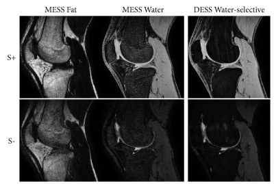 |
Extending DESS to MESS: A 5 minute knee protocol for water-fat separation and T2 mapping
Frank Zijlstra1 and Peter R Seevinck1
1Image Sciences Institute, UMC Utrecht, Utrecht, Netherlands
This study proposes a 5 minute knee protocol using an extension of the double-echo steady-state (DESS) sequence to include multiple readouts. This multiple-echo steady-state (MESS) sequence supports quantification of water, fat, and T2, in a single, efficient acquisition. These parameters may provide additional tissue-specific MRI biomarkers, for example in muscle and bone, on top of the T2 quantification of cartilage provided by the DESS sequence. In vivo results on 5 volunteers show robust water-fat separation and that T2 quantification using MESS corresponds well with quantification on water-selective DESS images.
|
|
0352.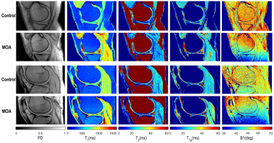 |
Rapid Simultaneous T1, T2, and T1ρ Relaxation Mapping of the knee joint with MR-Fingerprinting (MRF)
Azadeh Sharafi1, Marcelo V. W. Zibetti1, Gregory Chang1, Martijn A. Cloos1, and Ravinder R. Regatte1
1Radiology, NYU Langone Health, New York, NY, United States
Osteoarthritis of the knee, the most common joint disease, is a degenerative heterogeneous musculoskeletal disease which is mainly recognized by the progressive loss of hyaline articular cartilage (1). Spin-lattice relaxation in the rotating frame (T1ρ) and spin-spin relaxation (T2) have been shown to be sensitive to the biochemical changes associated with osteoarthritis progression including: loss of proteoglycans, increased water content, and disruption of collagen and anisotropy (1, 2). In this study, we propose a novel MR fingerprinting sequence for in-vivo simultaneous T1, T2, and T1ρ relaxation mapping of knee joint at 3T.
|
|
0353.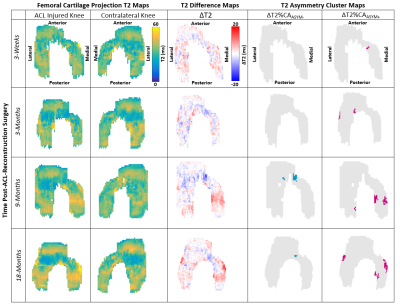 |
Bilateral Femoral Cartilage T2 Asymmetry Analysis for the Detection of Early Osteoarthritic Degeneration
Marianne S Black1,2, Katherine A Young1, Akshay S Chaudhari1, Feliks Kogan1, Bragi Sveinsson3, Emily J McWalter4, Garry E Gold1,5, Marc E Levenston1,2, and Brian A Hargreaves1,5,6
1Radiology, Stanford University, Stanford, CA, United States, 2Mechanical Engineering, Stanford University, Stanford, CA, United States, 3Massachusetts General Hospital, Boston, MA, United States, 4Mechanical Engineering, University of Saskatchewan, Saskatoon, SK, Canada, 5Bioengineering, Stanford University, Stanford, CA, United States, 6Electrical Engineering, Stanford University, Stanford, CA, United States
There is a pressing need for a single-time-point quantitative measure capable of predicting osteoarthritic change. Bilateral knee imaging with T2 cluster asymmetry analysis is a promising approach to achieve this goal. This study examines T2 cluster asymmetry in ACL-injured subjects and controls. ACL-injured subjects showed elevated T2 cluster asymmetry 9-months following reconstruction surgery relative to the controls in the superficial half of cartilage. This novel approach for analyzing T2 relaxation times in femoral cartilage shows promise in detecting changes that may be indicative of early osteoarthritis onset.
|
0354.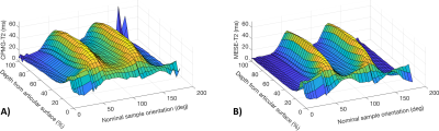 |
Collagen fiber anisotropy and orientation mapping of articular cartilage via T2 relaxation anisotropy
Henri Leskinen1, Nina E. Hänninen1,2, and Mikko J. Nissi1,2
1University of Eastern Finland, Kuopio, Finland, 2University of Oulu, Oulu, Finland
Number of studies have investigated the orientation dependence of T2 relaxation in articular cartilage and, more importantly, connected the orientation dependence to the properties of the collagen fiber network in cartilage. The dependence arises from the non-averaging secular part of the dipolar coupling, which in turn has been attributed to the water-orienting properties of the collagenous network. Using high angular resolution sample rotation, this study aimed to measure the in-plane fiber angle, collagen anisotropy as well as the isotropic and anisotropic components of T2 relaxation in cartilage. Potential for extracting physically meaningful properties of cartilage from multi-orientation measurements was demonstrated.
|
|
0355.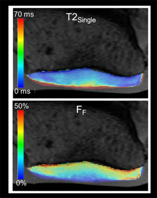 |
Correlation Between Single-Component and Bi-Component T2 Parameters and Proteoglycan Content and Mechanical Properties of Cartilage
Matthew Grondin1, Fang Liu2, Sami Faruqui2, Alexei Samsonov2, Wan-Ju Li3, Corinne Henak1, and Richard Kijowski2
1Mechanical Engineering, University of Wisconsin-Madison, Madison, WI, United States, 2Radiology, University of Wisconsin-Madison, Madison, WI, United States, 3Orthopedic Surgery, University of Wisconsin-Madison, Madison, WI, United States
Multi-component Driven Equilibrium Single Pulse Observation of T1 and T2 (mcDESPOT) was used to measure single-component T2 relaxation time (T2Single) and the fraction of the fast relaxing macromolecular bound water component (FF) of 24 human patellar cartilage samples at 3.0T. The cartilage samples underwent mechanical testing to measure linear modulus and energy dissipation and biochemical analysis to measure proteoglycan content. There were significant (p<0.01) and moderate positive correlations between FF and proteoglycan content, linear modulus, and energy dissipation of cartilage. There were non-significant (p=0.06-0.21) and low negative correlations between T2Single and proteoglycan content, linear modulus, and energy dissipation of cartilage.
|
|
|
0356.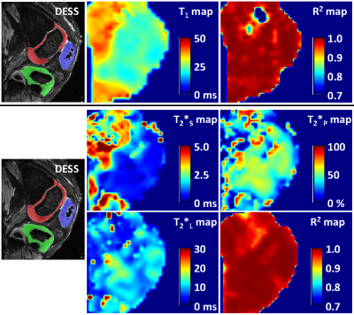 |
Ex Vivo Evaluation of Sodium Relaxation Times in Pediatric Articular-Epiphyseal Cartilage on a Whole-body 10.5T MR System – Initial Results
Stefan Zbyn1,2, Kai D. Ludwig1,2, Lauren Watkins3, Alexandra R. Armstrong4, Russell L. Lagore1,2, Amanda Nowacki1, Marc A. Tompkins5, Ferenc Toth4, Gregor Adriany1,2, Kevin G. Shea6, Garry Gold7, Armin M. Nagel8, Cathy S. Carlson4,
Gregory J. Metzger1,2, and Jutta M. Ellermann1,2
1Center for Magnetic Resonance Research, University of Minnesota, Minneapolis, MN, United States, 2Department of Radiology, University of Minnesota, Minneapolis, MN, United States, 3Department of Bioengineering, Stanford University, Stanford, CA, United States, 4Department of Veterinary Clinical Sciences, University of Minnesota, St. Paul, MN, United States, 5Department of Orthopaedic Surgery, University of Minnesota, Minneapolis, MN, United States, 6Department of Orthopaedic Surgery, Stanford Children's Hospital, Palo Alto, CA, United States, 7Department of Radiology, Stanford University, Stanford, CA, United States, 8Institute of Radiology, University Hospital Erlangen, Friedrich-Alexander-Universität Erlangen-Nürnberg, Erlangen, Germany
Sodium imaging is quantitative technique sensitive to changes in cartilage glycosaminoglycan content. Changes in cartilage matrix, due to maturation or degeneration, may influence sodium relaxation times which can lead to incorrect sodium concentration estimates when not addressed. This ex vivo study employs pediatric knee specimens to evaluate the relationship between sodium relaxation parameters and compositional changes in the developing cartilage matrix. Our preliminary evaluation suggests that cartilage maturation is accompanied by a decrease in sodium T1 and the short T2* component. Sodium concentrations in studies comparing healthy, diseased or immature cartilage should be corrected for possible changes in relaxation times.
|
0357.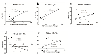 |
UTE-based biomarkers are selectively sensitive to enzymatic proteoglycan and collagen degradation in human articular cartilage
Lidi Wan1,2, Xin Cheng3, Adam C Searleman1, Yajun Ma1, Jonathan H Wong1,4, Mark E Murphy5, Jiang Du1, Guangyu Tang2, and Eric Y Chang1,4
1Department of Radiology, UC San Diego, San Diego, CA, United States, 2Department of Radiology, Shanghai Tenth People's Hospital, Shanghai, China, 3Division of Histology and Embryology, Jinan University, Guangzhou, China, 4Radiology Service, VA San Diego Healthcare System, San Diego, CA, United States, 5Orthopaedic Surgery Service, VA San Diego Healthcare System, San Diego, CA, United States
A panel of quantitative UTE techniques have been developed to assess articular cartilage. Osteoarthritis (OA) is a multifactorial disease characterized primarily by degeneration and loss of hyaline articular cartilage. This study investigated whether quantitative 3D UTE-Cones-based biomarkers are sensitive to proteoglycan (PG) loss and collagen degradation induced by enzyme in human cartilage, and also to determine the specificity of these biomarkers in quantitative cartilage imaging.
|
 Watch the Video