
Scientific Session
Emerging Methods and Machine Learning in Musculoskeletal MRI
Session Topic: Emerging Methods and Machine Learning in Musculoskeletal MRI
Session Sub-Topic: Musculoskeletal Emerging Methods
Oral
Musculoskeletal
| Monday Parallel 4 Live Q&A | Monday, 10 August 2020, 14:30 - 15:15 UTC | Moderators: James MacKay |
Session Number: O-36
0254.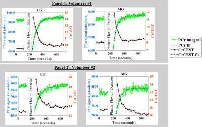 |
Intermuscular Variability of Phosphocreatine Recovery Constants in Exercised Muscle Measured using 31PMRS and CrCEST at 7.0T
Dushyant Kumar1, Ravi Prakash Reddy Nanga1, Deepa Thakuri1, Neil Wilson2, Hari Hariharan1, and Ravinder Reddy1
1Radiology, University of Pennsylvania, Philadelphia, PA, United States, 2Siemens Medical Solutions USA Inc, Malvern, PA, United States
Synopsis: As a noninvasive imaging biomarker, phosphorous magnetic resonance spectroscopy (31PMRS) has traditionally been used to measure the metabolic response of exercised skeletal muscle in humans and has contributed immensely to the vital understanding of muscle energetics. However, due to lack of spatial resolution in 31PMRS, it is difficult to resolve the intermuscular variabilities of creatine kinase kinetics. In this study we demonstrate that with proper placement of surface coil in a mild exercise study, the recovery constant for PCr determined from 31PMRS matches well with recovery constant measured from CrCEST using a volume coil for the same muscle group.
|
|
0255.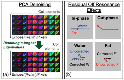 |
Noise and Bias Reduction in Two-Point Dixon Peripheral Nerve Imaging and Muscle Denervation Assessment
Ek T Tan1, Julia Sternberg1, Bin Lin1, Hollis G Potter1, and Darryl B Sneag1
1Radiology and Imaging, Hospital for Special Surgery, New York, NY, United States
High resolution, T2-weighted two-point Dixon is an effective technique for MR neurography (MRN) and can potentially also provide quantitative assessment of muscle ‘edema’ and fatty infiltration, which occur in acute and chronic muscle denervation, respectively. However, low SNR and residual off-resonance can introduce severe bias that impedes accurate interpretation. This study demonstrated that principal component analysis (PCA) denoising and off-resonance correction methods significantly improved proton density fat fraction (PDFF) measurements in 28 patient datasets and in signal simulations. Reduced bias allowed for further application of a proposed water-weighted processing enhances nerve conspicuity by suppressing perineural fat signal.
|
|
0256.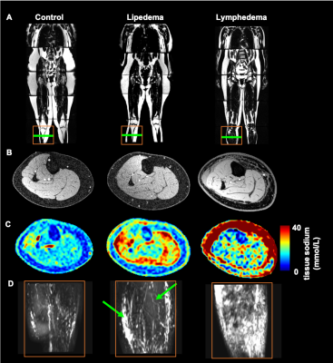 |
Lymphatic insufficiency observed by noninvasive MR lymphangiography and multi-nuclear 23Na-MRI in patients with lymphedema and lipedema
Rachelle Crescenzi1, Paula M.C. Donahue2,3, Kalen J Petersen1, Maria Garza1, Kelsey Guerreso1, Yu Luo1, Joshua A. Beckman4, and Manus J. Donahue1,5,6
1Radiology, Vanderbilt University Medical Center, Nashville, TN, United States, 2Dayani Center for Health and Wellness, Vanderbilt University Medical Center, Nashville, TN, United States, 3Physical Medicine and Rehabilitation, Vanderbilt University Medical Center, Nashville, TN, United States, 4Cardiovascular Medicine, Vanderbilt University Medical Center, Nashville, TN, United States, 5Neurology, Vanderbilt University Medical Center, Nashville, TN, United States, 6Psychiatry, Vanderbilt University Medical Center, Nashville, TN, United States
The lymphatic system comprises a central component of the circulatory system, yet imaging approaches to visualize lymphatics remain underdeveloped. We utilized MR lymphangiography and sodium MRI to confirm lymphatic impairment in patients with lymphedema of known causes, and in patients with the adipose disorder lipedema of unknown etiology. We report distinct profiles on MR lymphangiography that correlate with tissue sodium and fat deposition. Results provide evidence of lymphatic involvement in lipedema that informs disease mechanisms related to swelling, and more broadly relates to lymphatic clearance dysfunction in a range of diseases where sodium and fat are implicated.
|
|
 |
0257.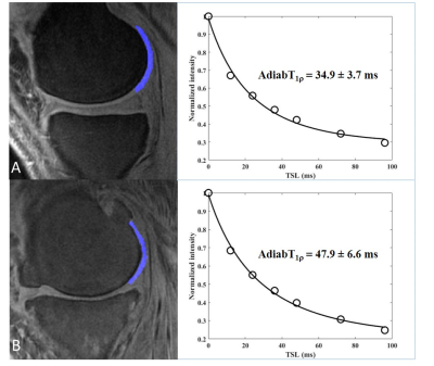 |
Quantitative Assessment of Articular Cartilage Degeneration Using 3D Ultrashort Echo Time Cones Adiabatic T1ρ (3D UTE Cones AdiabT1ρ) Imaging
Mei Wu1,2, Yanping Xue1, Yajun Ma1, Claire Tang1, Meghan Shen1, Saeed Jerban1, Eric Y Chang1,3, and Jiang Du1
1Department of Radiology, University of California, San Diego, San Diego, CA, United States, 2Department of Radiology, Guangzhou First People’s Hospital, School of Medicine, South China University of Technology, Guangzhou, China, 3Radiology Service, VA San Diego Healthcare System, San Diego, CA, United States
The study protocol included three-dimensional Ultrashort Echo Time Cones actual flip angle imaging (3D UTE-Cones-AFI) for T1 measurement and UTE-Cones with adiabatic T1ρ (AdiabT1ρ) preparation for AdiabT1ρ measurement. We applied the 3D UTE-Cones AdiabT1ρ sequence to healthy volunteers and patients with different degrees of OA for a systematic evaluation of its clinical performance. Results showed that the 3D UTE-Cones AdiabT1ρ sequence could be used for high resolution imaging and quantitative assessment of the knee cartilage, and that the AdiabT1ρ biomarker showed a significant positive relationship with WORMS.
|
|
0258.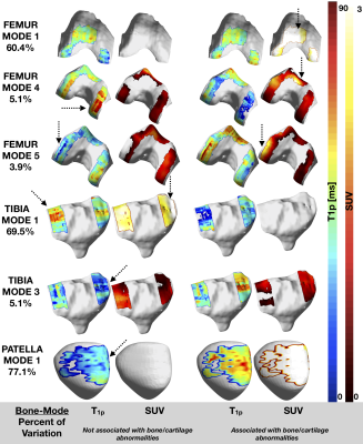 |
Principal Component Analysis of Simultaneous PET/MRI Reveals Patterns of Cartilage-Bone Interactions in Osteoarthritis
Radhika Tibrewala1, Valentina Pedoia1, Matthew Bucknor1, and Sharmila Majumdar1
1Radiology and Biomedical Imaging, University of California San Francisco, San Francisco, CA, United States
Osteoarthritis (OA) is a joint disorder, consisting of cartilage degeneration and metabolic bone changes, which have previously been correlated by using [18F]-NaF PET/MRI in the knee. However, these correlations were derived using averaging methods in areas of [18F] uptake in the bone and surrounding cartilage T1ρ/T2 mean values, which could miss potential multifaceted mechanisms that are not spatially correlated in the joint. The goal of this study is to find complex patterns in OA by building a cartilage-bone interface and using principal component analysis to find cartilage-bone interactions and find associations with known manifestations of OA.
|
|
0259.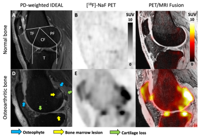 |
Evaluating the Relationship Between Dynamic [18F]-Sodium Fluoride Uptake Parameters and MRI Knee Osteoarthritic Findings
Lauren Watkins1, James MacKay2,3, Bryan Haddock4, Valentina Mazzoli5, Scott Uhlrich6, Garry Gold5, and Feliks Kogan5
1Bioengineering, Stanford University, Stanford, CA, United States, 2Radiology, University of East Anglia, Norwich, United Kingdom, 3Radiology, University of Cambridge, Cambridge, United Kingdom, 4Department of Clinical Physiology, Nuclear Medicine and PET, Copenhagen University Hospital, København, Denmark, 5Radiology, Stanford University, Stanford, CA, United States, 6Mechanical Engineering, Stanford University, Stanford, CA, United States
Abnormal bone physiology is a potential mechanism for the progression of knee osteoarthritis. Molecular information derived from PET imaging has shown promise in early detection of bone metabolic abnormalities. Here we investigated kinetic parameters of PET tracer ([18F]-NaF) uptake in subjects with knee osteoarthritis and evaluated the relationship between kinetic tracer uptake parameters and structural MRI findings. The kinetic parameters for [18F]-NaF delivery and uptake to regions of bone containing osteophytes, bone marrow lesions, and adjacent to cartilage lesions identified on MRI were significantly different compared to normal-appearing bone, suggesting strong spatial relationships between structural damage and bone metabolic abnormalities.
|
0260.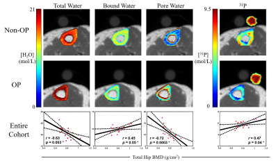 |
Quantitative Measurements of Bone Water and 31P in Postmenopausal Women: A Preliminary Study
Brandon Clinton Jones1, Cheng-Chieh Cheng1, Xia Zhao1, Mona Al Mukaddam2, Peter J Snyder2, Chamith S Rajapakse1, Hee Kwon Song1, and Felix W Wehrli1
1Radiology, University of Pennsylvania, Philadelphia, PA, United States, 2Medicine, University of Pennsylvania, Philadelphia, PA, United States
Seven osteoporotic treatment-naïve and 13 non-osteoporotic postmenopausal women have been examined in an ongoing MRI study to evaluate cortical bone properties. 1H dual-echo UTE and 1H IR-prepared rapid-UTE sequences were used for evaluation of pore and bound water concentrations in the tibial cortex, and a 31P PETRA-ZTE sequence for quantification of cortical bone mineralization. Elevated total water and pore water were found in the osteoporotic group (p=0.036, p=0.032), whereas 31P and bound water concentrations were not significantly different. Since pore water is a known surrogate of bone porosity, our preliminary results suggest it may be useful in evaluating bone health.
|
|
 |
0261.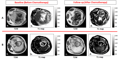 |
Can Tumor T1 Serve as Early Response Imaging Biomarker for Neoadjuvant Chemotherapy in Osteosarcoma? A Preliminary Study
Esha Baidya Kayal1, Nikhil Sharma1, Sameer Bakhshi2, Raju Sharma3, Devasenathipathy Kandasamy3, and Amit Mehndiratta1,4
Video Permission Withheld
1Centre for Biomedical Engineering, Indian Institute of Technology, Delhi, New Delhi, India, 2Department of Medical Oncology, Dr. B.R. Ambedkar Institute-Rotary Cancer Hospital (IRCH), All India Institute of Medical Sciences, New Delhi, New Delhi, India, 3Radio Diagnosis, All India Institute of Medical Sciences, New Delhi, New Delhi, India, 4Department of Biomedical engineering, All India Institute of Medical Sciences, New Delhi, New Delhi, India
Histopathological examination is the current gold standard for evaluating tumor response to anti-cancer therapy; though it is possible only after surgery. Non-invasive imaging biomarkers of tumor response to therapy may be useful in optimizing existing treatments improving overall outcome. The spin-lattice relaxation time of water protons (T1) reflects therapeutic changes in tumor and is thus a generic marker of tumor response to therapy. Experimental results showed T1 relaxation time reduces upon successful chemotherapy and thus, a change in tumor T1 may be a non-invasive imaging marker of chemo-sensitivity and chemotherapy response.
|
|
0262.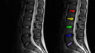 |
Accelerated T2 Mapping of the Lumbar Intervertebral Discs: Robust Quantification in Clinically Feasible Acquisition Times
Marcus Raudner1, Markus Schreiner1,2, Tom Hilbert3,4,5, Tobias Kober3,5,6, Anna Szelenyi7, Vladimir Juras1, and Siegfried Trattnig1
1High Field MR Centre, Department of Biomedical Imaging and Image-guided Therapy, Medical University of Vienna, Vienna, Austria, 2Department of Orthopedics and Trauma Srugery, Medical University of Vienna, Vienna, Austria, 3Advanced Clinical Imaging Technology, Siemens Healthcare, Lausanne, Switzerland, 4Department of Radiology, Lausanne University Hospital and University of Lausanne, Lausanne, Switzerland, 5LTS5, Ecole Polytechnique Fédérale de Lausanne (EPFL), Lausanne, Switzerland, 6Department of Radiology, University Hospital and University of Lausanne, Lausanne, Switzerland, 7Department of Biomedical Imaging and Image-guided Therapy, Medical University of Vienna, Vienna, Austria
The physiological biochemical state of the intervertebral disc (IVD) allows for passive water-storing capabilities because of a high concentration of glycosaminoglycans (GAG) and an inherent structural integrity. T2 mapping can quantitatively assess the IVD’s water content and GAG concentration, but a typical 2D multi echo spin echo (MESE) sequence suffers from clinically prohibitive scan times. To resolve this, we compared the MESE sequence against GRAPPATINI, an accelerated sequence combining parallel imaging and a model-based reconstruction for T2 quantification which uses undersampling to shorten the scan time from 13:18 to 2:27 minutes.
|
 Watch the Video