
Scientific Session
Cancer Imaging: Pre- & Post-Treatment
Session Topic: Cancer Imaging: Pre- & Post-Treatment
Session Sub-Topic: Cancer Imaging: Physiology & Metabolism
Oral
Cancer
| Monday Parallel 5 Live Q&A | Monday, 10 August 2020, 13:45 - 14:30 UTC | Moderators: Derek Johnson & Harish Poptani |
Session Number: O-38
0125.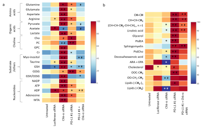 |
The immune checkpoint PD-L1 alters choline kinase expression and metabolism in triple negative breast cancer cells
Jesus Pacheco-Torres1, Marie-France Penet1,2, Flonne Wildes1, Yelena Mironchik1, Balaji Krishnamachary1, and Zaver M Bhujwalla1,2,3
1The Russell H Morgan Department of Radiology and Radiological Science, The Johns Hopkins University, School of Medicine, Baltimore, MD, United States, 2Sidney Kimmel Comprehensive Cancer Center, The Johns Hopkins University, School of Medicine, Baltimore, MD, United States, 3Radiation Oncology and Molecular Radiation Sciences, The Johns Hopkins University, School of Medicine, Baltimore, MD, United States
Expression of programmed death-ligand 1 (PD-L1) plays a significant role in creating an immune suppressive tumor microenvironment. We investigated the relationship between the aberrant choline metabolism observed in most cancers and PD-L1 expression in triple negative human MDA-MB-231 breast cancer cells. Using siRNA to downregulate Chk-a or PD-L1 or both, we identified a close inverse interdependence between Chk-α and PD-L1. We identified, for the first time, the role of PD-L1 in cancer cell metabolism. These results have significant implications for therapy and provide new insights into the relationship between metabolism and immune resistance in these breast cancer cells.
|
|
0126.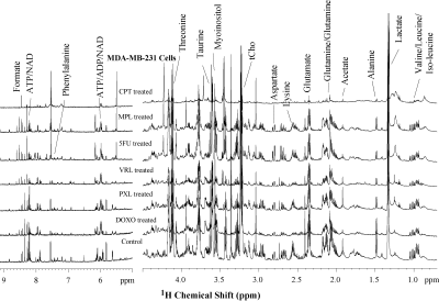 |
Chemotherapeutic drugs profoundly alter the metabolism of triple negative breast cancer cells
Kanchan Sonkar1, Caitlin M. Tressler1, and Kristine Glunde1,2
1The Russell H. Morgan Department of Radiology and Radiological Science, Johns Hopkins University School of Medicine, Baltimore, MD, United States, 2The Sidney Kimmel Comprehensive Cancer Center, Johns Hopkins University School of Medicine, Baltimore, MD, United States
Triple negative breast cancer (TNBC) is a highly aggressive form of cancer that poses severe health care problem as no targeted therapeutics are available for its treatment. TNBC is treated with chemotherapeutic agents, including doxorubicin, paclitaxel, vinorelbine, 5-fluorouracil, melphalan and cisplatin , which are either used alone or in various combinations. Studies investigating the metabolic effects of chemotherapy in TNBC are still limited. Here we have used high-resolution 1H MRS to study the metabolic profiles of TNBC cell lines MDA-MB-231 and SUM159 treated with these chemotherapeutic agents as compared to untreated controls.
|
|
 |
0127.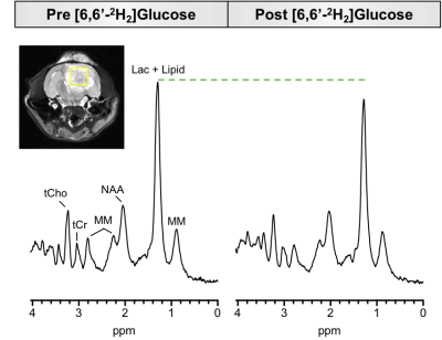 |
Detecting glycolytic metabolism in glioblastoma using a new 1H MRS and [6,6'-2H2]glucose infusion based approach
Laurie J Rich1, Puneet Bagga1, Gabor Mizsei1, Mitchell D Schnall1, John A Detre2, Mohammad Haris3, and Ravinder Reddy1
1Radiology, University of Pennyslvania, Philadelphia, PA, United States, 2Neurology, University of Pennyslvania, Philadelphia, PA, United States, 3Research Branch, Qatar University, Doha, Qatar
A key hallmark of malignant tissues is a metabolic shift from oxidative phosphorylation to glycolytic metabolism, leading to increased lactate production. Probing the kinetics of lactate production in vivo may play a key role in studying disease mechanisms and developing biomarkers of treatment response. Here, we developed a new approach for studying glycolytic metabolism in glioblastoma by combining 1H MRS with infusion of deuterated glucose. Infusion of [6,6'-2H2]glucose leads to downstream deuterium labeling of lactate, resulting in a reduction in the 1.33 ppm lactate peak on 1H MRS and making it is possible to monitor the metabolic turnover of lactate.
|
0128.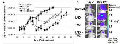 |
Metabolic modulation towards improved outcome in human glioblastoma model
Kavindra Nath1, David Nelson1, Jeffrey Roman1, Sofya Osharovich1, Saad Sheikh2, Stepan Orlovskiy1, Stephen Pickup1, Dennis Leeper3, Yancey Gillespie4, Corrine Griguer5, Jay Dorsey2, Mary Putt6, and Jerry Glickson1
1Radiology, University of Pennsylvania, Philadelphia, PA, United States, 2Radiation Oncology, University of Pennsylvania, Philadelphia, PA, United States, 3Radiation Oncology, Thomas Jefferson University, Philadelphia, PA, United States, 4Neurosurgery, University of Alabama, Birmingham, AL, United States, 5Radiation Oncology, University of Iowa, Iowa City, IA, United States, 6Biostatistics and Epidemiology, University of Pennsylvania, Philadelphia, PA, United States
Standard of care for glioblastoma multiforme (GBM) patients, the Stupp protocol, involves radiotherapy concurrent with adjuvant temozolomide (TMZ) chemotherapy. Lonidamine (LND), an inhibitor of monocarboxylate transporters, mitochondrial pyruvate carrier and mitochondrial complex I & II, is shown to potentiate TMZ chemotherapy inhibiting the growth of U251 glioblastoma cells orthotopically implanted in mice. LND effects measured in vivo by 31P and 1H MRS in subcutaneous U251 glioblastoma xenografts showed a sustained and tumor-selective decrease in intracellular pH, decrease in bioenergetics (βNTP/Pi) and an increase in lactate. Selective tumor acidification and deenergization induced by LND potentiated the TMZ response in U251 glioblastoma xenografts.
|
|
 |
0129.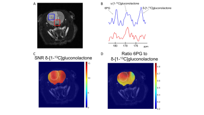 |
Hyperpolarized δ-[1-13C]gluconolactone monitors TERT-induced elevation in pentose phosphate pathway flux in brain tumors in vivo
Georgios Batsios1, Pavithra Viswanath1, Celine Taglang1, Robert Flavell1, Joseph Costello2, Russell O Pieper2, Peder Larson1, and Sabrina Ronen1
1Radiology and Biomedical Imaging, UCSF, San Francisco, CA, United States, 2Neurological Surgery, UCSF, San Francisco, CA, United States
Telomerase reverse transcriptase (TERT) expression is essential for tumor proliferation and is also an attractive therapeutic target for gliomas. Imaging TERT can help monitor tumor development and response to therapy. TERT expression has previously been shown to enhance glucose flux via the pentose phosphate pathway in low grade glioma cells expressing TERT. Here, we show that hyperpolarized δ-[1-13C]gluconolactone metabolism to 6-phospho-[1-13C]gluconate is significantly higher in tumor compared to contralateral normal brain in TERT-expressing low-grade oligodendrogliomas, pointing to the utility of hyperpolarized δ-[1-13C]gluconolactone for non-invasive in vivo assessment of this critical tumor hallmark in gliomas.
|
0130.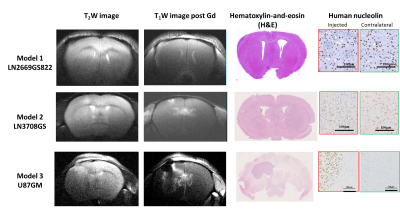 |
Hyperpolarized 13C-glucose MRS: a potential biosensor to visualize the infiltrative front in GBM
Mor Mishkovsky1, Olga Gusyatiner2, Bernard Lanz1, Cristina Cudalbu3, Irene Vassallo2, Marie-France Hamou2, Jocelyne Bloch2, Arnaud Comment4, Rolf Gruetter1,3,5,6, and Monika Hegi2
Video Permission Withheld
1Laboratory for Functional and Metabolic Imaging (LIFMET), Ecole Polytechnique Fédérale de Lausanne (EPFL), Lausanne, Switzerland, 2Department of Clinical Neurosciences, Lausanne University Hospital and University of Lausanne, Lausanne, Switzerland, 3Centre d'Imagerie Biomédicale (CIBM), Ecole Polytechnique Fédérale de Lausanne (EPFL), Lausanne, Switzerland, 4General Electric Healthcare, Chalfont St Giles, United Kingdom, 5Department of Radiology, University of Geneva (UNIGE), Geneva, Switzerland, 6Department of Radiology, University of Lausanne (UNIL), Lausanne, Switzerland
Glioblastoma (GBM) is the most malignant primary brain tumor in adults. Aberrant glucose metabolism is considered a hallmark of cancer, via the so-called ‘Warburg Effect’, however recent studies show distinct metabolic profile associated with the invasive phenotype in GBM, indicating active glucose oxidation. Hyperpolarized (HP) endogenous compounds, provides real-time metabolic information which is related to enzymatic activity. The aim of the present study was to apply HP 13C-glucose MRS in patient-derived GBM models and to investigate glucose metabolism in the infiltrative front of GBM, which potentially would enable to differentiate the invasive front of GBM from normal brain.
|
|
0131.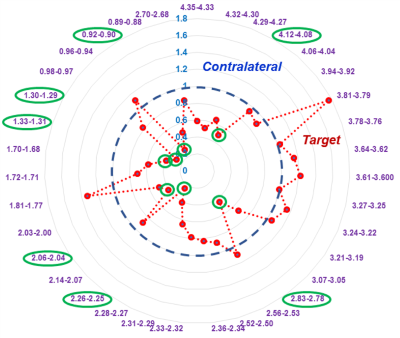 |
Metabolomic Characterization of Human Prostate Cancer with Tissue from MRI/US Fusion Biopsy
Leo L Cheng1, Lindsey Vandergrift1, Andrew Gusev1, Shulin Wu1, Mukesh Harisinghani1, Chin-Lee Wu1, and Adam Feldman1
1MGH/Harvard, Boston, MA, United States
Prostate cancer (PCa) clinic is challenged by heterogeneously distributed and clinical insignificant diseases. Multiparametric (mp)-MRI, with a PI-RADS score, correlated to clinically significant cancer and its morphological variations to establish a biopsy Target, and ultrasound fusion-guided biopsy guided to the targeted area has increased detection of clinically significant cancer. We studied PI-RADS score according to tissue MRS-based metabolomics. Metabolic differences between Target and contralateral cores, regardless if Targets were Ca-positive or not, support the assumption that targeted areas fundamentally and metabolomically differ from non-targeted areas.
|
|
0132.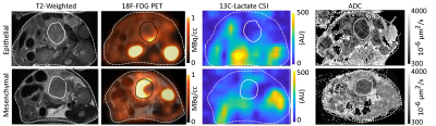 |
Differentiation of Murine Pancreatic Tumours at 7 T with Hyperpolarized 13C-Pyruvate-Lactate MRSI, 18F-FDG PET, and DWI
Geoffrey J. Topping1, Irina Heid2, Moritz Mayer2, Lukas Kritzner2, Florian Englert2, Martin Grashei1, Christian Hundshammer1, Katja Steiger3, Katja Peschke4, Markus Schwaiger1, Maximilian Reichert4, Franz Schilling1, and Rickmer Braren2
1Department of Nuclear Medicine, Klinikum rechts der Isar, Technical University of Munich, Munich, Germany, 2Institute of Radiology, Klinikum rechts der Isar, Technical University of Munich, Munich, Germany, 3Institute of Pathology, Technical University of Munich, Munich, Germany, 4Internal Medicine II, Klinikum rechts der Isar, Technical University of Munich, Munich, Germany
Multimodal imaging for characterization of pancreatic tumour cellularity and metabolism has potential to guide treatment. Murine orthotopically transplanted tumours were imaged with DWI, 13C-pyruvate CSI, and 18F-FDG PET, and endogenous tumours with DWI and CSI. Transplanted epithelial and mesenchymal tumours had similar cellularity, shown by ADC, but different metabolism, with higher mesenchymal AUC ratios and SUV. Compared with other endogenous tumour growth patterns, classical ductal had lower tumour cellularity (higher ADC), while solid had higher and more-variable AUC ratios. The combination of these methods can characterize tumour metabolism, including correcting for tumour cellularity, better than CSI alone.
|
|
|
0133.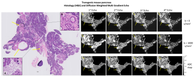 |
Identification of pancreatic intraepithelial neoplasia in the mouse pancreas with MR Microscopy
Carlos Bilreiro1, Rui V. Simões1, Francisca F. Fernandes1, Mireia Castillo-Martin1, Kevin Harkins2, Mark Does2, Celso Matos1, and Noam Shemesh1
1Champalimaud Research, Champalimaud Centre for the Unknown, Lisbon, Portugal, 2Department of Biomedical Engineering, Vanderbilt University, Nashville, TN, United States
Survival in pancreatic cancer resides on an early diagnosis, for which current imaging methods are insufficient. Here, we investigated which MRI contrast can reflect pancreatic pre-neoplastic lesions, particularly, pancreatic intraepithelial neoplasia (PanIN). To this end, we developed an ultrafast DWI-MGE pulse sequence and performed MR microscopy on pancreas extracted from transgenic mice with PanIN lesions (along with controls), and validated our findings using histology. PanIN lesions were clearly detected in the transgenic mice and differentiated from inflammatory changes at b=1000 sec/mm2 and long TE. Our findings are encouraging for future detection of PanIN in vivo.
|
 Watch the Video