
Scientific Session
Cancer Imaging: Pre- & Post-Treatment
Session Topic: Cancer Imaging: Pre- & Post-Treatment
Session Sub-Topic: Cancer Imaging: Perfusion & Diffusion
Oral
Cancer
| Monday Parallel 5 Live Q&A | Monday, 10 August 2020, 13:45 - 14:30 UTC | Moderators: Shu Xing |
Session Number: O-39
0134.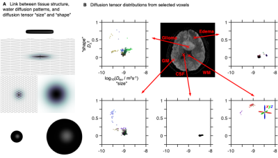 |
Diffusion tensor distribution imaging of brain tumor microstructure and heterogeneity
João de Almeida Martins1, Samo Lasic1, Yuan Zheng2, Qing Wei2, Sirui Li3, Wenbo Sun3, Haibo Xu3, Karin Bryske1, and Daniel Topgaard1,4
1Random Walk Imaging, Lund, Sweden, 2United Imaging Healthcare, Shanghai, China, 3Zhongnan Hospital of Wuhan University, Wuhan, China, 4Lund University, Lund, Sweden
For voxels containing multiple cell or tissue types, DTI metrics are challenging to interpret in terms of specific microstructural properties. We address this problem by adapting encoding and inversion strategies from solid-state and low-field NMR to determine diffusion tensor distributions (DTDs) with dimensions corresponding to cell densities, shapes, and orientations. Three patients with glioma, meningioma, or cerebral cyst underwent 5 min DTD imaging giving 15 distinct parameter maps for quantitative analysis and intuitive microsctructural interpretation. The DTD-derived metrics showed good agreement with expected tissue properties and structural insights not accessible with DTI.
|
|
 |
0135.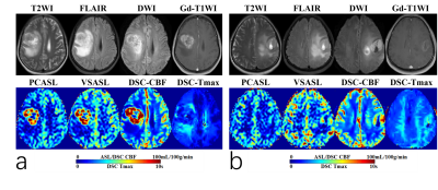 |
Perfusion Measurement in Brain Gliomas Using Velocity-Selective Arterial Spin Labeling: Comparison with PCASL and DSC Perfusion
Yaoming Qu1, Zhibo Wen1, and Qin Qin2,3
1Imaging diagnostic department, Zhujiang hospital of southern medical university, Guangzhou, China, 2The Russell H. Morgan Department of Radiology and Radiological Science, Johns Hopkins University School of Medicine, Baltimore, MD, United States, 3F.M. Kirby Research Center for Functional Brain Imaging, Kennedy Krieger Institute, Baltimore, MD, United States
Velocity-selective arterial spin labeling (VSASL) employing Fourier-transform based velocity-selective pulse trains is an emerging method for quantifying cerebral blood flow (CBF) with high sensitivity to perfusion signal. Its utility was assessed for glioma patients at 3T by a comparison between pseudo-continuous ASL with DSC-PWI. We demonstrated the existence of various and prolonged arterial transit time (ATT) in high-grade gliomas. Detecting by the dependence of the CBF based on Tmax, lesser sensitivity to ATT in VSASL was reported. VSASL showed great promise for accurate quantification of CBF and could potentially improve the diagnostic performance of ASL in preoperative grading of gliomas.
|
0136.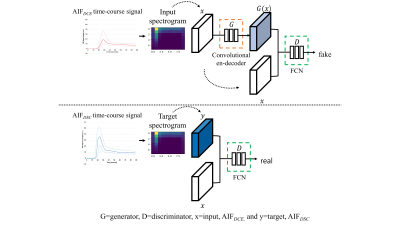 |
Improving the reliability of pharmacokinetic parameters in dynamic contrast-enhanced MRI in gliomas: Deep learning approach
Kyu Sung Choi1, Sung-Hye You2, Yoseob Han1, Jong Chul Ye1, Seung Hong Choi3, and Bumseok Jeong1
1Korea Advanced Institute for Science and Technology, Daejeon, Korea, Republic of, 2Korea University College of Medicine, Seoul, Korea, Republic of, 3Seoul National University Hospital, Seoul, Korea, Republic of
AIFDCE has been known to be sensitive to noise, because of the relatively weak T1 contrast-enhanced MR signal intensity (SI) compared to the T2* SI of DSC-MRI, leading to PK parameters – Ktrans, Ve, and Vp – with low reliability. In this study, we developed a neural network model generating an AIF similar to the AIF obtained from DSC-MRI – AIFgenerated DSC – and demonstrated that the accuracy and reliability of Ktrans and Ve derived from AIFgenerated DSC can be improved compared to those from AIFDCE without obtaining DSC-MRI, not leading to an additional
deposition of gadolinium in the brain.
|
|
0137.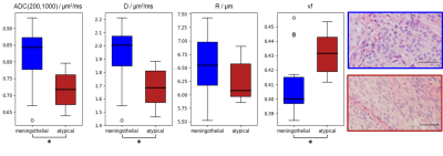 |
Characterizing the ADC-microstructure relationship in meningiomas through computational modelling.
Giulia Buizza1, Chiara Paganelli1, Lorenzo Preda2, Francesca Valvo2, Daniel C. Alexander3, Guido Baroni1,2, and Marco Palombo3
1CartCasLab at Department of Electronics, Information and Bioengineering, Politecnico di Milano, Milan, Italy, 2National Center Of Oncological Hadrontherapy (CNAO), Pavia, Italy, 3Centre for Medical Image Computing and Dept of Computer Science, University College London, London, United Kingdom
Tumour microstructure can be probed with diffusion-weighted MRI (DW-MRI), but the clinically-adopted apparent diffusion coefficient (ADC) lacks a clear link to microstructure. Aim of this work was to detail the ADC-microstructure relationship using a computational framework. Relying on a sparse representation of simulated DW-MRI data, we estimated diffusivity (D), cell radius (R) and volume fraction (vf) for 27 low and high-grade meningioma patients, which significantly differed in ADC, D and vf. Preliminary results showed the potential of the proposed framework for meningioma grading and proton-therapy response assessment, although extension to richer data and histological validation need to be further addressed.
|
|
0138.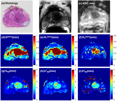 |
Parametric maps from the two-tissue compartment model for prostate DCE-MRI: compared with the standard Tofts model in diagnosis of cancer
Xiaobing Fan1, Xueyan Zhou2, Aritrick Chatterjee1, Aytekin Oto1, and Gregory S. Karczmar1
1Radiology, The University of Chicago, Chicago, IL, United States, 2Harbin University, Harbin, China
We compared standard Tofts model with a two-tissue compartment model (2TCM) of dynamic contrast enhanced (DCE) MRI for diagnosis of prostate cancer. The 2TCM has one slow and one fast exchanging compartment. The standard Tofts model parameters (Ktrans and kep) were compared with the 2TCM parameters (Kitrans and kiep, i=1,2). There was a strong correlation between Ktrans and K1trans for cancer, but weak correlation between kep and k1ep.
This demonstrated that the Tofts model often does not fit contrast agent concentration curves accurately, and the 2TCM can provide new diagnostic information with fewer false positives in diagnosis of prostate cancer.
|
|
 |
0139.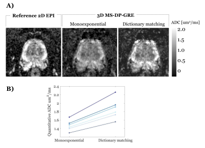 |
3D multi-shot diffusion imaging of the prostate with inter-shot correction and dictionary-based ADC matching
Elisa Roccia1, Radhouene Neji1,2, Thomas Benkert3, Berthold Kiefer3, Vicky Goh4, and Isabel Dregely1
1Biomedical Engineering Department, School of Biomedical Engineering and Imaging Sciences, King's College London, London, United Kingdom, 2Siemens Healthcare Limited, Frimley, United Kingdom, 3Siemens Healthcare GmbH, Erlangen, Germany, 4Cancer Imaging Department, School of Biomedical Engineering and Imaging Sciences, King's College London, London, United Kingdom
Diffusion imaging is a key contrast in assessing prostate cancer. However, current single-shot EPI-based techniques are often distorted and fundamentally limited in resolution. The aim of this study is to develop multi-shot diffusion-prepared gradient echo imaging to obtain accurate 3D ADC maps in the prostate. We developed a 3D Cartesian centric trajectory with self-navigation, and a shot rejection approach to correct for inter-shot magnitude errors. We used a custom dictionary of acquisition specific signal evolutions to estimate ADC. We have shown in simulations and in vivo that accurate ADC values could be recovered.
|
|
0140.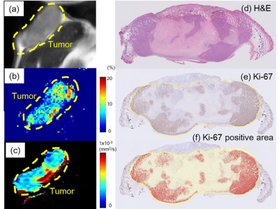 |
Investigation of breast cancer microstructure and microvasculature from time-dependent DWI and CEST in correlation with histology
Yuko Someya1, Mami Iima1,2, Hirohiko Imai3, Akihiko Yoshizawa4, Yuji Nakamoto1, Masako Kataoka1, Hiroyoshi Isoda1, and Kaori Togashi1
1Diagnostic Imaging and Nuclear Medicine, Kyoto University Graduate School of Medicine, Kyoto, Japan, 2Clinical Innovative Medicine, Institute for Advancement of Clinical and Translational Science, Kyoto University Hospital, Kyoto, Japan, 3Kyoto University Graduate School of Informatics, Kyoto, Japan, 4Diagnostic Pathology, Kyoto University Graduate School of Medicine, Kyoto, Japan
The association of time-dependent DWI (shifted ADC [sADC], IVIM, and non-Gaussian DWI) at different diffusion times and CEST (MTRasym, APT signal intensity) parameters was investigated with histological biomarkers in a breast cancer xenograft model. ADC values decreased with increased diffusion times. sADC values at a diffusion time=5ms had significant negative correlation with Ki-67 (r=−0.63, P<0.05). MTRasym had a significant positive correlation with Ki-67 positive area (r=0.73, P<0.05). Significant association was found between fIVIM and microvessel density (r=0.80, P<0.01). These results indicate their utility for investigating microstructure and microcirculation of breast cancers without using contrast agents.
|
|
0141.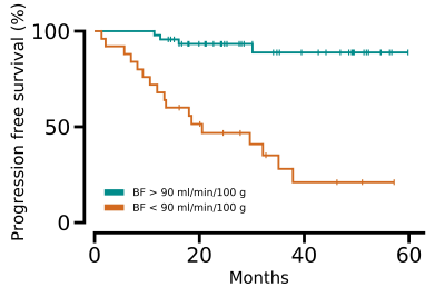 |
Perfusion MRI related to survival, treatment response and sex differences in rectal cancer
Kine Mari Bakke1,2, Sebastian Meltzer1, Endre Grøvik3, Anne Negård4,5, Stein Harald Holmedal4, Kjell-Inge Gjesdal4, Atle Bjørnerud2,3, Anne Hansen Ree1,5, and Kathrine Røe Redalen6
1Department of Oncology, Akershus University Hospital, Lørenskog, Norway, 2Department of Physics, University of Oslo, Oslo, Norway, 3Department of Diagnostic Physics, Division of Radiology and Nuclear Medicine, Oslo University Hospital, Oslo, Norway, 4Department of Radiology, Akershus University Hospital, Lørenskog, Norway, 5Institute of Clinical Medicine, University of Oslo, Oslo, Norway, 6Department of Physics, Norwegian University of Science and Technology, Trondheim, Norway
Three different MRI methods for obtaining perfusion related parameters were compared and evaluated as biomarkers in a study of 94 rectal cancer patients. The methods were dynamic contrast enhanced (DCE) MRI and dynamic susceptibility contrast (DSC) MRI analysed from a multi-echo dynamic EPI sequence, as well as intravoxel incoherent motion (IVIM) MRI analysed from a diffusion weighted sequence with 7 b-values. Tumour blood flow from DSC MRI was correlated to D* from IVIM MRI as well as Ktrans and vp from DCE MRI. Blood flow was also related to progression free survival, overall survival, treatment response and sex differences.
|
|
0142.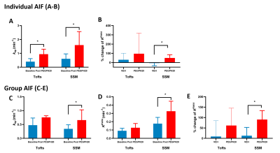 |
How the choice of PK model and AIF affect DCE-MRI detection of pancreatic cancer responses to stroma-directed drug?
Jianbo Cao1, Stephen Pickup1, Peter O’Dwyer2,3, Mark Rosen1,3, and Rong Zhou1,3
1Department of Radiology, University of Pennsylvania, Philadelphia, PA, United States, 2Pancreatic Cancer Research Center, University of Pennsylvania, Philadelphia, PA, United States, 3Abramson Cancer Center, University of Pennsylvania, Philadelphia, PA, United States
Pancreatic ductal adenocarcinoma (PDA) is characterized by a dense stroma, which poses a substantial barrier to drug penetration and motivates development of stroma-directed interventions. We aim to test the utility of DCE-MRI to predict PDA responses to such treatment. We compared individual versus group-arterial input function approach and metric including Ktrans, kep and Vp derived from three commonly used pharmacokinetic models. Our data provides rationale for choice of PK model and AIF approach which lead to quantitative DCE-MRI marker of optimal sensitivity and specificity for detection of PDA responses to human hyaluronidase that reduces PDA stroma.
|
 Watch the Video