
Scientific Session
Cancer Imaging: Pre- & Post-Treatment
Session Topic: Cancer Imaging: Pre- & Post-Treatment
Session Sub-Topic: Cancer Imaging: Treatment Planning & Response Assessment
Oral
Cancer
| Monday Parallel 5 Live Q&A | Monday, 10 August 2020, 13:45 - 14:30 UTC | Moderators: Sola Adeleke & Arvind Pathak |
Session Number: O-40
 |
0143.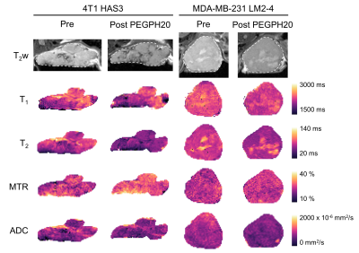 |
Breast tumour response to PEGPH20-induced stromal modulation assessed by multiparametric MRI
Emma L. Reeves1, Jin Li1, Jessica K. R. Boult1, Barbara Blouw2, David Kang2, Jeffrey C. Bamber1, Yann Jamin1, and Simon P. Robinson1
1Radiotherapy and Imaging, Institute of Cancer Research, London, United Kingdom, 2Halozyme Therapeutics, San Diego, CA, United States
Degradation of hyaluronan by PEGPH20 can improve stromal-dense tumour response to therapy. Given PEGPH20 treatment is associated with a reduction in tumour water content, we hypothesised that T1, T2, MTR and ADC may inform on PEGPH20 response. MRI was performed before and after PEGPH20 treatment in 4T1 HAS3 and MDA-MB-231 LM2-4 orthotopic breast tumours. T1, T2, and ADC significantly decreased, and MTR significantly increased following PEGPH20 treatment in 4T1 HAS3 tumours. PEGPH20 significantly decreased ADC but did not change T1, T2 or MTR in MDA-MB-231 LM2-4 tumours. These data suggest that ADC can detect breast tumour response to PEGPH20.
|
0144. |
The Role of Iron Chelation in the Tumour Microenvironment of Triple-Negative Breast Cancer
Paola Porcari1, Ellen Ackerstaff1, Dov P Winkleman1, Suresh Veeraperumal1, Natalia Kruchevsky1, H. Carl Lekaye1, and Jason A. Koutcher1,2,3,4
1Medical Physics, Memorial Sloan Kettering Cancer Center, New York, NY, United States, 2Department of Medicine, Memorial Sloan Kettering Cancer Center, New York, NY, United States, 3Molecular Pharmacology Program, Memorial Sloan Kettering Cancer Center, New York, NY, United States, 4Weill Cornell Medical College, Cornell University, New York, NY, United States
Intracellular iron, essential for cancer cell proliferation and metabolism, is modified in cancer cells. Triple-negative breast cancers are metastatic cancers associated with a high recurrence rate, poor prognosis and lack of effective targeted therapies. We are investigating the potential of Deferiprone, a clinically approved intracellular iron chelator for non-cancer related diseases, to improve chemotherapeutic treatment response in triple-negative breast cancer by altering iron-dependent proliferation and metabolism. The effectiveness of Deferiprone to impair triple-negative breast cancer cell growth and affect cellular metabolism was evaluated by monitoring live cells, exposed to Deferiprone, in an MR-compatible cell bioreactor using multi-nuclear MRS.
|
|
0145.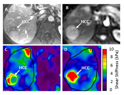 |
Immune Checkpoint Blockade (ICB) Response Evaluation with MRI/MR Elastography (MRE) in Surgical and Non-Surgical Patients with HCC
Aliya Qayyum1, Rony Avritscher2, Rizwan Aslam1, Jingfei Ma3, Mark Pagel4, Jia Sun5, Yehia Ibrahim Mohammed6, Manal Hassan7, Hesham Amin8, Asif Rashid9, Sunyoung Lee6, Robert A Wolff6, James C Yao6, Richard L Ehman10,
Gabriel Daniel Duda11, and Ahmed Omar Kaseb12
1Radiology, MD Anderson, Houston, TX, United States, 2Interventional Radiology, MD Anderson, Houston, TX, United States, 3Imaging Physics, MD Anderson, Houston, TX, United States, 4Cancer Systems Imaging, MD Anderson, Houston, TX, United States, 5Biostatistics, MD Anderson, Houston, TX, United States, 6GI Medical Oncology, MD Anderson, Houston, TX, United States, 7Epidemiology, MD Anderson, Houston, TX, United States, 8Hemopathology, MD Anderson, Houston, TX, United States, 9Pathology, MD Anderson, Houston, TX, United States, 10Radiology, Mayo Clinic, Rochester, MN, United States, 11Radiation Oncology, Massachusetts General Hospital, Boston, MA, United States, 12MD Anderson, Houston, TX, United States
Newer systemic treatments for advanced hepatocellular carcinoma (HCC) include immune checkpoint blockade (ICB) which act through increasing cytotoxic T-cell mediated response to tumor. There is a lack of biomarkers of ICB response and treatment outcomes are not correlated with change in tumor size. We evaluated MRI imaging features of HCC and change in tumor stiffness after 6 weeks immunotherapy in surgical and non-surgical patients. An increase in HCC stiffness on MRE after 6 weeks treatment was significantly correlated with treatment response. Longitudinal measurement of tumor stiffness on MRE provides a novel technique for early immunotherapy response assessment.
|
|
0146.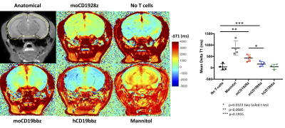 |
Imaging blood-brain barrier disruption caused by CD19 based CAR-T cell immunotherapy
Puneet Bagga1, Stephen Pickup1, Denis Migliorini2, Neil E Wilson1, Mohammad Haris3,4, Suyash Mohan1, Avery D Posey2, and Ravinder Reddy1
1Department of Radiology, University of Pennsylvania, Philadelphia, PA, United States, 2Center for Cellular Immunotherapies, University of Pennsylvania, Philadelphia, PA, United States, 3Sidra Medicine, Doha, Qatar, 4LARC, Qatar University, Doha, Qatar
With the advent of clinically effective CD19 based chimeric antigen receptor (CAR) T cell immunotherapies, there are incidents of associated neurotoxicity. In this study, we report the use of gadolinium enhanced MRI to image BBB disruption caused by CD1928z CAR-T cell on target action against brain pericytes of immunodeficient non-tumor bearing NSG mice. Pericytes are mural cells that wrap endothelial cells and are critical for maintaining blood-brain-barrier (BBB) integrity and express CD19. The MRI results were also found to corroborate with the Evans blue dye BBB permeability assay.
|
|
0147.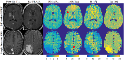 |
Impact of Single Fraction Stereotactic Radiosurgery vs. Hypofractionated Radiation Therapy on CEST and MT Parameters of Brain Metastases
Hatef Mehrabian1, Wilfred W Lam1, Hany Soliman1,2,3, Sten Myrehaug2,3, Arjun Sahgal1,2,3, and Greg J Stanisz1,4
1Physical Sciences, Sunnybrook Research Institute, Toronto, ON, Canada, 2Radiation Oncology, Sunnybrook Health Sciences Centre, Toronto, ON, Canada, 3Radiation Oncology, University of Toronto, Toronto, ON, Canada, 4Medical Biophysics, University of Toronto, Toronto, ON, Canada
Brain metastases are treated with single fraction stereotactic radiosurgery (sf-SRS) or hypofractionated radiation therapy (HFTR). CEST was previously shown to identify responders to sf-SRS within one-week post-treatment. This study investigated the differences in CEST and MT properties of brain metastases treated with sf-SRS and 5 fraction HFRT (5f-HFRT) one week after treatment. We observed statistically significantly larger reduction in CEST properties of tumours treated with sf-SRS compared to those treated with 5f-HFRT. However, changes in MT properties of the two cohort were similar. Such differences should be considered when evaluating response of brain metastases to radiotherapy using CEST and MT.
|
|
 |
0148.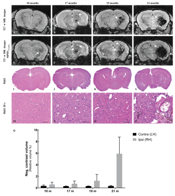 |
Low dose brain irradiation leads to delayed neuro-inflammation
Dina Sikpa1, Jérémie P. Fouquet1, Luc Tremblay1, Réjean Lebel1, Benoit Paquette1, and Martin Lepage1
1Université de Sherbrooke, Sherbrooke, QC, Canada
We studied the late radiation effect of a low radiation dose on the healthy mouse brain using MRI and histology. MRI enables the visualisation of early inflammation and late radiation necrosis. Histological analysis confirmed tissue damage and revealed that cellular (astrocytes, microglia) and molecular activation (ICAM-1, VCAM-1) as a result of neuro-inflammation precedes the formation of the necrotic core.
|
|
0149.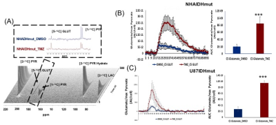 |
Hyperpolarized [1-13C]/[5-13C] glutamate as a metabolic imaging marker of IDH1 mutant glioma response to temozolomide therapy
Elavarasan Subramani1, Chloe Najac1, Georgios Batsios1, Marina Radoul1, Pavithra Viswanath1, Abigail Molloy1, Donghyun Hong1, Anne Marie Gillespie1, Russell O. Pieper2,3, Joseph Costello2, and Sabrina M Ronen1,3
1Department of Radiology and Biomedical Imaging, University of California San Francisco, San Francisco, CA, United States, 2Department of Neurological Surgery, Helen Diller Research Center, University of California San Francisco, San Francisco, CA, United States, 3Brain Tumor Research Center, University of California San Francisco, San Francisco, CA, United States
Temozolomide (TMZ) is most commonly used for the treatment of primary glioblastoma but is now being considered for the treatment of low-grade glioma that harbor mutations in the cytosolic isocitrate dehydrogenase 1 (IDH1) gene. Though the treatment of IDH1 mutant patients with TMZ improves survival, there is a need for complementary metabolic imaging approaches to help in assessing early response to therapy. Hyperpolarized 13C magnetic resonance spectroscopy-based metabolic profiling of mutant IDH1 cells treated with TMZ revealed that [1-13C]/[5-13C] glutamate production from [1-13C] α-ketoglutaric acid/[2-13C] pyruvate could serve as translatable biomarkers of response to therapy.
|
0150.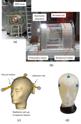 |
MRI-guided real-time 4D Radiation Dosimetry at an MRI-Linac using Polymer Gel Dosimeters
Yves De Deene1, Morgan Wheatley2, Gary Liney3, David Waddington4, Urszula Jelen3, and Bin Dong3
1Engineering, Macquarie University, North Ryde - Sydney, Australia, 2Macquarie University, North Ryde - Sydney, Australia, 3Ingham Institute, Liverpool, Australia, 4The University of Sydney, Sydney, Australia
4D radiation dosimetry using a highly radiation-sensitive polymer gel dosimeter with real-time quantitative MRI readout is presented as a technique to acquire the accumulated radiation dose distribution during image guided radiotherapy (IGRT) in an MRI-Linac. Optimized T2 weighted TSE scans are converted to quantitative R2 maps and subsequently to radiation dose maps. A further increase in temporal resolution using a keyhole imaging approach is proposed. The potential use of real-time 4D radiation dosimetry for safeguarding image guided radiotherapy (IGRT) of moving and deforming targets in an MRI-Linac will be discussed.
|
|
0151.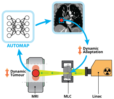 |
Towards Real-Time Beam Adaptation on an MRI-Linac using AUTOMAP
David Waddington1,2, Nicholas Hindley1, Neha Koonjoo2,3, Tess Reynolds1, Bo Zhu2,3, Chiara Paganelli4, Matthew Rosen2,3,5, and Paul Keall1
1ACRF Image X Institute, Faculty of Medicine and Health, The University of Sydney, Sydney, Australia, 2A. A. Martinos Center for Biomedical Imaging, Charlestown, MA, United States, 3Department of Physics, Harvard University, Cambridge, MA, United States, 4Dipartimento di Elettronica, Informazione e Bioingegneria, Politecnico di Milano, Milan, Italy, 5Harvard Medical School, Boston, MA, United States
MRI-Linacs are new cancer treatment machines integrating radiotherapy with MRI. Dynamically adapting the radiation beam on the basis of MR-detected anatomical changes (e.g. respiratory and cardiac motion) promises to increase the accuracy of MRI-Linac treatments. A key challenge in real-time beam adaptation is accurately reconstructing images in real time. Historically, reconstruction of data acquired with accelerated techniques, such as compressed sensing, has been very slow. Here, we use AUTOMAP, a machine-learning framework, to quickly and accurately reconstruct radial MRI data simulated from a digital thorax phantom. These results will guide development of real-time adaptation technologies on MRI-Linacs.
|
|
0152.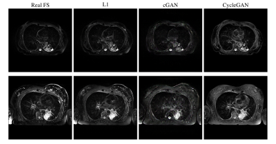 |
Retrospective Fat Suppression for Lung Radiotherapy Planning with Deep Learning Convolutional Neural Networks
Benjamin C Rowland1, Steven Jackson2, David Cobben1,2, Hanna Maria Hanson1, Ahmed Saleem1, Kathryn Banfill2, Lisa McDaid2, Michael Dubec2, and Marcel van Herk1
1University of Manchester, Manchester, United Kingdom, 2The Christie NHS Trust, Manchester, United Kingdom
We investigated three different Deep Learning techniques for performing retrospective fat suppression in T2 weighted imaging of lung cancer. The methods considered were two U-nets, using an L1 cost function or a conditional GAN, and a CycleGAN. The networks were trained on 900 images and then 16 test images were scored by 3 oncologists and a research radiographer. The L1 U-net and CycleGAN were scored at 73% and 72% respectively, relative to a gold standard of 80% for prospectively fat saturated images, and the scorers indicated they would be happy to use the generated images for radiotherapy target delineation.
|
 Watch the Video