
Scientific Session
Cancer Imaging: Machine Learning & Advanced Imaging
Session Topic: Cancer Imaging: Machine Learning & Advanced Imaging
Session Sub-Topic: Cancer Imaging: Non-Proton & Exogenous Contrast
Oral
Cancer
| Monday Parallel 5 Live Q&A | Monday, 10 August 2020, 14:30 - 15:15 UTC | Moderators: Matthew Grech-Sollars & Esther Warnert |
Session Number: O-41
0270.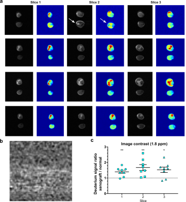 |
Deuterium MRI imaging of xenografted tumors following in vivo deuterated water labeling
Julian C. Assman1, Jeffrey R. Brender2, Don E. Farthing1, Keita Saito2, Kathrynne A. Warrick1, Natella Maglakelidze1, Hellmut R. Merkle3, Murali C. Krishna2, Ronald E. Gress1, and Nataliya P. Buxbaum1
1Experimental Transplantation and Immunotherapy Branch, National Cancer Institute, National Institutes of Health, Bethesda, MD, MD, United States, 2Radiation Biology Branch, Center for Cancer Research, National Cancer Institute, National Institutes of Health, Bethesda, MD, MD, United States, 3Laboratory for Functional and Molecular Imaging, National Institute of Neurological Disorders and StrokNational Institutes of Health, Bethesda, MD, MD, United States
Water is a substrate for many biochemical reactions. If D2O is ingested, it will be incorporated into proliferating cells. We hypothesized that rapidly proliferating cancer cells would become preferentially labeled with 2H which would allow visualization by deuterium MRI following a short in vivo D2O labeling period. We initiated systemic D2O labeling in HT-29 and MiaPaCa-2 xenograft models and performed deuterium MRI following 7 and 14 days of in vivo tumor growth and labeling. We show that small tumors could be distinguished from normal tissue by the incorporation D2O into lipids with a greater sensitivity and selectivity than anatomical MRI.
|
|
0271.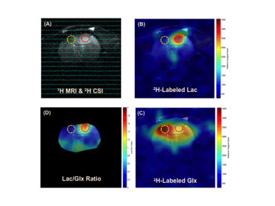 |
Dynamic Deuterium MRS Imaging of Brain Tumor with Enhanced Sensitivity and Spatiotemporal Resolution
Xiao-Hong Zhu1, Tao Wang1, Yibo Zhao2,3, Yudu Li2,3, Rong Guo2,3, Yi Zhang1, Walter Low4, Zhi-Pei Liang2,3, and Wei Chen1
Video Permission Withheld
1CMRR, Department of Radiology, University of Minnesota, Minneapolis, MN, United States, 2Beckman Institute for Advanced Science and Technology, University of Illinois at Urbana-Champaign, Urbana, IL, United States, 3Departments of Electrical and Computer Engineering, University of Illinois at Urbana-Champaign, Urbana, IL, United States, 4Department of Neurosurgery, University of Minnesota, Minneapolis, MN, United States
Noninvasive MR-based metabolic imaging of brain tumor may offer new tools for clinic diagnosis and monitoring of tumor growth or assessment of treatment efficacy. One potential candidate is the dynamic deuterium MRS (DMRS) imaging technique recently developed. To reach its full potential, we integrated advanced data processing with D-MRSI to enhance its sensitivity or spatiotemporal resolution. We demonstrated in this pilot study that quantitative “Warburg Effect” map and kinetic time courses of deuterated metabolites can be achieved with good spatiotemporal scales in rat brain tumor using Deep-SPICE based deuterium MRSI, which could potentially be applied to brain tumor patients.
|
|
0272.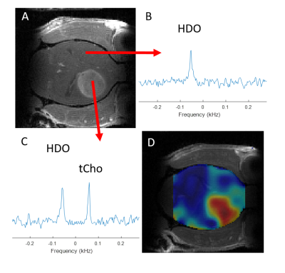 |
Mapping of exogenous choline uptake in brain tumors in vivo using Deuterium Metabolic Imaging (DMI)
Kevan L. Ip1, Monique A. Thomas1, Akshay Khunte1, Kevin L. Behar2, Robin A. de Graaf1, and Henk M. De Feyter1
1Dept. of Radiology and Biomedical Imaging, Yale University, New Haven, CT, United States, 2Dept. of Psychiatry, Yale University, New Haven, CT, United States
A high level of intracellular choline is an established marker of malignancy in brain tumors. Here we investigate the uptake of exogenous choline in vitro using high resolution 1H NMR into rodent glioblastoma cell lines. To map bloodborne uptake in vivo, we used the novel technique Deuterium Metabolic Imaging (DMI), combined with intravenous infusion of [2H9]-choline in two orthotopic rat (RG2) and mouse (GL261) models of glioblastoma. DMI-based metabolic maps revealed high uptake of choline in the tumors, in a stark image contrast with normal-appearing brain, illustrating the potential of [2H9]-choline chloride as metabolic imaging agent.
|
|
0273.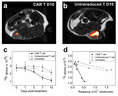 |
In vivo fluorine-19 MRI of intracellular oximetry response to chimeric antigen receptor T cell therapy against glioma
Fanny Chapelin1, Wenlian Zhu2, Benjamin Leach2, Hideho Okada3, and Eric Ahrens2
1Biomedical Engineering, University of Kentucky, Lexington, KY, United States, 2Radiology, University of California San Diego, La Jolla, CA, United States, 3Neurological Surgery, University of California San Francisco, San Francisco, CA, United States
We explore the temporal dynamics of tumor and T cell intracellular partial pressure of oxygen (pO2) in a murine flank glioma model receiving chimeric antigen receptor (CAR) T cell therapy. Tumor cells or T cells are intracellularly labeled with perfluorocarbon nanoemulsion prior to injection. 19F MRI relaxation rate measurements are used to elucidate intracellular pO2 in vivo. The tumor pO2 peaks at 3 days post-infusion, commensurate with CAR T cell infiltration and tumor cell killing. Moreover, the absolute 19F signal scales with tumor burden. Overall, 19F pO2 MRI measurements can assay cell-mediated apoptosis and provide insight into effector cell function.
|
|
 |
0274.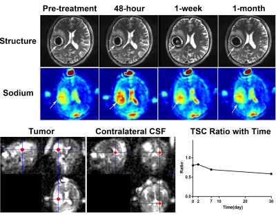 |
Quantitative Sodium MRI at 7T for the Early Efficacy Evaluation of Stereotactic Radiotherapy for Intracranial Tumors
Zihao Zhang1,2,3, Lichao Huang4, Xubin Chai1,2,3, Zhentao Zuo1,2,3, Zhe Wang1,2,3, Jing An5, Longsheng Pan4, and Yan Zhuo1,2,3
1State Key Laboratory of Brain and Cognitive Science, Institute of Biophysics, Chinese Academy of Sciences, Beijing, China, 2University of Chinese Academy of Sciences, Beijing, China, 3CAS Center for Excellence in Brain Science and Intelligence Technology, Beijing, China, 4Department of Neurosurgery, PLA General Hospital, Beijing, China, 5Siemens Shenzhen Magnetic Resonance Ltd., Shenzhen, China
Follow-up efficacy evaluation of stereotactic radiotherapy (SRT) helps to provide individualized treatment for intracranial tumors. However, biological changes at cytological level happen earlier than structural MRI can detect. Tissue sodium concentration (TSC) is sensitive to changes of tissue metabolic state and cell membrane integrity. In this study, we used sodium MRI at 7T to noninvasively quantify TSC and monitor the evolution of intracranial tumors after SRT. The results demonstrated that quantitative sodium MRI at 7T can reflect the efficacy of SRT, predict the volume change of tumor, and has the potential of finding the relapse of tumors in early stage.
|
0275.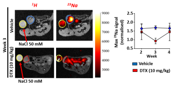 |
Utilising preclinical 23Na MRI to probe the ionic microenvironment of breast cancer: developing novel diagnostics to probe treatment efficacy.
Andrew D James1,2, Theresa K Leslie1,2, Marie-Christine BD Labarthe3, Michaela Nelson1, Frank Riemer4, Gabrielle Baxter5, Joshua D Kaggie5, Fiona J Gilbert5, William Brackenbury1,2, and Aneurin J Kennerley2,3
1Biology, University of York, York, United Kingdom, 2York Biomedical Research Institute, University of York, York, United Kingdom, 3Chemistry, University of York, York, United Kingdom, 4MMIV, Haukeland University Hospital, Bergen, Norway, 5Department of Radiology, University of Cambridge, Cambridge, United Kingdom
Here we applied 23Na MRI, as part of a multiparametric imaging approach to measure ionic sodium concentration ([Na+]) levels in a longitudinal in-vivo mouse model of breast cancer. We investigated tumour [Na+] in response to neoadjuvant chemotherapy and ion channel inhibitors as a novel therapeutic means of reducing metastasis. Results show that [Na+] is decreased in tumour-bearing mice receiving standard chemotherapy. Data suggest that elevated tumour [Na+] in breast cancer may represent a potential imaging biomarker for malignancy and response to chemotherapy.
|
|
 |
0276.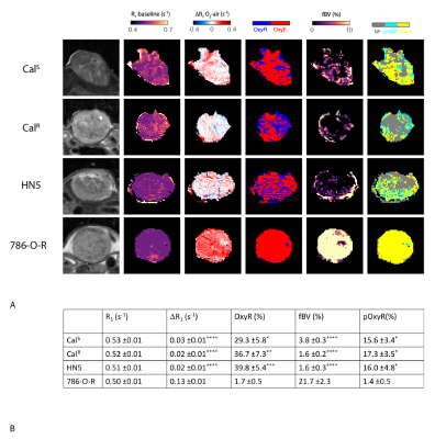 |
Imaging hypoxia in head and neck cancer xenografts with oxygen-enhanced MRI
Elise Lepicard1, Jessica Boult1, Yann Jamin1, Konstantinos Zormpas-Petridis1, Adam Featherstone2, Carol Box1, Rafal Panek3, James O'Connor2, and Simon Robinson1
1Radiotherapy & Imaging, Institute of Cancer Research, Sutton, United Kingdom, 2Centre for Imaging Sciences, University of Manchester, Manchester, United Kingdom, 3Nottingham University Hospital, Nottingham, United Kingdom
Oxygen-enhanced (OE)-MRI was used to map and quantify hypoxia in head and neck squamous cell carcinoma xenografts, a tumour type in which hypoxia adversely affects patient prognosis. Application of a refined OE-MRI protocol revealed a markedly high proportion of voxels refractory to hyperoxia-induced changes in R1, shown to be hypoxic in imaging-aligned tissue sections stained for the hypoxia marker pimonidazole.
|
0277. |
USPIO-enhanced MRI for pre-operative lymph node staging after neoadjuvant chemoradiotherapy in esophageal cancer
Didi de Gouw1, John Hermans2, Bastiaan Klarenbeek 1, Marnix Maas2, Atsushi Nakamoto3, Tom Scheenen2, and Camiel Rosman1
1surgery, Radboudumc, Nijmegen, Netherlands, 2radiology and nuclear medicine, Radboudumc, Nijmegen, Netherlands, 3radiology, Osaka University Graduate School of Medicine, Osaka, Japan
Lymph node dissections during esophagectomy may be omitted or minimized in patients with esophageal cancer without or with limited lymph node metastases, thereby reducing associated morbidity. A promising technique to detect lymph node metastases is T2*-weighted MRI after the injection of ultrasmall superparamagnetic iron oxide nanoparticles (USPIO, ferumoxtran-10). The aim of this study is to evaluate the feasibility of USPIO-enhanced MRI in the detection of locoregional lymph node metastases in patients with esophageal cancer whom underwent nCRT, and to study the effect of nCRT on the evaluation of USPIO-enhanced MRI.
|
|
0278.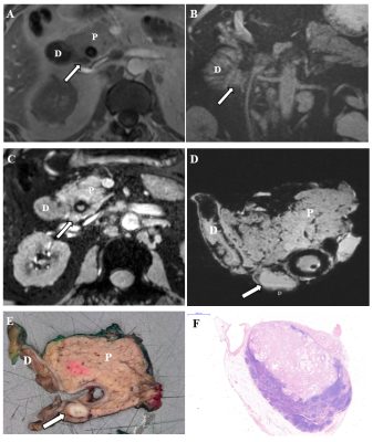 |
USPIO-enhanced MRI for pre-operative lymph node staging in patients with pancreatic and periampullary carcinoma: a feasibility study
Geke Litjens1, Atsushi Nakamoto2, Lodewijk Brosens3,4, Erwin van Geenen5, Marnix Maas1, Mathias Prokop1, Tom Scheenen1, Patrik Zámecnik1, Kees van Laarhoven6, Jelle Barentsz1, and John Hermans1
1Radiology and Nuclear Medicine, Radboudumc, Nijmegen, Netherlands, 2Diagnostic and Interventional Radiology, Osaka University Graduate School of Medicine, Suita, Japan, 3Pathology, Radboudumc, Nijmegen, Netherlands, 4Pathology, UMC Utrecht, Utrecht, Netherlands, 5Gastroenterology and Hepatology, Radboudumc, Nijmegen, Netherlands, 6Surgery, Radboudumc, Nijmegen, Netherlands
Detecting lymph node metastases is important but challenging in patients with pancreatic or periampullary carcinoma. USPIO-MRI is a promising tool to detect lymph node metastases. In 13 patients we detected on USPIO-MRI 86/307 suspect lymph nodes (28/78 regional and 58/229 distant). All patients with suspect regional lymph nodes had positive regional lymph nodes at histopathology. In evaluation of paraaortic lymph nodes discrimination between ganglions and lymph nodes showed to be important. Node-to-node analysis and follow-up of this study will give more accurate information on the value of USPIO-MRI for the detection of lymph node metastases in these patients.
|
 Watch the Video