
Scientific Session
MRS: New Developments, Applicatons, & Fighting the Noise
Session Topic: MRS: New Developments, Applicatons, & Fighting the Noise
Session Sub-Topic: MRS Applications
Oral
Spectroscopy
| Monday Parallel 5 Live Q&A | Monday, 10 August 2020, 15:15 - 16:00 UTC | Moderators: Nathalie Just & Gulin Oz |
Session Number: O-42
0378.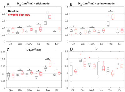 |
Diffusion of brain metabolites highlights altered brain microstructure in chronic hepatic encephalopathy
Cristina Cudalbu1, Katarzyna Pierzchala1,2,3, Dunja Simicic1,2, Graham Knott4, Stephanie Clerc-Rosset4, Bernard Lanz2, and Ileana Jelescu1
1Centre d'Imagerie Biomedicale, École Polytechnique Fédérale de Lausanne, Lausanne, Switzerland, 2Laboratory for functional and metabolic imaging, École Polytechnique Fédérale de Lausanne, Lausanne, Switzerland, 3Service of Clinical Chemistry, University of Lausanne and University Hospital of Lausanne, Lausanne, Switzerland, 4Biological Electron Microscopy Facility, École Polytechnique Fédérale de Lausanne, Lausanne, Switzerland
In chronic hepatic encephalopathy (HE), high ammonium delivery to the brain is causing the accumulation of glutamine (Gln) and gradual release of other osmolytes. We aimed to follow the longitudinal evolution of brain Gln and other metabolite properties in chronic-HE using diffusion-weighted spectroscopy (DW-MRS) and evaluate the potential changes in diffusion behavior which might provide information on Gln localization and potential microstructural alterations during chronic-HE. Increased diffusivity and reduced kurtosis in BDL rats, showcased by DW-MRS analysis, are fully consistent with a less complex microstructure and swollen soma as highlighted by fluorescence and electron microscopy leading to increased molecule mobility.
|
|
 |
0379.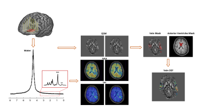 |
Simultaneous High-Resolution 3D MRSI and Oxygen Extraction Fraction Mapping in Acute Stroke Using SPICE
Tianxiao Zhang1, Tianyao Wang2, Zengping Lin1, Rong Guo3,4, Yudu Li3,4, Yibo Zhao3,4, Ziyu Meng1,3, Jun Liu2, Danhong Wu5, Zheng Jin6, Xin Yu7, Zhi-Pei Liang3,4, and Yao Li1
1Institute for Medical Imaging Technology, School of Biomedical Engineering, Shanghai Jiao Tong University, Shanghai, China, 2Radiology Department, The Fifth People's Hospital of Shanghai, Fudan University, Shanghai, China, 3Beckman Institute for Advanced Science and Technology, University of Illinois at Urbana-Champaign, Urbana, IL, United States, 4Department of Electrical and Computer Engineering, University of Illinois at Urbana-Champaign, Urbana, IL, United States, 5Neurology Department, The Fifth People's Hospital of Shanghai, Fudan University, Shanghai, China, 6Shanghai Minhang Hospital of Integrated Traditional Chinese and Western Medicine Hospital, Shanghai, China, 7Department of Biomedical Engineering, Case Western Reserve University, Cleveland, OH, United States
Mapping the concurrent changes in oxygen extraction fraction (OEF) and neurometabolic markers could provide a powerful tool for evaluation of brain tissue viability after stroke. In this work, we investigated the feasibility of fast simultaneous 3D brain OEF and neurometabolic imaging noninvasively in acute ischemic stroke using SPICE. We achieved concurrent mapping of OEF (1.2×1.2×1.2 mm3 nominal resolution) and MRSI (2.0×3.0×3.0 mm3 nominal resolution) within a 7-minute scan. Our experimental results demonstrated the feasibility of mapping OEF and neurometabolic alterations in acute stroke.
|
0380.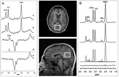 |
Documentation of Anti-glutamatergic Effect of N-Acetylcysteine Treatment with 1H MRS Monitoring of Cortical Glutathione and Glutamate In Vivo
Dikoma C. Shungu1, Xiangling Mao1, Michelle Blate2, Diana Vu2, Guoxin Kang1, Halinder S. Mangat3, Claire Henchcliffe3, Bejamin Natelson2, and Nora Weiduschat1
1Radiology, Weill Cornell Medicine, New York, NY, United States, 2Icahn School of Medicine at Mount Sinai, New York, NY, United States, 3Neurology, Weill Cornell Medicine, New York, NY, United States
N-acetylcysteine (NAC), a glutathione (GSH) synthesis precursor, is thought to have anti-glutamatergic properties for which direct in vivo evidence is lacking. In this study, the postulated anti-glutamatergic properties of NAC were investigated by using 1H MRS to monitor changes in brain levels of both GSH and glutamate (Glu) in response to 4 weeks of NAC supplementation in patients with chronic fatigue syndrome (CFS) and healthy volunteers (HV). Following NAC treatment, GSH levels increased significantly in CFS and numerically in HV, while Glu decreased significantly in both groups compared to baseline – a finding that supports NAC as an anti-glutamatergic agent.
|
|
 |
0381.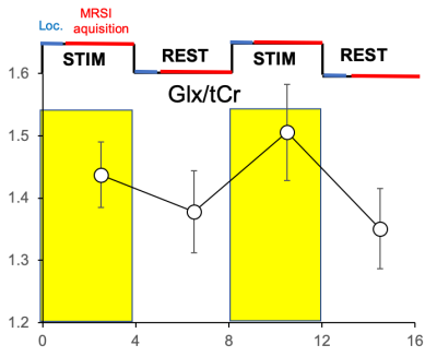 |
Functional spectroscopic imaging (fMRSI) detects metabolite changes in the activated primary sensorimotor cortex at 7T
Petr Bednarik1, Lukas Hingerl1, Dario Goranovic1, Alena Svatkova1, Pedro de Lima Cardoso1, Siegfried Trattnig1, Rupert Lanzenberger2, and Wolfgang Bogner1
1Department of Medical Imaging and Image-guided Therapy, Medical University of Vienna, Vienna, Austria, 2Department of Psychiatry and Psychotherapy, Medical University of Vienna, Vienna, Austria
Functional single-voxel MRS (fMRS) was capable to sensitively detect metabolite responses to sensory stimulation, but suffered from large partial volume effects, that questioned the clinical utility of fMRS. Free-induction decay (FID)-MRSI promises to possess sufficient SNR to reach the sensitivity of SV-MRS and overcome its limitations by selective mapping the volume of interest with multiple voxels and thus, with higher spatial resolution, minimize the partial volume issue. Concentric-ring-trajectories (CRT)-based 3D FID-MRSI showed sufficient sensitivity and temporal stability to detect functional glutamate changes in the dominant sensorimotor region with expected most robust metabolite responses during finger tapping task.
|
|
0382.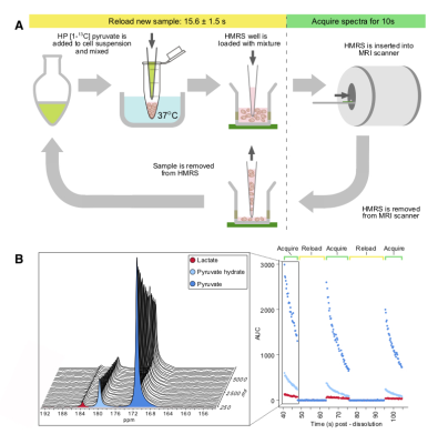 |
Multi-sample measurement of pyruvate/lactate flux in melanoma cells using an HP micromagnetic resonance spectrometer and D2O solvation
Hannah J. Lees1, Micaela Millan1, Fayyaz Ahamed2, Roozbeh Eskandari1, Kristin L. Granlund1, Sangmoo Jeong1, and Kayvan R. Keshari1
1Memorial Sloan Kettering Cancer Center, NEW YORK, NY, United States, 2University of California, Berkeley, Berkeley, CA, United States
The pyruvate-lactate flux, kPL, shows promise as a biomarker of cancer presence and aggressiveness, and assessment of kPL in patient-derived cells may be a useful tool to assess treatment response for advanced personalized medicine. Here we present a novel experimental protocol for the real-time measurement of pyruvate-lactate metabolic flux in multiple mass-limited cell suspension samples using a single dissolution, thereby increasing efficiency and providing greater control of the methodological variability associated with HP experiments. We then applied this protocol to the measurement of pyruvate-lactate flux in melanoma cells for the assessment of treatment response to BRAF inhibition.
|
|
0383. |
Penumbra Identification in Acute Stroke Using Fast 3D 1H-MRSI
Yao Li1, Zengping Lin1, Tianyao Wang2, Tianxiao Zhang1, Rong Guo3,4, Yudu Li3,4, Yibo Zhao3,4, Ziyu Meng1,3, Jun Liu2, Xin Yu5, and Zhi-Pei Liang3,4
1Institute for Medical Imaging Technology, School of Biomedical Engineering, Shanghai Jiao Tong University, Shanghai, China, 2Radiology Department, The Fifth People's Hospital of Shanghai, Fudan University, Shanghai, China, 3Beckman Institute for Advanced Science and Technology, University of Illinois at Urbana-Champaign, Urbana, IL, United States, 4Department of Electrical and Computer Engineering, University of Illinois at Urbana-Champaign, Urbana, IL, United States, 5Department of Biomedical Engineering, Case Western Reserve University, Cleveland, OH, United States
Impaired metabolism was a key factor in the definition of ischemic penumbra. 1H-MRSI has been recognized as a potentially powerful tool for metabolic imaging of stroke. In this proof of concept clinical study, we explored the potential of fast 3D high-resolution 1H-MRSI to investigate brain neurometabolic changes at tissue-level in acute stroke. In a 6-min scan, we obtained N-acetylaspartate (NAA) and lactate (Lac) maps simultaneously. Our experimental results showed different NAA and Lac concentrations between hypoperfused tissue recruited to final infarct and that survived, indicating an improved delineation of penumbra by incorporating the tissue neuronal damage and acidosis information.
|
0384. |
Detection of Carbonic Anhydrase Activity in the Frontal Lobe of Human Brain
Shizhe Li1, Li An1, Christopher Johnson1, Maria Ferraris-Araneta1, Milalynn Victorino1, Jyoti Tomar1, and Jun Shen1
1National Institutes of Health, Bethesda, MD, United States
This study demonstrates the feasibility of detecting carbonic anhydrase activity in the frontal lobe of the human brain. Upon saturation of carbon dioxide, a very large magnetization transfer effect catalyzed by carbonic anhydrase was measured in the frontal lobe of healthy human subjects using 13C MRS with oral administration of [U-13C6]glucose. The results showed that it is feasible to examine carbonic anhydrase activity using magnetization transfer 13C MRS in the frontal cortex—where structural lesions, disturbed function, and morphology are strongly associated with many psychiatric symptoms.
|
|
|
0385.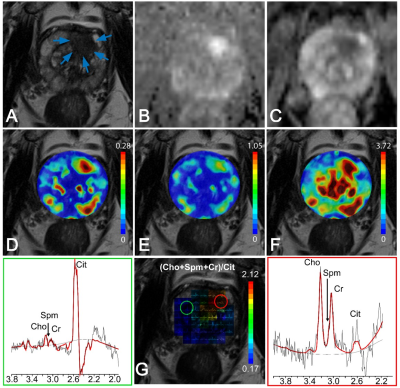 |
MRSI of the prostate revisited: The potential role of GOIA-sLASER in multiparametric MRI of central gland prostate cancer
Neda Gholizadeh1, Peter B Greer2,3, John Simpson2,3, Jonathan Goodwin2,3, Peter Lau4,5, Arend Heerschap6, and Saadallah Ramadan1,5
1Health Science, The University of Newcastle, Newcastle, Australia, 2Radiation Oncology, Calvary Mater Newcastle, Newcastle, Australia, 3Physics and mathematics, The University of Newcastle, Newcastle, Australia, 4Radiology, Calvary Mater Newcastle, Newcastle, Australia, 5Imaging Centre, Hunter Medical Research Institute (HMRI), Newcastle, Australia, 6Radiology and Nuclear Medicine, Radboud University Medical Center, Nijmegen, Nijmegen, Netherlands
Due to histological heterogeneity of the central gland, accurate detection of central gland prostate cancer remains a challenge. A reliable and non-invasive imaging technique could increase the sensitivity and specificity for identification of central gland lesions missed by PI-RADS V2 or biopsies. This study evaluates the diagnostic performance of individual and combined parameters of an mp-MRI exam, employed for PI-RADS evaluations (T2WI, DWI, DCE) and advanced GOIA-sLASER MRSI using an external phased-array coil for central gland prostate cancer detection, localization and grading. The results demonstrate that MRSI using GOIA-sLASER considerably improves central gland prostate cancer detection and localization.
|
0386.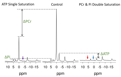 |
Phosphate Metabolite T1 Relaxation Times, ATP Hydrolysis Flux and Creatine Kinase Reaction Kinetics in the Human Skeletal Muscle.
Adil Bashir1, Jianyi Zhang2, and Thomas S Denney 1
1Electrical and Computer Engineering, Auburn University, Auburn, AL, United States, 2Biomedical Engineering, The University of Alabama at Birmingham, Birmingham, AL, United States
Long TR and low concentration makes the quantification of inorganic phosphate (Pi) difficult in 31P magnetization saturation transfer experiments. An indirect method to measure Adenosine Triphosphate (ATP) turnover was demonstrated in animal studies, which does not require quantification of Pi. We demonstrate the application and validation of this technique in human studies. We measured the fluxes of ATP production and hydrolysis reactions using the indirect approach and validated the results against direct measurements. We also report the intrinsic T1 of Phosphocreatine, ATP and Pi in skeletal muscle at 7T. This will facilitate future studies of impaired bioenergetics in vivo.
|
|
0387.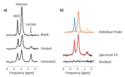 |
Deuterium (2H) magnetic resonance spectroscopy for monitoring chemotherapeutic response in vitro
Josephine L Tan1,2, Daniel Djayakarsana1,2, Rachel W Chan2, Colleen Bailey1,2, and Angus Z Lau1,2
1Medical Biophysics, University of Toronto, Toronto, ON, Canada, 2Physical Sciences, Sunnybrook Research Institute, Toronto, ON, Canada
Deuterium (2H) magnetic resonance spectroscopy (MRS) is a novel metabolic imaging method that can measure aberrant glucose metabolism in cancer. In this abstract, we use 2H MRS to measure metabolic changes in acute myeloid leukemia (AML) cells after treatment with cisplatin. We show that this method is sensitive to differences in lactate levels, produced via glycolysis of deuterium-enriched glucose at 7T. These studies demonstrate the potential of 2H MRS to monitor chemotherapeutic response.
|
 Watch the Video