
Scientific Session
MRS and Molecular Imaging, Development and Applications
Session Topic: MRS and Molecular Imaging, Development and Applications
Session Sub-Topic: Molecular Imaging: Non-Hyperpolarized
Oral
Molecular Imaging
| Tuesday Parallel 5 Live Q&A | Tuesday, 11 August 2020, 14:30 - 15:15 UTC | Moderators: Jeff Bulte & Lingzhi Hu |
Session Number: O-47
 |
0635.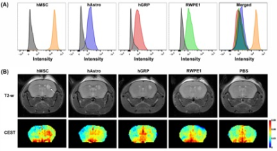 |
Label-free Tracking of Transplanted Mesenchymal Stem Cells Using manCEST MRI
Yue Yuan1, Congxiao Wang1, Jia Zhang1, and Jeff W.M. Bulte1
Video Permission Withheld
1The Russell H. Morgan Department of Radiology and Radiological Science, The Johns Hopkins University School of Medicine, Baltimore, Maryland, USA., Baltimore, MD, United States
Human mesenchymal stem cells (hMSCs) overexpress high-mannose-type (HM) N-glycans on their membrane surface. Taking advantage of the five exchangeable hydroxyl groups on mannose that provide CEST MRI contrast, we present a label-free method for tracking hMSCs in vivo. The mannose-sensitive CEST (manCEST) signal of hMSCs was clearly distinguishable from the surrounding host tissue and stood out against several other transplanted cell lines tested.
|
 |
0636.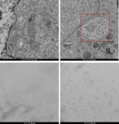 |
Bright-Ferritin: A novel MRI gene reporter complex for sensitive and longitudinal cell tracking
Daniel Andrzej Szulc1,2, Xavier Alexander Lee2,3, Hai-Ying Mary Cheng4,5, and Hai-Ling Margaret Cheng1,2,6
1Institute of Biomaterials and Biomedical Engineering, University of Toronto, Toronto, ON, Canada, 2Translational Biology & Engineering Program, Ted Rogers Centre for Heart Research, Toronto, ON, Canada, 3Department of Physiology, University of Toronto, Toronto, ON, Canada, 4Biology, University of Toronto Mississauga, Toronto, ON, Canada, 5Department of Cell & Systems Biology, University of Toronto, Toronto, ON, Canada, 6Edward S. Rogers Sr. Department of Electrical & Computer Engineering, University of Toronto, Toronto, ON, Canada
Tissue engineering with transplanted cells has the potential to repair and regenerate almost every tissue and organ of the body. One major obstacle of cell therapies is the inability to longitudinally assess injected cells. Non-invasive imaging with contrast-enhanced MRI is highly suited for this task but is limited with current methods. In this study, we report a novel method for producing bright endogenous cellular contrast through a genetic MRI reporter that results in the formation of in situ ferritin-manganese nanoparticles. The signal produced by these cells is significantly higher than traditional iron labelled ferritin-overexpressing cells and manganese-permeable cell lines.
|
|
0637.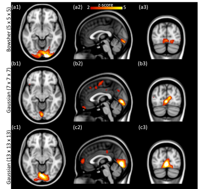 |
MRI-assisted high temporal resolution dynamic FDG-PET imaging for assessing brain functions
Viswanath Pamulakanty Sudarshan1,2,3, Shenpeng Li4, Anthony Fernandez1, Phillip Ward4,5, Sharna Jamadar4,5, Gary Egan4,5, Suyash Awate3, and Zhaolin Chen4
1Monash University, Clayton, Australia, 2IITB Monash Research Academy, Mumbai, India, 3Indian Institute of Technology, Bombay, Mumbai, India, 4Monash Biomedical Imaging, Clayton, Australia, 5Turner Institute for Brain and Mental Health, Clayton, Australia
Simultaneous positron emission tomography (PET) and magnetic resonance imaging (MRI) provide complementary structural and functional information. Recent developments in continuous infusion functional PET (fPET) have shown promising results to track dynamic changes in brain metabolism. Although fPET provides opportunities to investigate functional metabolism in the brain, the temporal resolution still remains a major challenge compared to functional MRI (fMRI). In this work, we use anatomical MRI information modeled as a Bowsher prior to improve the sensitivity of fPET at higher temporal resolution. We validate our MRI-assisted fPET analysis framework using both in-silico and in-vivo experiments.
|
|
0638.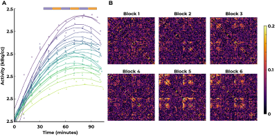 |
Multimodal resting-state functional and metabolic connectivity with simultaneous MR-PET
Phillip G.D. Ward1,2,3, Xingwen Liang1, Gary F Egan1,2,3, and Sharna D Jamadar1,2,3
1Monash Biomedical Imaging, Monash University, Melbourne, Australia, 2Turner Institute for Brain and Mental Health, School of Psychological Sciences, Monash University, Melbourne, Australia, 3Australian Research Council Centre of Excellence for Integrative Brain Function, Melbourne, Australia
Metabolic connectivity measured using FDG-PET has been proposed as a biomarker for disease, however static FDG-PET cannot provide subject-level measures of connectivity. We applied constant infusion functional FDG-fPET to measure subject-level metabolic connectivity simultaneously with BOLD-fMRI connectivity. Group-average FDG-fPET and BOLD-fMRI connectivity profiles showed similarities and differences. FDG-fPET and BOLD-fMRI connectivity was most similar in superior cortex, and least similar in subcortical regions. Group-average FDG-fPET within-subject connectivity showed little similarity with static FDG-PET connectivity. Our new method opens up the opportunity for new metabolic neuroimaging biomarkers for disease, as well as approaches for multimodality MR-PET imaging.
|
|
0639.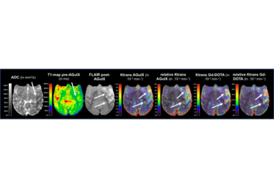 |
Quantitative multiparametric PET-MRI of blood-brain barrier damage after stroke recanalization: nanoparticles versus small contrast agent
Justine Debatisse1,2, Omer Eker3,4, Oceane Wateau5, Tae-Hee Cho1,6, Marlene Wiart1, Nicolas Costes7, Ines Merida7, Christelle Leon1, Jean-Baptiste Langlois7, Thomas Troalen2, Christian Tourvielle7, Thibault Iecker7, Didier Le Bars7,
Sophie Lancelot7, Norbert Nighoghossian1,6, Michel Ovize1,8, Hugues Contamin5, Francois Lux9, Olivier Tillement9, and Emmanuelle Canet Soulas1
Video Permission Withheld
1CarMeN lab, U1060 INSERM, University of Lyon, Lyon, France, 2Siemens Healthcare SAS, Saint-Denis, France, 3Interventional Neuroradiology, Hospices Civils de Lyon, Lyon, France, 4CREATIS lab, UMR CNRS 5220, INSERM U1206, INSA, University of Lyon, Villeurbanne, France, 5Cynbiose SAS, Marcy l'Etoile, France, 6Neurology, Hospices Civils de Lyon, Lyon, France, 7CERMEP, Lyon, France, 8Cardiology, Hospices Civils de Lyon, Lyon, France, 9ILM, CNRS UMR5306, University of Lyon, Villeurbanne, France
Quantification of blood-brain barrier (BBB) leakage is of main interest in the stroke field to identify patients susceptible to develop hemorrhage and to identify the therapeutic window for neuroprotective drugs administration. We used small (Gd-DOTA) and medium size (nanoparticles AGuIX) contrast agents (CA) to quantify BBB permeability 60-to-90 minutes post-recanalization in a model of stroke using dynamic contrast-enhanced (DCE) MRI. We confirmed 1) Early BBB leakage with both CA and 2) BBB opening to nanoparticles at post-recanalization provides opportunity for selective neuroprotection drug delivery.
|
|
0640.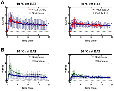 |
Estimation of brown adipose tissue perfusion by DCE-MRI improves measurement of oxidative metabolism by 11C-acetate PET in a rat model
Gabriel Richard1, Christophe Noll1, Mélanie Archambault1, Luc Tremblay1, Serge Phoenix1, Samia Ait-Mohand1, Réjean Lebel1, Brigitte Guérin1, André C. Carpentier1, and Martin Lepage1
1Université de Sherbrooke, Sherbrooke, QC, Canada
Brown adipose tissue (BAT) oxidative metabolism can be measured by 11C-acetate PET with a 3-tissue pharmacokinetic model. However, this model can have trouble distinguishing between increased oxidation and increased blood volume, both of which occur in active BAT. A sequential DCE-MRI and 11C-acetate PET protocol was performed in male Wistar rats with and without BAT activation. DCE-MRI perfusion measures were comparable to those obtained previously with 68Ga-DOTA PET. Incorporating the DCE-MRI blood volume information into the 11C-acetate model revealed higher oxidation in activated BAT than indicated by the unconstrained model.
|
|
0641.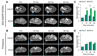 |
MR Molecular Imaging of EDB-Fibronectin for Precision Imaging of Oral Squamous Cell Carcinoma
Ryan Hall1, Nadia Ayat1, Peter Qiao1, Amita Vaidya1, and Zheng-Rong Lu1
1Department of Biomedical Engineering, Case Western Reserve University, Cleveland, OH, United States
Oral squamous cell carcinoma (OSCC) has maintained poor prognosis due to its aggressive nature and lack of targetable biomarkers for early and accurate detection. We have developed a targeted MRI contrast agent specific to extradomain-B fibronectin (EDB-FN), an extracellular matrix protein closely associated with tumor aggressiveness. Immunohistochemical analysis revealed strong EDB-FN expression in OSCC with minimal expression in normal tongue tissue. MR molecular imaging with our targeted contrast agent demonstrated the ability for differential contrast enhancement of aggressive OSCC tumors, underscoring the potential for using EDB-FN as a targetable biomarker for precision molecular imaging of OSCC.
|
|
0642.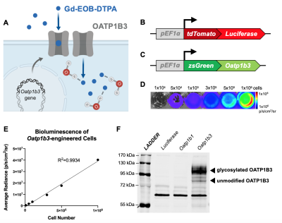 |
Whole-body profiling of early cancer metastasis using multimodality reporter gene imaging
Nivin N Nyström1,2, Timothy J Scholl2,3, and John Andrew Ronald1,2,4
1Medical Biophysics, University of Western Ontario, London, ON, Canada, 2Medical Imaging Laboratories, Robarts Research Institute, London, ON, Canada, 3Medical Biophysics, Robarts Research Institute, London, ON, Canada, 4Lawson Health Research Institute, London, ON, Canada
Organic anion-transporting polypeptide 1b3 (Oatp1b3) is a protein derived from the human liver that is capable of taking up Gd-EOB-DTPA, a clinical contrast agent, into cells. We synthetically express the Oatp1b3 gene on breast cancer cells and are able to track them throughout the bodies of preclinical animal models with high sensitivity and resolution as they metastasize. In the future, we hope to develop Oatp1b3 as a tool to track the activation and location of gene and cellular therapies in patients on MRI.
|
 Watch the Video