
Scientific Session
Quantitative Tissue Properties
Session Topic: Quantitative Tissue Properties
Session Sub-Topic: Emerging Trends in QSM
Oral
Contrast Mechanisms
| Monday Parallel 1 Live Q&A | Monday, 10 August 2020, 14:30 - 15:15 UTC | Moderators: Pascal Spincemaille |
Session Number: O-48
 |
0153.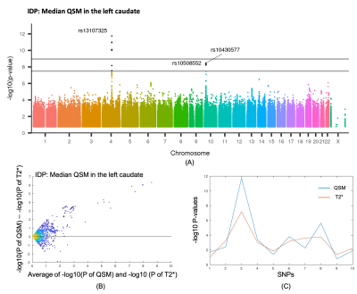 |
Quantitative susceptibility mapping in UK Biobank brain imaging: pipeline and preliminary results in 2400 subjects
Chaoyue Wang1, Stephen M. Smith1, Fidel Alfaro-Almagro1, Cristiana Fiscone2, Richard Bowtell2, Lloyd T. Elliott3, Karla L. Miller1, and Benjamin C. Tendler1
1Wellcome Centre for Integrative Neuroimaging, FMRIB, Nuffield Department of Clinical Neurosciences, University of Oxford, Oxford, United Kingdom, 2Sir Peter Mansfield Imaging Centre, School of Physics and Astronomy, University of Nottingham, Nottingham, United Kingdom, 3Department of Statistics and Actuarial Science, Simon Fraser University, Vancouver, BC, Canada
UK Biobank aims to scan 100,000 participants and its brain protocol acquires susceptibility-weighted MRI (swMRI). To date, only the swMRI magnitude data were processed to produce T2* maps. The aim of this work is to develop a robust processing pipeline for QSM using the acquired swMRI phase data. We ran this pipeline on 2408 volunteers and report some preliminary results, including age-dependent curves and genetic associations. Significant correlations were found between susceptibility and age in subcortical structures. QSM discovered replicable genetic associations previously identified in T2*. Our results suggest that there is unique information in susceptibility maps compared to T2*.
|
|
0154. |
Eliminating chemical shift and relaxation effects in QSM using SMURF imaging
Beata Bachrata1,2,3, Bernhard Strasser1,2,4, Wolfgang Bogner1,2, Albrecht Ingo Schmid1,5, Siegfried Trattnig1,2,3, and Simon Daniel Robinson1,2,6,7
1High Field MR Centre, Medical University of Vienna, Vienna, Austria, 2Department of Biomedical Imaging and Image-guided Therapy, Medical University of Vienna, Vienna, Austria, 3Christian Doppler Laboratory for Clinical Molecular MR Imaging, Vienna, Austria, 4Athinoula A. Martinos Center for Biomedical Imaging, Department of Radiology, Massachusetts General Hospital, Harvard Medical School, Boston, MA, United States, 5Center for Medical Physics and Biomedical Engineering, Medical University of Vienna, Vienna, Austria, 6Centre for Advanced Imaging, The University of Queensland, Brisbane, Australia, 7Department of Neurology, Medical University of Graz, Graz, Austria
The accuracy of Quantitative Susceptibility Mapping in fatty regions is adversely affected by the chemical shift effects and by the relaxation rate differences between fat and water. We propose using a recently developed water-fat separation technique based on multi-band principles, Simultaneous Multiple Resonance Frequency (SMURF) imaging, to correct for these effects. SMURF achieves clean water-fat separation in the head-and-neck, allowing the generation of recombined water-fat images fully corrected for chemical shift and relaxation effects. This makes bias-free Quantitative Susceptibility Mapping possible in body regions containing significant amounts of fat, with the free selection of echo-times, receiver bandwidths and flip angles.
|
1635.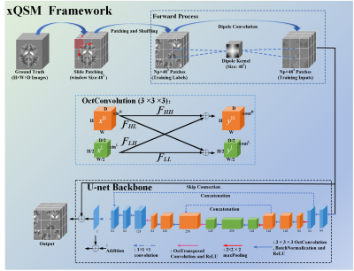 |
xQSM: a deep learning QSM network using Octave Convolution
Yang Gao1, Xuanyu Zhu1, Stuart Crozier 1, Feng Liu1, and Hongfu Sun1
1University of Queensland, Brisbane, Australia
Deep learning frameworks are emerging methods for solving ill-posed inverse problems in medical imaging, including Quantitative Susceptibility Mapping (QSM). Previously, U-net has been successfully trained on susceptibility maps to learn the dipole inversion process; however, susceptibility contrast loss was observed in iron-rich deep grey matter regions. In this study, we propose an enhanced deep learning network “xQSM” using the state-of-the-art Octave Convolution, which shows more accurate susceptibility contrasts than the original U-net in both simulated and in vivo datasets.
|
|
 |
0155.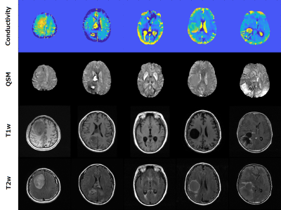 |
Morphology Enabled Quantitative Conductivity–Susceptibility Mapping with B1 and B0 Estimation from Complex Multi-echo Gradient Echo Signal
Motofumi Fushimi1,2, Thanh Nguyen2, and Yi Wang2,3
1Graduate School of Information Science and Technology, The University of Tokyo, Tokyo, Japan, 2Radiology, Weill Cornell Medical College, New York, NY, United States, 3Biomedical Engineering, Cornell University, Ithaca, NY, United States
We propose a simultaneous conductivity and susceptibility reconstruction method by estimating B1 phase and B0 distributions from a multi-echo gradient echo (mGRE) signal. B1 phase and B0 maps are simultaneously determined by applying nonlinear least squares method on the complex signal equation of the mGRE signal. The poor conditioned inversion of field (B1/B0) to source (conductivity/susceptibility) is regularized using anatomical information. This morphology enabled quantitative conductivity and susceptibility mapping (QCSM) was performed on healthy subjects and patients with brain tumors. Our preliminary in-vivo experiments demonstrated that the proposed QCSM method can reconstruct conductivity and susceptibility from a single mGRE acquisition.
|
0156.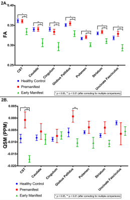 |
7 Tesla Diffusion Tensor Imaging and Quantitative Susceptibility Mapping of Huntington’s Disease
Paul Rowley1,2, Melanie Morrison, PhD1, Yicheng Chen1, Angela Jakary1, Michael Geschwind, MD, PhD3, Alexandra Nelson, MD, PhD3, Duan Xu, PhD1, Christopher Hess, MD, PhD1, and Janine Lupo, PhD1
1Radiology & Biomedical Imaging, University of California, San Francisco, San Francisco, CA, United States, 2University of Wisconsin School of Medicine and Public Health, Madison, WI, United States, 3Neurology, University of California, San Francisco, San Francisco, CA, United States
Ultra-high-field 7 Tesla (7T) MRI was acquired to examine and compare white matter microstructure and quantitative susceptibility in patients with premanifest (PM) and early manifest (EM) Huntington’s disease (HD) and age-matched healthy control (HC) subjects. Tract-averaged and along-tract fractional anisotropy (FA) and susceptibility (PPM) were calculated to determine the spatial spread of disease along motor tracks originating from the striatum and ending in the cortex. HC and PM patients demonstrated different areas of significantly increased susceptibility compared to EM at the tract-averaged level as well as significant focal along-tract variations in FA and susceptibility which were undetected by tract-averaged analysis.
|
|
|
0157.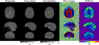 |
Simultaneous QSM and MR Elastography of the Brain Using Spiral Staircase
Xi Peng1 and James G. Pipe1
1Department of Radiology, Mayo Clinic, Rochester, MN, United States
This work presents a new feasibility to extract tissue susceptibility from gradient-echo MRE data and enables simultaneous QSM and MRE in a single scan. The proposed method builds on a new spiral staircase acquisition which enables high resolution often required by QSM using inherently improved through-plane parallel imaging. In-plane parallel imaging, constrained reconstruction and deblurring method are also integrated to generate high quality spiral images for QSM and MRE processing. In vivo experiment results demonstrate the capability of proposed method in producing high quality tissue susceptibility along with shear stiffness maps from a single 5-minute scan.
|
|
0158.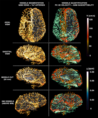 |
Multimodal quantitative arterial-venous segmentation of the human brain at 7T: structure, susceptibility and flow
Michaël Bernier1,2, Berkin Bilgic1,2, Saskia Bollmann1,2, Nina E. Fultz1,3, and Jonathan R. Polimeni1,2,4
1Athinoula A. Martinos Center for Biomedical Imaging, Massachusetts General Hospital, Charlestown, MA, United States, 2Radiology, Harvard Medical School, Boston, MA, United States, 3Engineering, Boston University, Boston, MA, United States, 45Division of Health Sciences and Technology, Massachusetts Institute of Technology, Cambridge, MA, United States
Vascular imaging acquisition techniques to extract veins and arteries are not impervious to flaws: venography by susceptibility weighted imaging is prone to blooming effects and false-negatives, and angiography from time-of-flight imaging is affected by veins detection and false-negatives. They also fail to provide quantitative measures of vascular physiology such as flow and susceptibility important for understanding the origin of vascular-based biases. Thus, we aimed to employ multi-orientation quantitative susceptibility mapping, multi-echo time-of-flight and quantitative phase-contrast to more accurately detect and quantify the susceptibility and flow along the vascular tree, paving the way for a joint anatomical/physiological vascular atlas at 7T.
|
|
0159. |
High-Resolution QSM for Simultaneous QSM/MRSI
Rong Guo1,2, Yudu Li1,2, Yibo Zhao1,2, Tianyao Wang3, Yao Li4,5, Brad Sutton1,2,6, and Zhi-Pei Liang1,2
1Department of Electrical and Computer Engineering, University of Illinois at Urbana-Champaign, Urbana, IL, United States, 2Beckman Institute for Advanced Science and Technology, University of Illinois at Urbana-Champaign, Urbana, IL, United States, 3Radiology Department, The Fifth People's Hospital of Shanghai, Shanghai, China, 4School of Biomedical Engineering, Shanghai Jiao Tong University, Shanghai, China, 5Med-X Research Institute, Shanghai Jiao Tong University, Shanghai, China, 6Department of Bioengineering, University of Illinois at Urbana-Champaign, Urbana, IL, United States
In this work, we present a new method to achieve high-resolution QSM for simultaneous QSM/MRSI experiments. This work extends SPICE with a novel data acquisition scheme that provides larger k-space coverage for the unsuppressed water signals. A union-of-subspaces model incorporating sensitivity encodings (parallel imaging) and pre-determined spatiospectral features is used to solve the underlying image reconstruction problem. High-resolution capability (on the order of 1.0 × 1.0 × 1.2 mm3) for QSM has been demonstrated in 3D in vivo simultaneous QSM/MRSI experiments.
|
|
0160.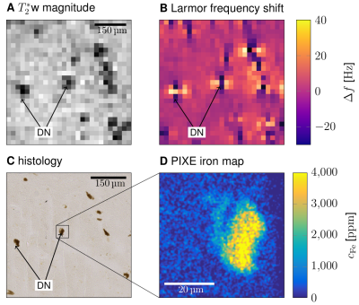 |
Magnetic properties of dopaminergic neurons in human substantia nigra quantified with MR microscopy
Malte Brammerloh1,2, Evgeniya Kirilina1,3, Renat Sibgatulin4, Karl-Heinz Herrmann4, Tilo Reinert1, Carsten Jäger1,5, Primož Pelicon6, Primož Vavpetič6, Kerrin J. Pine1, Andreas Deistung7, Markus Morawski5, Jürgen R. Reichenbach4, and Nikolaus Weiskopf1,2
1Department of Neurophysics, Max Planck Institute for Human Cognitive and Brain Sciences, Leipzig, Germany, 2Faculty of Physics and Earth Sciences, Leipzig University, Leipzig, Germany, 3Center for Cognitive Neuroscience Berlin, Freie Universität Berlin, Berlin, Germany, 4Medical Physics Group, University Hospital Jena, Jena, Germany, 5Paul Flechsig Institute of Brain Research, Leipzig, Germany, 6Microanalytical Center, Department for Low and Medium Energy Physics, Jožef Stefan Institute, Ljubljana, Slovenia, 7Department of Radiology, University Hospital Halle, Halle, Germany
MRI-based quantification of dopaminergic neurons (DN) and their neuromelanin (NM) in substantia nigra (SN) has great potential to serve as a specific biomarker for neurodegeneration in movement disorders. We used 22-µm-resolution post mortem MR microscopy combined with ion beam microscopy to characterize the magnetic properties of DN. MR microscopy visualized individual DN and provided 3D cellular maps of the entire SN. Static dephasing was determined as main effective transverse relaxation mechanism of DN. We characterized the susceptibility of iron in DN and estimated that the contribution of DN to R2* and QSM may also be detected with in vivo MRI.
|
 Watch the Video