
Scientific Session
Quantitative Tissue Properties
Session Topic: Quantitative Tissue Properties
Session Sub-Topic: Magnetic Resonance Elastography
Oral
Contrast Mechanisms
| Monday Parallel 1 Live Q&A | Monday, 10 August 2020, 14:30 - 15:15 UTC | Moderators: Ziying Yin |
Session Number: O-49
 |
0161.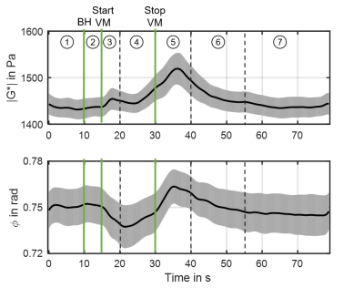 |
Real-time MR elastography of the human brain reveals short-term cerebral autoregulation in response to the Valsalva maneuver.
Helge Herthum1, Mehrgan Shahryari1, Gergely Bertalan1, Carsten Warmuth1, Stefan Hetzer2, Jürgen Braun3, and Ingolf Sack1
1Department of Radiology, Charité Universitätsmedizin Berlin, Berlin, Germany, 2Bernstein Center for Computational Neuroscience, Berlin, Germany, 3Institute of Medical Informatics, Berlin, Germany
Real-time MR elastography (rt-MRE) with 4.9Hz-frame rate was developed for in-vivo brain stiffness quantification during short-term tissue mechanical adaptation due to cerebral autoregulation. Six healthy participants performed a 15s-Valsalva maneuver with 50s recovery period following 10s resting period and 5s deep inspiration during continuous rt-MRE. 387 maps of tissue stiffness and fluidity were generated depicting a significant increase of stiffness due to Valsalva and an overshoot of stiffness by 3.4% fading out within 7s after the maneuver. rt-MRE is potentially sensitive to several diseases associated with cerebral autoregulation and reveals new insights into brain viscoelasticity changes on short time scales.
|
|
0162. |
Fast Whole-Brain MR Elastography Using 3D GRE Multishot Variable Density Spiral Staircase Acquisition
Xi Peng1, Yi Sui1, Sandeep Ganji2, Ashley Anderson2, John Huston1, Richard Ehman1, and James Pipe1
1Department of Radiology, Mayo Clinic, Rochester, MN, United States, 2Philips Healthcare, Gainesville, FL, United States
This work reports a new method for fast whole-brain MRE using a 3D gradient-echo multishot variable-density spiral staircase acquisition. The proposed method enables high-resolution MRE by exploiting the inherently improved through-plane parallel imaging. The displacement-SNR is further enhanced by exploiting a motion encoding gradient of a 2 wave-period duration. Constrained reconstruction and deblurring method are used to generate high-quality spiral images. By integrating all these features, the proposed technique provides flexible trade-off among SNR, resolution, spatial blurring and scan time. In vivo experiments have demonstrated the capability of the proposed method for high-SNR high-resolution MRE in less than 5 minutes.
|
0163.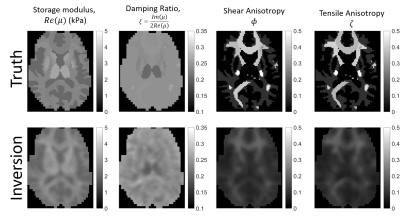 |
Model-based heterogenous transverse isotropic MR elastography inversion for brain tissue with aligned fiber tracts
Matthew Mcgarry1, Elijah Van Houten2, Damian Sowinski1, Philip Bayly3, Daniel Smith4, Curtis Johnson4, John Weaver1,5, and Keith Paulsen1,5
1Dartmouth College, Hanover, NH, United States, 2Université de Sherbrooke, Sherbrooke, QC, Canada, 3Washington University in St Louis, St Louis, MO, United States, 4University of Delaware, Newark, DE, United States, 5Dartmouth-Hitchcock Medical Center, Lebanon, NH, United States
An implementation of a transverse isotropic model with fiber directions defined by DTI was added to our finite element model-based nonlinear inversion MRE platform. The algorithm can recover accurate images of complex valued shear modulus, shear anisotropy, and tensile anisotropy from a realistic brain simulation. In vivo application to multi-excitation brain MRE data produced promising results, maintaining high quality images for the base shear modulus and damping ratio, while recovering additional images of anisotropy which may be useful for characterizing diseases affecting white matter tracts or muscle.
|
|
 |
0164.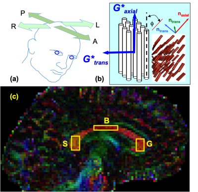 |
Variation of In Vivo Anisotropic MRE Metrics in Corpus Callosum: Effect of Aging
Nicolas R Gallo1, Stacey M Cahoon1, Aaron T Anderson2, Noel M Naughton3, Assimina A Pelegri4, and John G Georgiadis1
1Biomedical Engineering, Illinois Institute of Technology, Chicago, IL, United States, 2Beckman Institute, Urbana, IL, United States, 3Department of Mechanical Science and Engineering, University of Illinois at Urbana-Champaign, Urbana, IL, United States, 4Department of Mechanical and Aerospace Engineering, Rutgers University, New Brunswick, NJ, United States
Healthy aging involves local variations in viscoelastic shear properties of the brain. We employ high-resolution, multi-excitation MRE and a novel anisotropic inversion scheme (iTI) to extract local shear anisotropic moduli in vivo. The ratio of transverse to axial moduli, a new MRE metric, remains greater than 1 along the splenium, body and genu regions of the corpus callosum for both young and old subjects. This metric peaks in the body region and decreases with age throughout the corpus callosum.
|
0165.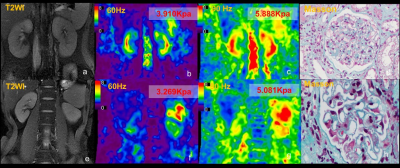 |
Preliminary application of Three-Dimensional Multifrequency MR Elastography for Chronic Kidney Disease
Shan Pi1, Jonathan M. Scott2, Yin Li3, Hui Peng3, Huiquan Wen1, Matthew C. Murphy2, Jingbiao Chen1, Meng Yin2, Jun Chen2, Kevin J. Glaser2, Rchard L. Ehman2, and Jin Wang1
Video Permission Withheld
1Department of Radiology, the Third Affiliated Hospital, Sun Yat-sen University, Guang Zhou, China, 2Department of Radiology, Mayo Clinic, Rochester, Micronesia, 3Department of Nephrology, the Third Affiliated Hospital, Sun Yat-sen University, Guang Zhou, China
Chronic kidney disease (CKD) is increasing in incidence and prevalence worldwide and early detection of CKD is a major challenge. MR elastography (MRE) is a noninvasive technique capable of quantifying the mechanical properties of tissue that has shown potential for assessing kidney diseases. MRE using 60-Hz and 90-Hz vibration frequencies can provide potential quantitative biomarkers for evaluating kidney function and biopsy score in CKD patients.
|
|
0166.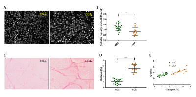 |
Evolution of tumour mechanical properties under static preload as potential biomarker of solid stress
Gwenaël Pagé1, Laurent Besret2, Marion Tardieu1, Maïlys Vidal1, Bernard Van Beers1,3, and Philippe Garteiser1
1Laboratory of Biomarkers in Imaging, Center of Research on Inflammation, UMR 1149 Inserm-Université de Paris, Paris, France, 2Sanofi R&D, Vitry-sur-Seine, France, 3Department of Radiology, Beaujon University Hospital Paris Nord, Clichy, France
The purpose of this study was to assess in two different human liver tumours the correlation between tumour solid stress and changes of mechanical properties under preload. MR elastography acquisitions were performed at different pressure levels by externally compressing the tumour with an inflatable balloon. Reference values for tumour fluid pressure and solid stress were acquired with a catheterized pressure transducer. The results, obtained in two liver tumour types with largely different basal mechanical properties, show that the evolution of tumour elasticity under preload is correlated with the tumour solid stress and could be a potential biomarker of tumour pressure.
|
|
0167.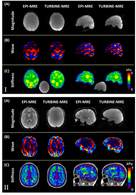 |
TURBINE-MRE: A 3D Hybrid Radial-Cartesian EPI Acquisition for MR Elastography
Yi Sui1, Arvin Forghanian-Arani1, Joshua D. Trzasko, 1, Matthew C. Murphy1, Phillip J. Rossman1, Kevin J. Glaser1, Kiaran P. McGee1, Armando Manduca2, Richard L. Ehman1, Philip A. Araoz1, and John III Huston1
1Radiology, Mayo Clinic, Rochester, MN, United States, 2Physiology and Biomedical Engineering, Mayo Clinic, Rochester, MN, United States
This study demonstrates the technical feasibility of a 3D radially batched internal navigator echo magnetic resonance elastography (TURBINE-MRE) technique in the brain. The highly efficient TURBINE-MRE approach allows for a true 3D wave displacement field to be acquired over the entire human brain volume in approximately 1.5 minutes.
|
|
|
0168.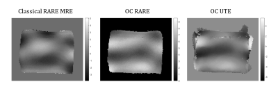 |
Ultra-short echo time Magnetic Resonance Elastography
Pilar Sango Solanas1, Kevin Tse Ve Koon1, Eric Van Reeth1, Cyrielle Caussy2, and Olivier Beuf1
1Univ Lyon, INSA‐Lyon, Université Claude Bernard Lyon 1, UJM-Saint Etienne, CNRS, Inserm, CREATIS UMR 5220, U1206, F‐69616, Lyon, France, Lyon, France, 2Département d’Endocrinologie, Diabète et Nutrition, Centre Hospitalier Lyon Sud, Hospices Civils de Lyon, CarMeN, INSERM U1060, INRA U1397, Lyon, France, Lyon, France
Magnetic Resonance Elastography (MRE) is a valuable technique to quantitatively characterize mechanical properties of tissues based on the properties of shear waves propagation. In this study, a radial acquisition MRE sequence potentially able to quantify viscoelastic parameters of tissues whose T2 values are very short is proposed. To this end, an optimal control-based RF pulse is applied with a constant gradient during the mechanical excitation to simultaneously perform spatially selective excitation and motion encoding. Acquisition is started right after, enabling a very short TE. Results on phantom experiments demonstrated the feasibility of our ultra-short echo time MRE technique.
|
|
0169.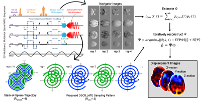 |
OSCILLATE: A Low-Rank Approach for Accelerated Magnetic Resonance Elastography
Grace McIlvain1, Alex M Cerjanic2, Anthony G Christodoulou3, Matthew DJ McGarry4, and Curtis L Johnson1
1Biomedical Engineering, University of Delaware, Newark, DE, United States, 2Bioengineering, University of Illinois, Urbana, IL, United States, 3Biomedical Imaging Research Institute, Cedars-Sinai Medical Center, Los Angeles, CA, United States, 4Thayer School of Engineering, Dartmouth College, Hanover, NH, United States
MR elastography (MRE) has emerged as a sensitive measure of brain health, but due to the need for repeated measures of wave motion, it is a fundamentally long scan. We have developed a method for accurate MRE using spatiotemporal undersampling and low rank joint reconstruction across all samples. We demonstrate the ability to collect accurate MRE data in half the time with under 2% stiffness error. This accelerated method will be used to scan challenging populations, such as those with developmental disabilities, as well as improve achievable resolution and feasibility of multi-frequency or multi-excitation methods.
|
|
0170.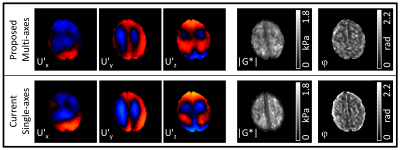 |
Accelerating DENSE MR elastography by including multi-axes motion encoding into the multiphase DENSE-MRE acquisition scheme
Johannes Strasser1, Martin Soellradl1, Christian Enzinger1, and Stefan Ropele1
1Department of Neurology, Medical University of Graz, Graz, Austria
In MR elastography, the propagation of three-dimensional wave motion is acquired to assess mechanical tissue properties. We here propose an accelerated approach of the multiphase DENSE-MRE acquisition scheme which additionally includes three-dimensional motion encoding besides the multiple phase offsets within one TR. In addition to phantom experiments, this multi-axes encoding concept was also investigated in the human brain in vivo. The gathered wave images and shear modulus maps are confirmed by three consecutive single-axes multiphase DENSE-MRE acquisitions for x-, y- and z-motion encoding direction. With this concept, the acquisition can be accelerated up to a factor of 3.
|
 Watch the Video