
Scientific Session
CEST, MT, Zero-TE and Relaxometry
Session Topic: CEST, MT, Zero-TE and Relaxometry
Session Sub-Topic: Chemical Exchange & Magnetisation Transfer: Mechanisms & Applications
Oral
Contrast Mechanisms
| Tuesday Parallel 1 Live Q&A | Tuesday, 11 August 2020, 14:30 - 15:15 UTC | Moderators: Rosa Tamara Branca |
Session Number: O-50
 |
0497.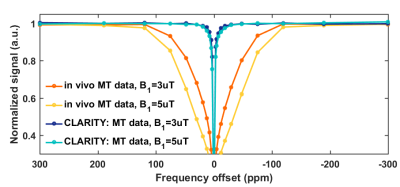 |
The CLARITY procedure of lipid removal from brain tissue sample reveals the lipid-origin of MT contrast in CEST imaging experiment
Anna Orzylowska1, Tymoteusz Słowik2, Agata Chudzik1, Anna Pankowska3, Wilfred W Lam4, and Greg J Stanisz1,4,5
1Department of Neurosurgery and Paediatric Neurosurgery, Medical University of Lublin, Lublin, Poland, 2Center of Experimental Medicine, Medical University of Lublin, Lublin, Poland, 3Department of Radiography, Medical University of Lublin, Lublin, Poland, 4Physical Sciences, Sunnybrook Research Institute, Toronto, ON, Canada, 5Department of Medical Biophysics, University of Toronto, Toronto, ON, Canada
The study compares the differences between Z-spectra derived from CEST imaging of rat brain in vivo and after post-mortem CLARITY lipids removal procedure. The lipids removal nulled-out MT macromolecular-originating signal measured with B1 saturation amplitudes of 3 and 5 µT as compared to in vivo, and resulted in negligible MT contribution to CEST Z-spectra acquired with B1s of 0.5 and 0.75 µT, as opposite to living tissue, where the MT effect was significant. Our results showed that the macromolecular MT contribution into in vivo Z-spectra originates mostly from lipids, since the CLARITY technique removed the MT component from the spectrum.
|
0498.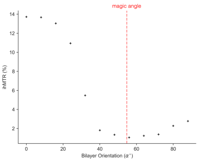 |
Orientation dependence of inhomogeneous magnetization transfer and dipolar order relaxation time in a phospholipid bilayer sample
Sarah Rosemary Morris1,2,3, Rebecca Frederick1, Alex L MacKay1,2,4, Cornelia Laule1,2,3,5, and Carl A. Michal1
1Physics & Astronomy, University of British Columbia, Vancouver, BC, Canada, 2Radiology, University of British Columbia, Vancouver, BC, Canada, 3International Collaboration on Repair Discoveries, Vancouver, BC, Canada, 4UBC MRI Research Centre, Vancouver, BC, Canada, 5Pathology & Laboratory Medicine, University of British Columbia, Vancouver, BC, Canada
Inhomogeneous magnetization transfer ratio (ihMTR) is reported to have significant orientation dependence in the brain, likely due to the anisotropy of dipolar couplings between methylene protons on the oriented lipids in myelin bilayers. We measured the orientation dependence of linewidth, dipolar relaxation time (T1D) and ihMTR in an aligned phospholipid bilayer sample at 9.4T. ihMTR was maximized when the bilayers were parallel to B0 and minimized near the magic angle (~54.7°) despite the fact that T1D is maximized there. This is in contrast to previous in vivo results which show maximal ihMTR for bilayers perpendicular to B0.
|
|
0499.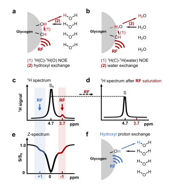 |
Understanding the magnetization transfer pathway for water-based detection of the aliphatic protons in glycogen
Yang Zhou1,2, Peter C.M. van Zijl1,2, Jiadi Xu1,2, and Nirbhay N. Yadav1,2
1The Russell H. Morgan Department of Radiology, The Johns Hopkins University School of Medicine, Baltimore, MD, United States, 2F.M. Kirby Research Center for Functional, Brain Imaging, Kennedy Krieger Institute, Baltimore, MD, United States
Recently a new MRI method was developed for the sensitivity enhanced detection of glycogen based on magnetization transfer between glycogen aliphatic protons and water, yet the mechanism of this transfer pathway is still not well understood. Here, we show that the magnetization transfer occurs via the relayed-NOE (rNOE) CEST effect. A theoretical model is proposed to quantitatively describe the rNOE signal in these magnetization transfer MRI experiments. This study provides insight into the rNOE mechanism that commonly occurs in magnetization transfer MRI on systems such as proteins and carbohydrate polymers.
|
|
0500. |
Fluid suppression in CEST imaging affects predominantly IDH-mutant 1p/19q retained gliomas with T2-FLAIR mismatch
Stefano Casagranda1, Laura Mancini2,3, Guillaume Gautier1, Philippe Peter1, Bruno Lopez1, Sebastian Brandner4,5, Enrico De Vita2,6, Xavier Golay2,3, and Sotirios Bisdas2,3
1Olea Medical, La Ciotat, France, 2Lysholm Dept of Neuroradiology, University College of London Hospitals NHS Foundation Trust, London, United Kingdom, 3Institute of Neurology UCL, London, United Kingdom, 4National Hospital for Neurology & Neurosurgery, University College of London Hospitals NHS Foundation Trust, London, United Kingdom, 5Department of Neurodegenerative Disease, Institute of Neurology UCL, London, United Kingdom, 6Biomedical Engineering Department, School of Biomedical Engineering and Imaging Sciences, King's College London, London, United Kingdom
CEST is a novel MR technique helpful for predicting IDH and 1p/19q status in gliomas. The asymmetry-based methods however are sensitive to fluid signal and recent studies have shown that a significant proportion of IDH-mutant 1p/19q retained gliomas have T2-FLAIR mismatch, indicating the presence of a more fluid microenvironment. This work shows how fluid-suppressed CEST imaging metrics have an impact on amide and amine signals in glioma, with highest effect on IDH-mutant 1p/19q retained with T2-FLAIR mismatch. The combined use of asymmetry-based and fluid-suppressed CEST metrics could be a valuable tool for glioma staging more robust than asymmetry-based metrics alone.
|
|
0501.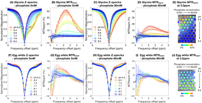 |
Influence of phosphate concentration on amine and amide chemical exchange saturation transfer (CEST) contrast
Jingwen Yao1,2,3, Chencai Wang1,2, and Benjamin M. Ellingson1,2,3
1Brain Tumor Imaging Laboratory (BTIL), Center of Computer Vision and Imaging Biomarker, David Geffen School of Medicine, UCLA, Los Angeles, CA, United States, 2Department of Radiological Sciences, David Geffen School of Medicine, UCLA, Los Angeles, CA, United States, 3Department of Bioengineering, Henry Samueli School of Engineering and Applied Science, UCLA, Los Angeles, CA, United States
Effects of the catalytic in vivo chemical environment have often been neglected or underestimated in CEST-MRI studies. Phosphate is the predominant exchange catalyst in intracellular fluid and is essential for biosynthesis and bioenergetics. In this study, we evaluated the influence of phosphate on amine and amide CEST contrast using Bloch-McConnell simulations applied to physical phantom data. We demonstrate that amine proton exchange is greatly catalyzed by phosphate, under a physiological concentration range. We propose that catalytic agents should be considered as confounding factors in future CEST-MRI researches. This new dimension may also benefit the development of novel phosphate-sensitive imaging method.
|
|
|
0502.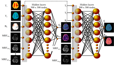 |
Early Detection of Tumor Apoptotic Response to Oncolytic Virotherapy using Deep CEST MR Fingerprinting
Or Perlman1, Hirotaka Ito2, Kai Herz3,4, Hiroshi Nakashima2, Moritz Zaiss3,5, E. Antonio Chiocca2, Christopher Nguyen1, Ouri Cohen6, Matthew S. Rosen1,7, and Christian T. Farrar1
1Athinoula A. Martinos Center for Biomedical Imaging, Massachusetts General Hospital and Harvard Medical School, Charlestown, MA, United States, 2Brigham and Women’s Hospital and Harvard Medical School, Boston, MA, United States, 3Magnetic Resonance Center, Max Planck Institute for Biological Cybernetics, Tübingen, Germany, 4IMPRS for Cognitive and Systems Neuroscience, University of Tübingen, Tübingen, Germany, 5Department of Neuroradiology, University Clinic Erlangen, Erlangen, Germany, 6Memorial Sloan Kettering Cancer Center, New York, NY, United States, 7Department of Physics, Harvard University, Cambridge, MA, United States
Oncolytic virotherapy (OV) is a promising treatment for high mortality cancers. To optimize the clinical outcome, non-invasive monitoring is essential. The goal of this work was to develop a deep-learning-based technique for quantitative and rapid molecular imaging of OV treatment response. Two CEST MR-fingerprinting protocols were sequentially implemented (105s each) and incorporated within a deep-reconstruction-network, trained to output the quantitative semi-solid and amide pool exchange parameters. The resulting molecular maps allowed early apoptosis detection in brain tumor OV mouse models. Clinical translation of CEST-MRF is demonstrated in a normal human subject and yielded parameters in good agreement with literature values.
|
0503.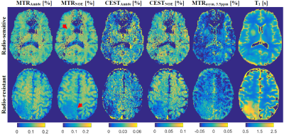 |
CEST and qMT Properties of Brain Metastases from Radio-resistant and Radio-sensitive Primary Tumours
Hatef Mehrabian1, Wilfred W Lam1, Hany Soliman1,2,3, Sten Myrehaug2,3, Arjun Sahgal1,2,3, and Greg J Stanisz1,4
1Physical Sciences, Sunnybrook Research Institute, Toronto, ON, Canada, 2Radiation Oncology, Sunnybrook Health Sciences Centre, Toronto, ON, Canada, 3Radiation Oncology, University of Toronto, Toronto, ON, Canada, 4Medical Biophysics, University of Toronto, Toronto, ON, Canada
Previous animal studies have shown differences in CEST properties of radio-resistant and radio-sensitive renal cell carcinoma xenografts. The current study probed the differences in CEST and MT properties of brain metastases from radio-resistant and radio-sensitive primary tumours. We observed significantly lower amount of magnetization transfer, RM0B/RA, and direct water saturation effect, 1/(RAT2A) in brain metastases from radio-resistant tumours compared to those from radio-sensitive tumours. However, the CEST effects of the two cohorts were not different. Such information should be considered when investigating brain metastases and their response to treatment.
|
|
 |
0504.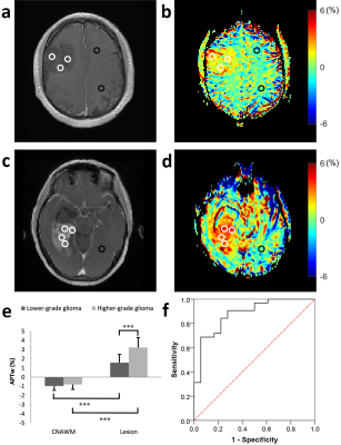 |
Prediction of prognostic characteristics in glioma patients using amide proton transfer imaging at 3 Tesla
Jie Liu1, Xiaofei Lv2, Chao Ke3, Zongwei Xu1, Long Qian4, Shijie Xu3, Xin Liu1, Hairong Zheng1, and Yin Wu1
1Paul C. Lauterbur Research Center for Biomedical Imaging, Shenzhen Institutes of Advanced Technology, Chinese Academy of Sciences, Shenzhen, China, 2Department of Medical Imaging, Sun Yat-Sen University Cancer Center, Guangzhou, China, 3Department of Neurosurgery, Sun Yat-Sen University Cancer Center, Guangzhou, China, 4GE Healthcare, Beijing, China
Early identification of glioma prognostic characteristics is of great clinical importance. This study aims to evaluate the feasibility of APT in the prediction of tumor grade, IDH mutation and MGMT promoter methylation status at 3 Tesla. A total of 50 patients were recruited. Results show that although APTw effect exhibits no substantial difference based on MGMT methylation status, it enables the discrimination of histopathological grade and IDH mutant status with AUCs higher than 0.88. The results suggest APTw is a valuable imaging biomarker for prediction of tumor prognostic parameters, that may benefit accurate diagnosis and prompt treatment decisions.
|
|
0505.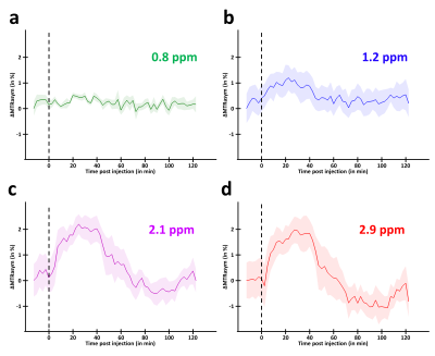 |
Unveiling the fate of glycolytic substrates using multi-spectral CEST: proof of concept with 2DG in the rat brain
Yohann Mathieu-Daudé1,2, Mélissa Vincent1,2, Julien Valette1,2, and Julien Flament1,2
1Molecular Imaging Research Center (MIRCen), Commissariat à l’Energie Atomique et aux Energies Alternatives (CEA), Fontenay-aux-Roses, France, 2UMR 9199, Neurodegenerative Diseases Laboratory, Centre National de la Recherche Scientifique (CNRS), Université Paris-Sud, Université Paris-Saclay, Fontenay-aux-Roses, France
2-Deoxy-D-glucose (2DG), an analogue of glucose similarly transported but with metabolism blocked after the first phosphorylation into 2DG-6-phosphate (2DG6P), has already been used to study glycolytic metabolism using gluCEST. However, origin of glucoCEST signal is still an open question important to be addressed. In this study, we measured for the first time variations of CEST signal in the rat brain at different resonance frequencies following 2DG injection. The richness of CEST signal can help assessing the fate of glycolytic substrates and would constitute a first step toward quantitative measurement of glucose metabolism using CEST method.
|
|
0506.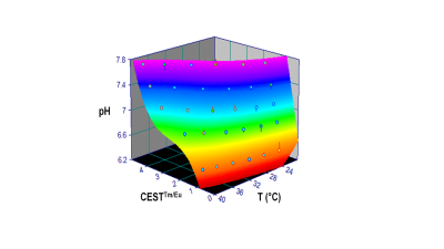 |
High-resolution pH imaging with ratiometric CEST and BIRDS using dual paramagnetic DOTA–tetraglycinate agents
Jelena Mihailovic1,2, Yuegao Huang1, John Walsh1, Daniel Coman1, Sara Samuel3, and Fahmeed Hyder1,3
1(1)Magnetic Resonance Research Center (MRRC), Yale University, New Haven, CT, United States, 2(2)Department of Diagnostic Radiology, Yale University, New Haven, CT, United States, 3Core Center for Quantitative Neuroscience with Magnetic Resonance (QNMR), Yale University, New Haven, CT, United States
Chemical Exchange Saturation Transfer (CEST) and Biosensor Imaging of Redundant Deviation in Shifts (BIRDS) biosensing methods differ respectively by detecting exchangeable and non-exchangeable protons on the agent. Given that CEST and BIRDS properties observed from the same paramagnetic agent are complimentary, we describe a novel approach for high-resolution pH imaging using dual agents of europium and thulium complexed with DOTA-tetraglycinate. In vitro results test the hypothesis that ratiometric paraCEST attributes are conserved when temperature from paraBIRDS is detected simultaneously, enabling absolute pH imaging. In vivo results in glioblastoma demonstrate feasibility of this dual paraCEST-paraBIRDS biosensing method for high-resolution pH imaging.
|
 Watch the Video