
Scientific Session
Perfusion and Permeability
Session Topic: Perfusion and Permeability
Session Sub-Topic: Arterial Spin Labelling Perfusion Imaging
Oral
Contrast Mechanisms
| Monday Parallel 1 Live Q&A | Monday, 10 August 2020, 13:45 - 14:30 UTC | Moderators: Thomas Lindner & Vasily Yarnykh |
Session Number: O-51
0017.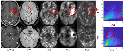 |
Differences in quantitative glioma perfusion imaging with ASL and DSC: validation with 15O-H2O PET
Jan Petr1, Niels Verburg2, Joost P.A. Kuijer3, Thomas Koopman3, Vera C. Keil4, Esther A.H. Warnert5, Frederik Barkhof3,6, Jörg van den Hoff1, Ronald Boellaard3, Philip C. de Witt Hamer2, and Henri J.M.M. Mutsaerts3,7
1Institute of Radiopharmaceutical Cancer Research, Helmholtz-Zentrum Dresden-Rossendorf, Dresden, Germany, 2Neurosurgical Center Amsterdam, Amsterdam University Medical Center, location VU, Amsterdam, Netherlands, 3Department of Radiology & Nuclear Medicine, Amsterdam University Medical Center, location VU, Amsterdam, Netherlands, 4Department of Neuroradiology, Bonn University Hospital, Bonn, Germany, 5Department of Radiology and Nuclear Medicine, Erasmus MC, Rotterdam, Netherlands, 6UCL Institutes of Neurology and Healthcare Engineering, London, United Kingdom, 7Ghent Institute for Functional and Metabolic Imaging (GIfMI), Ghent University, Ghent, Belgium
While agreement between ASL, DSC, and PET perfusion is well established in healthy volunteers, an analogous comparison in gliomas is still missing and more challenging. We compared ASL and DSC perfusion measurements with the gold-standard of 15O-H2O-PET perfusion measurements in eight patients diagnosed with gliomas. We showed the importance of normalization to the contralateral hemisphere, and identified several examples of different regional perfusion as assessed with ASL and DSC and interpreted them using the PET reference.
|
|
 |
0018.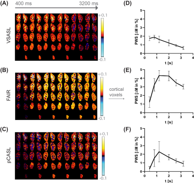 |
Exploring label dynamics of velocity selective arterial spin labeling in the kidney
Isabell K. Bones1, Suzanne L. Franklin1,2, Anita A. Harteveld1, Matthias J.P. van Osch2, Sophie Schmid2, Jeroen Hendrikse3, Chrit Moonen1, Marijn van Stralen1, and Clemens Bos1
1Center for Image Sciences, University Medical Center Utrecht, Utrecht, Netherlands, 2C.J.Gorter Center for High Field MRI, Department of Radiology, Leiden University Medical Center, Leiden, Netherlands, 3Department of Radiology, University Medical Center Utrecht, Utrecht, Netherlands
VSASL for renal application has constraints on the cut-off velocity, as low Vc in the presence of respiratory motion causes spurious labeling of moving tissue. With higher Vc, label could be generated more upstream in the vascular tree, potentially introducing ATT sensitivity. To study label dynamics of renal VSASL using a Vc compatible with free-breathing (Vc of 10cm/s), data at multiple time points were acquired. High ASL signal was already observed at early time points, thus supporting that spatially non-selective VSASL in the kidney generates label close to, or even inside the target tissue, also using a free-breathing compatible Vc.
|
 |
0019.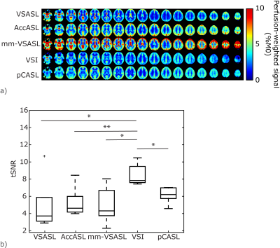 |
Multi-organ comparison of flow-based Arterial Spin Labeling techniques: brain and kidney perfusion imaging without transit time artefacts
Suzanne L. Franklin1,2,3, Isabell K. Bones2, Anita A. Harteveld2, Lydiane Hirschler1, Marijn van Stralen2, Anneloes de Boer2, Hans Hoogduin2, Matthias J.P. van Osch1,3, Sophie Schmid1,3, and Clemens Bos2
1C.J. Gorter Center for High Field MRI, Department of Radiology, Leiden University Medical Center, Leiden, Netherlands, 2Center for Image Sciences, University Medical Center Utrecht, Utrecht, Netherlands, 3Leiden Institute for Brain and Cognition, Leiden University, Leiden, Netherlands
Different flow-based arterial spin labeling (ASL)-techniques were proposed in recent years. In this multi-organ study four flow-based ASL-techniques were compared, with pCASL in brain, and with both pCASL and FAIR in kidney. ASL-techniques were compared based on temporal-SNR, sensitivity to perfusion changes (in brain) and robustness to respiratory motion (in kidney). In brain, Velocity-Selective Inversion showed superior temporal-SNR and sensitivity to perfusion changes. In kidney, flow-based ASL-techniques showed decreased temporal-SNR compared to FAIR, although their settings can be improved to increase robustness to B1-inhomogeneity. All ASL-techniques were relatively robust to respiratory motion, showing potential for free-breathing kidney-ASL at 3T.
|
|
0020.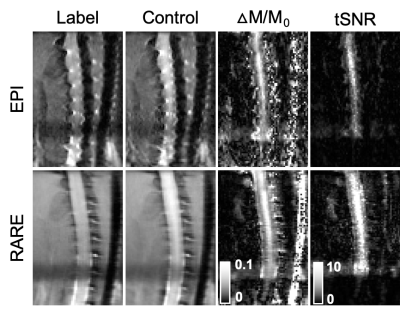 |
Preclinical Spinal Cord Perfusion Imaging with Pseudo-Continuous Arterial Spin Labeling
Briana Meyer1, Lydiane Hirschler2,3, Jan Warnking2, Emmanuel Barbier2, and Matthew Budde4
1Biophysics, Medical College of Wisconsin, Wauwatosa, WI, United States, 2Univ. Grenoble Alpes, Inserm, U1216, Grenoble Institut des Neurosciences, Grenoble, France, 3C.J. Gorter Center for High Field MRI, Radiology, Leiden University Medical Center, Leiden, Netherlands, 4Neurosurgery, Medical College of Wisconsin, Milwaukee, WI, United States
Pseudo-continuous arterial spin labeling (pCASL) to monitor spinal cord perfusion and hemodynamics has the potential to inform the clinical care of spinal cord injury and other disorders. This work demonstrates successful implementation and application of pCASL of the rodent cervical spinal cord at high field.
|
|
0021.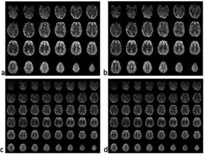 |
Super-Resolution Multi-band ASL using Slice Dithered Enhanced Resolution (SLIDER) Technique
Qinyang Shou1, Xingfeng Shao1, and Danny Wang1
1University of Southern California, Los Angeles, CA, United States
Arterial Spin Labelling (ASL) is a noninvasive imaging technique that can quantitatively measure Cerebral Blood Flow (CBF). However, existing ASL techniques generally have a low spatial resolution due to a relative low Signal-to-noise ratio (SNR). In this study, we develop a super-resolution ASL method by combining the Slice Dithered Enhanced Resolution (SLIDER) with multi-band ASL with optimized slice-dependent background suppression to enhance both the resolution and SNR. The reconstructed images achieve a resolution of isotropic 2x2x2 mm3, and show increased spatial and temporal SNR compared to standard high-resolution ASL images.
|
|
0022.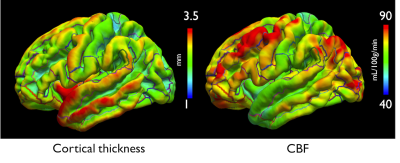 |
Regional and depth dependence of cortical blood-flow assessed with high-resolution Arterial Spin Labeling
Manuel Taso1, Fanny Munsch1, Li Zhao2, and David C. Alsop1
1Division of MRI research, Department of Radiology, Beth Israel Deaconess Medical Center, Harvard Medical School, Boston, MA, United States, 2Diagnostic Imaging and Radiology, Children's National Medical Center, Washington, DC, United States
Imaging cortical blood-flow using ASL is relevant to unravel the basis of brain functional autoregulation or response to stimuli, but challenging because of the usual compromise between brain coverage, SNR and spatial resolution in ASL. We here propose to push the limits of volumetric ASL resolution using sparse variable-density FSE and Compressed-Sensing to study the distribution of cortical flow in healthy volunteers. We show through a group surface-based analysis some regional variations in cortical flow, but also depth-dependence of cortical flow. We also propose a high-resolution average ASL perfusion-weighted template that could have benefits for large-scale group studies.
|
0023.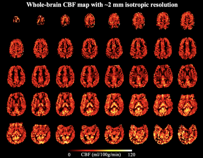 |
High-resolution whole brain ASL perfusion imaging at 7T with 12-fold acceleration and spatial-temporal regularized reconstruction
Xingfeng Shao1, Stefan M Spann2, Kai Wang1, Lirong Yan1,3, Stollberger Rudolf2, and Danny JJ Wang1,3
1Mark & Mary Stevens Neuroimaging and Informatics Institute, Keck School of Medicine, University of Southern California, Los Angeles, CA, United States, 2Institute of Medical Engineering, Graz University of Technology, Graz, Austria, 3Department of Neurology, University of Southern California, Los Angeles, CA, United States
Ultra-high field allows ASL to achieve higher spatial resolution due to increased SNR and prolonged T1 relaxation. We present a single-shot 3D GRASE pCASL technique with 12-fold acceleration using time-dependent 2D CAIPI sampling strategy, and reconstruction of the label/control time series with joint spatial and temporal total-generalized-variation (TGV) regularization. 2D CAIPI under-sampling pattern increases temporal incoherence between measurements which allows joint reconstruction of the highly accelerated ASL time series. Combining the advantages of ultra-high field strength, pTx coils, accelerated acquisition and advanced reconstruction, whole-brain CBF map with 2 mm isotropic resolution was obtained within 5 mins.
|
|
|
0024.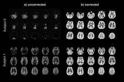 |
Optimization of Pseudo-Continuous Arterial Spin Labeling using Off-resonance Compensation Strategies at 7T
Gael Saib1, Alan Koretsky1, and S Lalith Talagala2
1NINDS/LFMI, National Institutes of Health, Bethesda, MD, United States, 2NINDS/NMRF, National Institutes of Health, Bethesda, MD, United States
Pseudo-continuous arterial spin labeling (PCASL) is very sensitive to off-resonance effects. This is especially a problem at higher fields (>3T). Off-resonance effects can be compensated by using an average or a vessel-specific correction integrated into the PCASL tagging/control pulse. Vessel-specific corrections can be performed using a prescan or a field map. In this study, we compared three off-resonance compensation strategies at 7T. Data showed that a large improvement (> 2 times) of the PCASL signal can be obtained with subject specific off-resonance correction with all 3 methods. The field map based method showed slightly better performance over the others.
|
|
0025.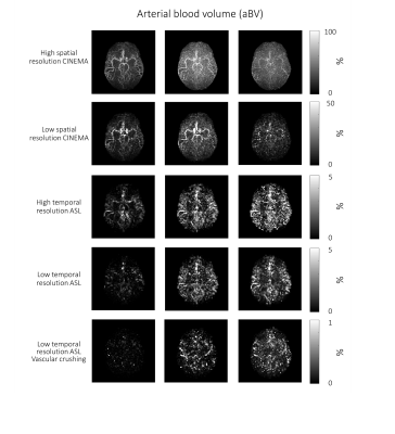 |
Validation of the estimation of the macrovascular contribution in multi-timepoint arterial spin labeling MRI using a two-component model
Merlijn van der Plas1, Sophie Schmid1, Martin Craig2, Michael Chappell2,3, and Matthias van Osch1
1Radiology, C.J. Gorter Center for High Field MRI, Leiden, Netherlands, 2Wellcome Centre for Integrative Neuroimaging, FMRIB, Nuffield Department of Clinical Neurosciences, University of Oxford, Oxford, United Kingdom, 3Institute of Biomedical Engineering, Research Council UK (EP/P012361/1), University of Oxford, Oxford, United Kingdom
A two-component kinetic model allows for the separation of the macrovascular and tissue signal. This model relies on the availability of multi-timepoint data and generates cerebral blood flow, arterial blood volume and arterial transit time maps. The goal of this study was to validate this separation of the macrovascular and tissue signal. A 4D-ASL angiography and densely sampled ASL data were acquired and fitted with different model settings. Fitting the 4D-ASL angiography with a macrovascular component showed the best fit for the model with gamma dispersion included but with limited freedom to change the dispersion parameters.
|
0026.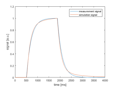 |
Towards patient specific dispersion correction for more accurate quantification in pCASL: modeling and experimental findings
Mareike Alicja Buck1 and Matthias Günther1,2
1Fraunhofer MEVIS, Bremen, Germany, 2MR-Imaging and Spectroscopy, Faculty 01 (Physics, Electrical Engineering), University Bremen, Bremen, Germany
This abstract compares quantified perfusion values of the standard model and a new dispersion model based on an AIF using the ASLIF-sequence. For different ATTs, the voxel´s signal was simulated using the dispersion model. The simulations show that the standard model overestimates the signal. This may result from lack of dispersion effects especially in the inflow phase of the labeled bolus. Consequently, the determined perfusion values vary for different ATTS. Thus, using an AIF based on an acquired patient specific reference bolus instead could improve the stability and robustness of quantified perfusion values.
|
 Watch the Video