
Scientific Session
Perfusion and Permeability
| Monday Parallel 1 Live Q&A | Monday, 10 August 2020, 13:45 - 14:30 UTC | Moderators: Amit Mehndiratta |
0007.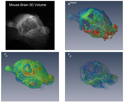 |
Whole tumor pharmacokinetic model analysis with 3D isotropic high resolution using 3D-UTE-GRASP sequence at 7T
Jin Zhang1, Karl Kiser1, Chongda Zhang1, Ayesha Bharadwaj Das1, and Sungheon Gene Kim1
1New York University School of Medicine, New York, NY, United States
Quantitative pharmacokinetic model parameter maps from dynamic contrast enhanced (DCE)-MRI can provide useful physiologically relevant information about tumor microenvironment, but often in low spatial resolution due to challenges in acquiring high resolution 3D data with high temporal resolution. The purpose of this study is to investigate the feasibility of generating the whole tumor high resolution pharmacokinetic model parameter maps with the 3D-UTE-GRASP1 sequence for both T1 mapping and dynamic scan.
|
|
 |
0008.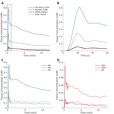 |
Hemodynamics and permeability of the windows of the brain: dynamic contrast-enhanced MRI of the circumventricular organs
Inge Verheggen1, Joost de Jong2, Martin van Boxtel1, Alida Postma2, Frans Verhey1, Jacobus Jansen2,3, and Walter Backes2
1Department of Psychiatry and Neuropsychology, Maastricht University, Maastricht, Netherlands, 2Department of Radiology and Nuclear Medicine, Maastricht University Medical Center, Maastricht, Netherlands, 3Department of Electrical Engineering, Eindhoven University of Technology, Eindhoven, Netherlands Circumventricular organs (CVOs), located around the ventricles without blood-brain barrier, maintain homeostasis between the blood, cerebrospinal fluid, and brain. Secretory CVOs are involved in peptide release and sensory CVOs regulate signal transmission. These organs can be an entrance point for pathogens. For the first time, physiological properties of the CVOs were assessed in vivo with dynamic contrast-enhanced (DCE) MRI. Assessing pharmacokinetics (leakage rate; blood perfusion; uptake capacity/retention) with DCE MRI in 20 healthy males, demonstrated that only secretory CVOs had noticeable stronger hemodynamics and higher permeability than normal-appearing brain matter. |
 |
0009.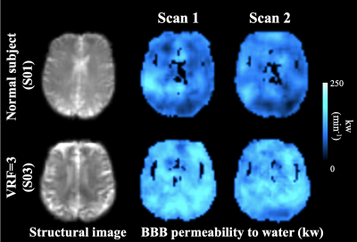 |
Comparison between blood-brain barrier permeability to water and gadolinium-based contrast agents in an elderly cohort
Xingfeng Shao1, Samantha Jenny Ma1, and Danny JJ Wang1,2
1Mark & Mary Stevens Neuroimaging and Informatics Institute, Keck School of Medicine, University of Southern California, Los Angeles, CA, United States, 2Department of Neurology, University of Southern California, Los Angeles, CA, United States
A diffusion-weighted arterial spin labeling (DW-ASL) technique has been proposed to non-invasively measure water exchange rate (kw) across the BBB. kw was compared with GBCAs permeability (Ktrans) in aged subjects at risk of small vessel disease. A positive correlation was found between kw and Ktrans only in the caudate, suggesting different BBB mechanisms probed by kw and Ktrans. Significant increase of kw was found in subjects with diabetes or high vascular risk while no Ktrans difference was observed. Water permeability could be a sensitive biomarker to study glymphatic function and vascular diseases before detectable BBB disruption occurs.
|
0010.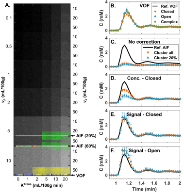 |
Partial Volume Correction of the Arterial Input Function with Surrounding Tissue Signal for Dynamic Contrast Enhanced MRI in the Brain
Benoît Bourassa-Moreau1, Réjean Lebel1, Guillaume Gilbert2, David Mathieu3, and Martin Lepage1
1Centre d’imagerie moléculaire de Sherbrooke, Département de médecine nucléaire et radiobiologie, Université de Sherbrooke, Sherbrooke, QC, Canada, 2MR Clinical Science, Philips Healthcare Canada, Markham, ON, Canada, 3Service de neurochirurgie, Département de chirurgie, Université de Sherbrooke, Sherbrooke, QC, Canada The arterial input function measured for brain dynamic contrast-enhanced MRI is contaminated by the signal contribution of surrounding tissues. This work corrects these partial volume effects on signal level by using the surrounding gray matter enhancement to discriminate pure arterial signal. The method also accounts for the high contrast agent concentration reached in arteries and veins that leads to signal non-linearity, saturation, and concurrent unwanted $$$T_2^*$$$ effects. This partial volume correction method is compared to concentration scaling on a digital reference object and on eight subjects. Better recovery of the arterial first pass and recirculation are shown. |
|
|
0011.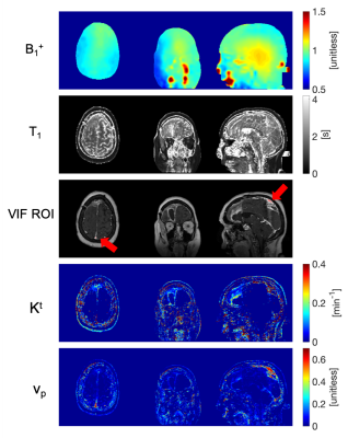 |
Pseudo Test-Retest Evaluation of Sparse DCE-MRI of Brain Tumor
Yannick Bliesener1, Robert Marc Lebel2,3, Jay Acharya4, Richard Frayne5, and Krishna Shrinivas Nayak1
1Department of Electrical and Computer Engineering, University of Southern California, Los Angeles, CA, United States, 2Applications and Workflow, GE Healthcare, Calgary, AB, Canada, 3Department of Radiology, University of Calgary, Calgary, AB, Canada, 4Department of Clinical Radiology, Keck School of Medicine of University of Southern California, Los Angeles, CA, United States, 5Departments of Radiology, and Clinical Neurosciences, University of Calgary, Calgary, AB, Canada
Brain DCE MRI suffers from poor spatial coverage, lack of standardization, and insufficient quantitative understanding of the extent of (physical) uncertainty in the measurements. Here, we attempt to overcome these by providing a fully automated high-resolution whole-brain DCE MRI pipeline with no user interaction. Prospective test-retest repeatability evaluation is challenging, therefore we employ a surrogate: multiple post-treatment time points in stable brain tumor patients. The proposed framework is able to yield consistent vascular input functions and tracer kinetic parameter histograms for repeated visits.
|
|
0012.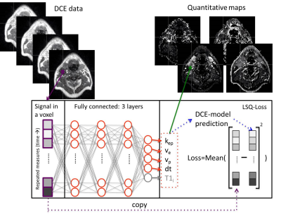 |
Unsupervised neural networks to improve quantitative DCE modelling
Oliver Gurney-Champion1, Matthew Orton1, Kevin Harrington1, Uwe Oelfke1, and Sebastiano Barbieri2
1The Institute of Cancer Research and Royal Marsden NHS Foundation Trust, London, United Kingdom, 2Centre for Big Data Research in Health, University of New South Wales, Sydney, Australia
We introduce a novel approach to fitting parameters from DCE MRI using an unsupervised neural network. The network is trained on in vivo data, with no ground truth, and is able to predicts DCE model parameters directly from the obtained MRI images. In simulations, our method outperformed the ordinary least squares fit approach in that it is more accurate and precise. In vivo, it produced substantially less noisy parameter maps than the current practise least-squares fit.
|
0013.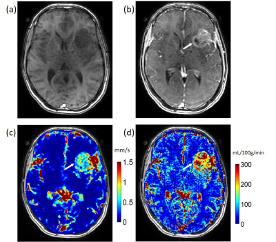 |
Quantitative Transport Mapping (QTM): Inverse Solution to a Voxelized Equation of Mass Flux of Contrast Agent in a Porous Tissue Model
Qihao Zhang1, Liangdong Zhou2, John Morgan3, Thanh D Nguyen4, Pascal Spincemaille3, and Yi Wang2
1Biomedical Engineeering, Cornell University, New York, NY, United States, 2Weill Cornell Medicine, New York, NY, United States, 3Radiology, Weill Cornell Medicine, New York, NY, United States, 4Weill Cornell Medicine College, New York, NY, United States
We purpose to calculate a tracer velocity field by solving the inverse problem of a voxelized transport equation for time resolved 3D (4D) dynamic contrast enhanced (DCE) data, which is termed as quantitative transport mapping (QTM). Using a porous medium model, the 4D imaging data is connected to the voxel-averaged transport equation of mass flux. The transport inverse problem is solved to estimate velocity and pseudo tortuosity. QTM provides the advantage of high accuracy in numerical validation and automated procession without manual input for in vivo DCE brain tumor data, compared to the traditional Kety’s method of perfusion quantification.
|
|
|
0014.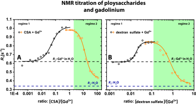 |
Gd3+ Deposition as an Underestimated Hazard? – Potential Masking of Gadolinium Long-Term Deposition in Biological Regimes
Patrick Werner1,2, Patrick Schuenke1, Antje Ludwig3, Daria Dymnikova4, Christian Teutloff4, Matthias Taupitz5, and Leif Schröder1
1Leibniz-Forschungsinstitut fuer Molekulare Pharmakologie (FMP), Berlin, Germany, 2BIOphysical Quantitative Imaging Towards Clinical Diagnosis (BIOQIC), Berlin, Germany, 3Center for Cardiovascular Research (CCR), Charite Berlin, Berlin, Germany, 4Freie Universität Berlin, Berlin, Germany, 5Department of Radiology, Charite Berlin, Berlin, Germany
Gd3+-ions can be released from GBCAs after in vivo application and polysaccharides like glycosaminoglycans are candidates for binding of released Gd3+-ions by acting as competing chelators. We showed that the chelation of Gd3+-ions to polysaccharides cause an increase of R1 due to the high relaxivity of such complexes. However, at high GAG/Gd3+ ratios and in cell experiments, we observed a decrease of R1 after the chelation of Gd3+. Our results demonstrate the importance of more in vivo-like setups for the investigation of gadolinium transchelation processes to prevent an underestimation of the amount of deposited gadolinium in biological tissues.
|
0015.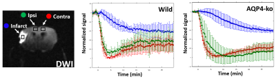 |
Distribution of intraperitoneally administered D2O in AQP4-knockout mouse brain after MCA occlusion
Obata Takayuki1, Takuya Urushihata1, Manami Takahashi1, Sayaka Shibata1, Nobuhiro Nitta1, Jeff Kershaw1, Yasuhiko Tachibana1, Masato Yasui2, Ichio Aoki1, Tatsuya Higashi1, Makoto Higuchi1, and Hiroyuki Takuwa1
1National Institute of Radiological Sciences, QST, Chiba, Japan, 2Department of Pharmacology, Keio University School of medicine, Tokyo, Japan
Using dynamic PDWI after intraperitoneal D2O injection, we observed a difference in the D2O distribution between aquaporin-4 knockout (AQP4-ko) and wild type (Wild) mice with MCA occlusion. The results suggest that blood flow changes and cell membrane water permeability have a complex relationship.
|
|
|
0016.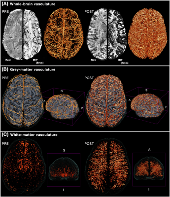 |
Human cerebral white-matter vasculature imaged using the blood-pool contrast agent Ferumoxytol: bundle-specific vessels and vascular density
Michaël Bernier1,2, Olivia Viessmann1,2, Ned Ohringer1, Jingyuan E. Chen1,2, Nina E. Fultz1,3, Rebecca Karp Leaf4, Lawrence L. Wald1,2,5, and Jonathan R. Polimeni1,2,5
1Athinoula A. Martinos Center for Biomedical Imaging, Massachusetts General Hospital, Charlestown, MA, United States, 2Radiology, Harvard Medical School, Boston, MA, United States, 3Engineering, Boston University, Boston, MA, United States, 4Division of Hematology, Massachusetts General Hospital, Boston, MA, United States, 55Division of Health Sciences and Technology, Massachusetts Institute of Technology, Cambridge, MA, United States
Ferumoxytol—a safe, superparamagnetic iron oxide nanoparticle that amplifies T2* dephasing in blood vessels—can be used as a powerful image contrast enhancement agent to aid vascular imaging. Combining this with an innovative vascular segmentation tool, here we evaluate how Ferumoxytol improves vascular detection throughout the brain using a region-based analysis of the gray-matter and a bundle-specific analysis of the white-matter. We report increases in white-matter vasculature specificity and uncover spatial patterns similar to white-matter tracts, therefore this work sheds new light on the possible existence and influence of a concurrent network of vasculature that follows the known fiber bundles.
|
 Watch the Video