
Scientific Session
Perfusion and Permeability
Session Topic: Perfusion and Permeability
Session Sub-Topic: Novel Spin Labelling Methods
Oral
Contrast Mechanisms
| Monday Parallel 1 Live Q&A | Monday, 10 August 2020, 13:45 - 14:30 UTC | Moderators: David Thomas |
Session Number: O-54
 |
0027.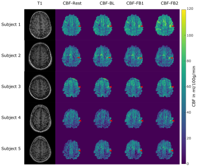 |
Self-Regulation of Brain Functions using Real-Time Neurofeedback Functional Arterial Spin Labeling
Stefan M Spann1, Doris Grössinger2, Christoph Stefan Aigner1,3, Josef Pfeuffer4, Guilherme Wood2,5, and Rudolf Stollberger1,5
1Institute of Medical Engineering, Graz University of Technology, Graz, Austria, 2Institute of Psychology, University of Graz, Graz, Austria, 3Physikalisch-Technische Bundesanstalt (PTB), Braunschweig and Berlin, Germany, 4Application Development, Siemens Healthcare, Erlangen, Germany, 5BioTechMed-Graz, Graz, Austria
Real-time neurofeedback (RT-NF) fMRI allows the subjects to regulate their own brain activity by providing them a neurofeedback. Functional ASL is perfectly suited for RT-NF studies due to the absolute quantification of activation related changes in the cerebral blood flow (CBF). In this study we implemented a real-time solution for ASL data processing and feedback generation which includes the following steps: data acquisition, image reconstruction, post-processing and neurofeedback presentation. The results of this RT-NF fASL study show that subjects were able to learn to regulate their own brain activation during a finger tapping experiment.
|
 |
0028.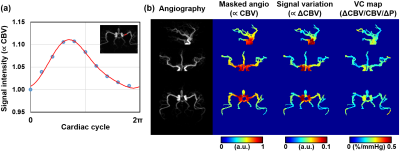 |
Non-contrast, high-resolution compliance mapping of intracranial vessels
Yang Li1, Michael Schär1, Dengrong Jiang1, and Hanzhang Lu1
1Johns Hopkins University Department of Radiology, Baltimore, MD, United States
Vascular compliance is an important predictor of cardiovascular disease and stroke. Here we proposed a technique to map vascular compliance along the entire intracranial arterial tree. We applied the ASL MRA to acquire k-space data in radial fashion. Then the k-space data was retrospectively grouped by cardiac phases and reconstructed by the GRASP algorithm. A series of cardiac-phase-resolved angiography images were obtained, allowing quantification of vascular compliance.
|
|
0029.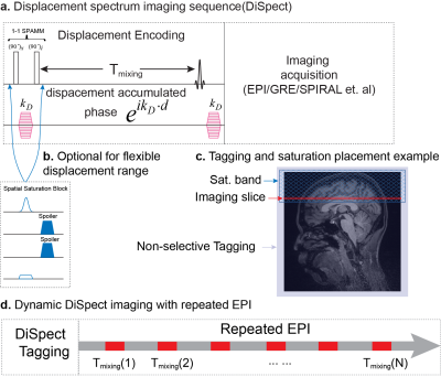 |
Retrospective Vessel Selective Perfusion Imaging with Displacement Spectrum Imaging (DiSpect) at Multiple Mixing Times
Ekin Karasan1, Michael Lustig1, and Zhiyong Zhang1,2
1Department of Electrical Engineering, University of California, Berkeley, CA, United States, 2Institute for Medical Imaging Technology, School of Biomedical Engineering, Shanghai Jiao Tong University, Shanghai, China
Displacement spectrum imaging (DiSpect) performs multiple scans with increasing displacement (DENSE) encodings. It can resolve the multi-dimensional spectrum of displacements that spins exhibit over the mixing time between tagging and imaging. This work presents two innovations: 1) Imaging dynamic displacement spectra by repeatedly imaging after tagging, each image corresponding to an increased mixing time. 2) Post acquisition, it is possible to retrospectively select source vessels from the displacement maps and display only their contribution to perfusion in the imaging slice. We demonstrate feasibility in flow phantom and in-vivo brain at 3T.
|
|
0030.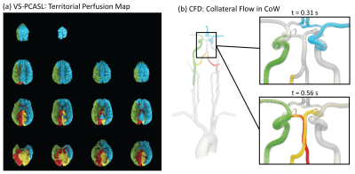 |
Calibration of patient-specific computational models of cerebral blood flow in cerebrovascular disease using arterial spin labeling
Jonas Schollenberger1, Luis Hernandez-Garcia1,2, and C. Alberto Figueroa1,3
1Biomedical Engineering, University of Michigan, Ann Arbor, MI, United States, 2fMRI Laboratory, University of Michigan, Ann Arbor, MI, United States, 3Surgery, University of Michigan, Ann Arbor, MI, United States
Collateral flow patterns in the circle of Willis play a major role in maintaining adequate blood supply to the brain in the presence of cerebrovascular occlusive disease. In this work, we present a strategy to quantify collateral flow by calibrating patient-specific computational fluid dynamic models of cerebral blood flow with perfusion data from arterial spin labeling. For a patient with right carotid stenosis, the collateral flow patterns in the circle of Willis obtained with the calibrated computational model show good agreement with territorial perfusion maps acquired with vessel-selective arterial spin labeling.
|
|
0031.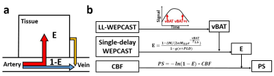 |
Improved accuracy of blood-brain barrier (BBB) assessment with water-extraction-with-phase-contrast-arterial-spin-tagging (WEPCAST) MRI
Zixuan Lin1, Dengrong Jiang1, Yang Li1, Pan Su1, Jay J. Pillai1, and Hanzhang Lu1
1Department of Radiology, Johns Hopkins University, Baltimore, MD, United States
A new scheme of water-extraction-with-phase-contrast-arterial-spin-tagging (WEPCAST) MRI was proposed for non-invasive assessment of blood-brain-barrier (BBB) permeability to water. In this scheme, venous bolus-arrival-time was measured first by Look-Locker WEPCAST and then applied to single-delay long-labeling-duration WEPCAST scan to estimate water extraction fraction. The results showed an improved accuracy for estimation of BBB permeability.
|
|
0032.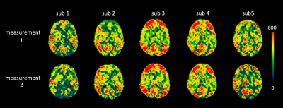 |
Assessing Repeatability of Blood Brain Barrier Permeability Measure Using Contrast-free MRI
Amnah Mahroo1, Nora-Josefin Breutigam1, and Matthias Günther1,2
1MR Physics, Fraunhofer Institute for Digital Medicine MEVIS, Bremen, Germany, 2MR-Imaging and Spectroscopy, University of Bremen, Bremen, Germany
Disrupted blood brain barrier (BBB) is reported to be one of the causes in various neuropathological diseases1. We assessed the quantified permeability of BBB using blood to tissue water exchange dynamics by employing multi-echo ASL sequence in five healthy individuals. A repeated measurement was conducted to assess the robustness and precision of the method. The average gray matter values were 357 ± 62 ms which are in-accordance with the literature reported values.
|
|
0033.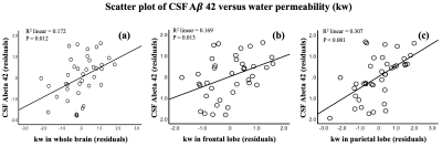 |
Water exchange across blood-brain barrier is associated with CSF Amyloid-β 42 and cognition in healthy older adults
Xingfeng Shao1, Brian T Gold2, and Danny JJ Wang1,3
1Mark & Mary Stevens Neuroimaging and Informatics Institute, Keck School of Medicine, University of Southern California, Los Angeles, CA, United States, 2Department of Neuroscience, College of Medicine, University of Kentucky, Lexington, KY, United States, 3Department of Neurology, University of Southern California, Los Angeles, CA, United States
Abnormally low CSF amyloid-β (Aβ)-42 level is an early biomarker of the Alzheimer’s Disease (AD), however lumbar puncture is required to collect CSF samples. A diffusion prepared arterial spin labeling technique was proposed to measure blood-brain barrier (BBB) water permeability (kw) non-invasively. Associations between water permeability and CSF Aβ42 levels in the “healthy” aging brains were studied. Significant positive correlations were found between kw and CSF Aβ42 and digit symbol scores, which suggests kw may serve as an early imaging marker of AD.
|
|
0034.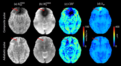 |
High Resolution Water Exchange Rate Mapping using 3D Diffusion Prepared Arterial Spin Labeled Perfusion MRI
Qihao Zhang1, Thanh D Nguyen2, Jana Ivanidze3, and Yi Wang3
1Cornell University, Ithaca, NY, NY, United States, 2Weill Cornell Medicine College, New York, NY, United States, 3Weill Cornell Medicine, New York, NY, United States
In this work, we propose an optimized acquisition for high resolution water exchange rate (kw) mapping, that uses adiabatic RF pulses, 3D fast spin echo readout, regularized inversion to a direct model of the water exchange rate, and fast T1 mapping. Feasibility and superior performance is shown in a regional based analysis in 6 healthy subjects.
|
|
0035.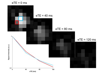 |
Oxygen Extraction Fraction Mapping using Remote Sensing: Spatially Encoded T2-Relaxation-Under-Spin-Tagging (SE-TRUST)
Caitlin O'Brien1, Thomas Okell1, Mark Chiew1, and Peter Jezzard1
1Wellcome Centre for Integrative Neuroimaging (FMRIB), University of Oxford, Oxford, United Kingdom
This work uses remote sensing methods to encode spatial information of venous blood spins in the brain into the longitudinal magnetisation. This information was then decoded remotely from the blood signal in the superior sagittal sinus. A T2-preparation module allowed venous blood T2 and therefore oxygen extraction fraction, to be mapped. An optimum inversion delay (TI) of 2s was found, and the sensitivity of the method to the spatial origins of the blood spins was verified. Low resolution venous T2 maps were obtained in two healthy volunteers. Average values were comparable to global T2 using conventional TRUST.
|
|
0036.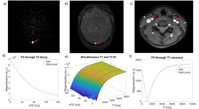 |
T1 and T2 relaxometry of arterial and venous blood: reliability of different methods
Koen P.A. Baas1, Bram F. Coolen2, Gustav J. Strijkers2, and Aart J. Nederveen1
1Radiology and Nuclear Medicine, Amsterdam UMC, Amsterdam, Netherlands, 2Biomedical Engineering & Physics, Amsterdam UMC, Amsterdam, Netherlands
We investigated the reliability of different T1 and T2 relaxometry methods for arterial and venous blood. While TRIR enables measurements of both venous blood T1 and T2, T2 estimates from TRIR showed poorer repeatability compared to TRUST. Moreover, significantly higher venous blood T2 values were observed using TRIR. Lastly, arterial blood T1 measurements showed a better repeatability compared to venous T1 measurements using TRIR. These findings advocate for the use of the arterial T1 measurements instead of venous T1 and the use of TRUST to measure venous blood T2.
|
 Watch the Video