
Scientific Session
Managing Motion and Artifacts
Session Topic: Managing Motion and Artifacts
Session Sub-Topic: Keep Still: Managing Motion in the Body
Oral
Acquisition, Reconstruction & Analysis
| Tuesday Parallel 4 Live Q&A | Tuesday, 11 August 2020, 13:45 - 14:30 UTC | Moderators: Claudia Prieto & Daniel Staeb |
Session Number: O-55
 |
0452.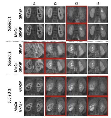 |
Bulk Motion Compensated Image Reconstruction for Renal Function Estimation with DCE-MRI
Jaume Coll-Font1,2, Onur Afacan1,2, Alto Stemmer3, Richard S. Lee2,4, Jeanne Chow1,2, Simon Warfield1,2, and Sila Kurugol1,2
1Radiology, Boston Children's Hospital, Boston, MA, United States, 2Harvard Medical School, Boston, MA, United States, 3Siemens Healthcare GmbH, Erlangen, Germany, 4Urology, Boston Children's Hospital, Boston, MA, United States
Dynamic Radial VIBE (DRV) DCE-MRI can provide high spatio-temporal resolution in the dynamic series of volumes used to evaluate the kidney function. However, bulk motion during the scan corrupts the volumes and deteriorates the quality of the kidney function estimation. We introduce a bulk-motion robust image reconstruction technique to mitigate the effects of motion. Our algorithm detects corrupted k-space data, reconstructs the volumes without signal dropout and aligns them. We applied this approach on non-sedated babies undergoing feed-and-wrap DCE-MRI with DRV. Our results show that our method improves the image quality and the estimation of the kidney function parameters.
|
|
0453.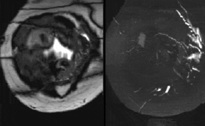 |
Motion corrected reconstruction of abdominal SWEEP data using local similarity graphs and deformable slice to volume registration
Laurence H Jackson1, Alena Uus1, Dafnis Batalle2,3, Jana M Hutter1, Thomas A Roberts1, Anthony N Price1, Alison Ho2,4, Laura McCabe2, Maria Deprez1, Lucy Chappell4, Mary Rutherford2, and Joseph V Hajnal1,2
1Biomedical Engineering, School of Biomedical Engineering & Imaging Sciences, Kings College London, London, United Kingdom, 2Centre for the Developing Brain, School of Biomedical Engineering & Imaging Sciences, Kings College London, London, United Kingdom, 3Department of Forensic and Neurodevelopmental Science, Institute of Psychiatry, Psychology & Neuroscience, Kings College London, London, United Kingdom, 4Department of Women and Children’s Health, School of Life Course Sciences, Kings College London, London, United Kingdom
In this work we introduce a novel pipeline for motion correction of SWEEP style acquisition data. The method utilizes local similarity graphs for efficient generation of static volumes by extracting the most coherent slices within a local neighborhood and interpolating over missing data. These static volumes are then used as registration targets for a patch-based deformable slice-to-volume registration. The pipeline produces highly coherent 3D volumes and is demonstrated in adult abdominal and fetal/placental imaging using 2D SWEEP bSSFP and SPGR acquisitions.
|
 |
0454.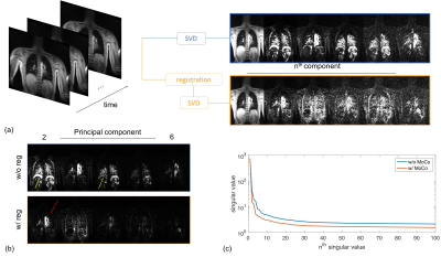 |
Motion Compensated Low-Rank(MoCoLoR) constrained reconstruction with application to motion resolved lung MRI
Xucheng Zhu1,2, Frank Ong3, Michael Lustig4, and Peder Larson1,2
1Radiology and Biomedical Imaging, University of California, San Francisco, San Francisco, CA, United States, 2UC Berkeley-UCSF Graduate Program in Bioengineering, University of California, San Francisco and University of California, Berkeley, Berkeley, CA, United States, 3Electrical Engineering, Stanford University, Stanford, CA, United States, 4Electrical Engineering and Computer Sciences, University of California, Berkeley, Berkeley, CA, United States
Respiratory motion is one of the most challenging problems in thoracic and abdominal MRI. Motion resolved reconstruction is introduced to reduce the respiratory motion effects by grouping the data to different motion states, then using compressed sensing techniques to reconstruct different motion states images. Spatio-temporal low-rank constrained reconstruction is one of the widely used techniques. In this work, we proposed a new method incorporating motion compensation into the low-rank model, called MoCoLoR. The proposed method is applied to high resolution free breathing lung MRI, and the results show that MoCoLoR outperforms the standard low-rank constrained reconstruction.
|
|
0455.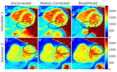 |
Pilot tone–based respiratory motion correction for 2D myocardial T1 mapping
Juliane Ludwig1, Kirsten Miriam Kerkering1, Peter Speier2, Frank Seifert1, Tobias Schaeffter1,3,4, and Christoph Kolbitsch1,3
Video Permission Withheld
1Physikalisch-Technische Bundesanstalt (PTB), Braunschweig and Berlin, Germany, 2Siemens Healthcare, Erlangen, Germany, 3Division of Imaging Sciences and Biomedical Engineering, King's College London, London, United Kingdom, 4Biomedical Engineering and Einstein Center Digital Future, Technische Universität Berlin, Berlin, Germany
Respiratory heart motion during T1 data acquisition can lead to strong motion artefacts, compromising the quality of reconstructed T1 maps. Commonly, breathhold techniques are used to minimize respiratory motion but they suffer from low scan efficiency and require patient cooperation. Here, we propose a Pilot tone-based respiratory motion correction approach for free-breathing myocardial T1 mapping. First, through-plane motion is corrected for by performing prospective slice tracking online during data acquisition. Second, in-plane motion is corrected for retrospectively by applying a phase shift to k-space data before image reconstruction. The feasibility of the proposed approach was demonstrated in four healthy volunteers.
|
0456.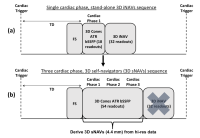 |
3D Self-Navigator Acquisition for Translational and Nonrigid Motion Correction in Multiphase Coronary MR Angiography
Kristin Quah1, Srivathsan P. Koundinyan1, Frank Ong1, Mario O. Malavé1, and Dwight Nishimura1
1Stanford University, Stanford, CA, United States
A whole-heart multiphase coronary angiography method has been developed that extracts a 3D navigator image every heartbeat from the 3D cones high-resolution imaging data. Such 3D self-navigators (sNAVs) enable direct beat-to-beat respiratory motion tracking of the heart for translational and nonrigid correction. 3D sNAVs are derived from the inner k-space region of phyllotaxis-ordered 3D cones interleaves collected over multiple cardiac phases. A multiscale low-rank method is used for reconstruction.
|
|
|
0457.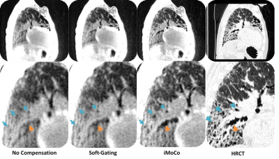 |
Motion Compensation in Pulmonary Ultra-short Echo Time MRI: Preliminary results in Idiopathic Pulmonary Fibrosis
Luis A Torres1, Xucheng Zhu2,3, Nathan Sandbo4, Mark L Shiebler4,5, Peder Larson2,3, and Sean B Fain1,5,6
1Dept. of Medical Physics, University of Wisconsin - Madison, Madison, WI, United States, 2Dept. of Radiology and Biomedical Imaging, University of California - San Francisco, San Francisco, CA, United States, 3UCSF/UC Berkeley Graduate Program in Bioengineering, University of California - San Francisco, San Francisco, CA, United States, 4Dept. of Medicine, University of Wisconsin - Madison, Madison, WI, United States, 5Dept. of Radiology, University of Wisconsin - Madison, Madison, WI, United States, 6Dept. of Biomedical Engineering, University of Wisconsin - Madison, Madison, WI, United States
Acquiring pulmonary MRI images without motion corruption is a challenging task. In this work, we evaluate several conventional and advanced retrospective motion compensation techniques in subjects with idiopathic pulmonary fibrosis (IPF). We evaluate the effectiveness of each technique using concomitantly acquired CT scans, contrast to noise, and sharpness measures. We find that registration-based techniques show a significant improvement in CNR and sharpness. We also observe significantly improved image quality when referenced side-by-side with CT. We conclude that registration-based techniques could be used to better resolve subtle fibrotic textures in IPF.
|
|
0458.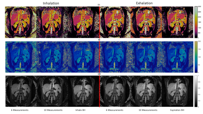 |
Free-Breathing Abdominal Magnetic Resonance Fingerprinting Using a Pilot Tone Navigator
Sherry Huang1, Rasim Boyacioglu2, Reid Bolding3, Yong Chen2, and Mark A. Griswold2
1Biomedical Engineering, Case Western Reserve University, Cleveland, OH, United States, 2Radiology, Case Western Reserve University, Cleveland, OH, United States, 3Physics, Case Western Reserve University, Cleveland, OH, United States
This study presents a novel free-breathing technique which addresses some of the difficulties in quantitative T1 and T2 mapping of the abdomen. The technique integrates Magnetic Resonance Fingerprinting (MRF) and pilot tone (PT) navigator to retrospectively provide simultaneous quantification of multiple tissue properties in the abdomen in inhalation and expiration states of the respiratory motion. The proposed method can be implemented with both 2D and 3D MRF acquisitions.
|
0459.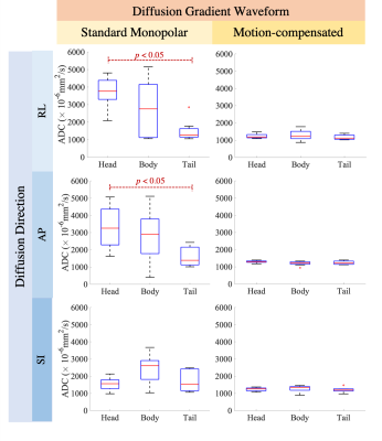 |
Characterization and Correction of Cardiovascular Pulsation Artifacts in Diffusion-Weighted Imaging of the Pancreas
Ruiqi Geng1,2, Yuxin Zhang1,2, Jitka Starekova1, Lloyd Estkowski3, and Diego Hernando1,2
1Department of Radiology, University of Wisconsin-Madison, Madison, WI, United States, 2Medical Physics, University of Wisconsin-Madison, Madison, WI, United States, 3MR, GE Healthcare, Waukesha, WI, United States
Diffusion-weighted imaging (DWI) of the abdomen faces multiple challenges, particularly artifacts induced by respiratory, peristaltic, and cardiovascular-related motions. Effects of cardiovascular pulsation on pancreas DWI have not been previously characterized. This motion introduces artifactual signal voids and unreliable apparent diffusion coefficient (ADC) values across the pancreas in DWI, regardless of cardiac phases and diffusion directions. Importantly, these artifacts can be addressed by motion-compensated diffusion gradient waveforms. These findings may facilitate the development of reliable and reproducible DWI of the pancreas.
|
|
0460.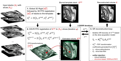 |
Non-rigid Motion Correction for Fetal Body MRI
Alena Uus1, Jacqueline Matthew1, Milou P. M. van Poppel1, Johannes Steinweg1, Laurence Jackson1, Mary Rutherford1, Joseph V. Hajnal1, and Maria Deprez1
1Biomedical Engineering Department, School of Biomedical Engineering and Imaging Sciences, King's College London, London, United Kingdom
Motion correction for fetal body MRI is particularly challenging due to non-rigid deformations of organs caused by bending and stretching. Rigid slice-to-volume registration (SVR) methods are efficient for 3D fetal brain reconstruction. However, for full body reconstruction, misregistration errors caused by deformable motion lead to degradation of features. We propose a novel deformable SVR (DSVR) method based on hierarchical deformable registration for reconstruction of 3D fetal trunk from multiple motion corrupted stacks. The method is quantitatively evaluated by comparison to the state-of-the-art methods on 20 iFIND fetal MRI datasets. Furthermore, DSVR reconstruction quality is assessed on 100 fetal MRI cases.
|
|
0461.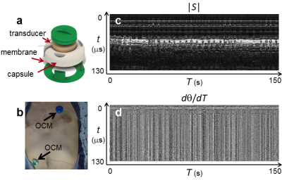 |
Motion monitoring using MR-compatible ultrasound-based sensors
Bruno Madore1, Frank Preiswerk1, Jeremy Bredfeldt1, Shenyan Zong1, and Cheng-Chieh Cheng1
1Brigham and Women's Hospital, Harvard Medical School, Boston, MA, United States
The purpose of this work is to develop MR-compatible sensors that can attach to the patient’s skin, to monitor breathing in a comprehensive manner. In contrast, alternatives such as MRI navigator echoes and optical tracking are typically rigidly fixed to walls or floors and as such cannot accompany a given patient through serial diagnostic and/or therapeutic procedures. We show here that these sensors capture breathing motion in much of its complexity, as validated against MRI and optical tracking data. These sensors could be used to help combine information from different modalities in a manner that takes internal motion into account.
|
 Watch the Video