
Scientific Session
Novel Pulse Sequences and Reconstruction Techiques
Session Topic: Novel Pulse Sequences and Reconstruction Techiques
Session Sub-Topic: Data Sampling & Spatial Encoding Techniques
Oral
Acquisition, Reconstruction & Analysis
| Tuesday Parallel 4 Live Q&A | Tuesday, 11 August 2020, 14:30 - 15:15 UTC | Moderators: Gigi Galiana & Jason Stockmann |
Session Number: O-63
 |
0614.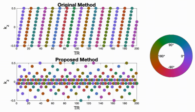 |
Improved 3D real-time MRI with Stack-of-Spiral (SOSP) trajectory and variable density randomized encoding of speech production
Ziwei Zhao1, Yongwan Lim1, Dani Byrd 2, Shrikanth Narayanan1, and Krishna Nayak1
1Ming Hsieh Department of Electrical and Computer Engineering, Viterbi School of Engineering, University of Southern California, Los Angeles, CA, United States, 2Department of Linguistics, Dornsife College of Letters, Arts and Sciences, University of Southern California, Los Angeles, CA, United States
3D real-time (RT) MRI is a useful tool in speech production research, as it enables full visualization of the dynamics of vocal tract shaping during natural speech. Limited spatial and temporal resolution, and a tradeoff between them, is however common in highly accelerated MRI. In this work, we demonstrate improved spatio-temporal resolution by using variable density randomized stack-of-spiral sampling and a constrained reconstruction. We can capture rapid movement of articulators, specifically lips and tongue body movements at both normal and rapid speech rates, yielding a substantial improvement over prior approaches in measuring fine details of human speech production.
|
0615.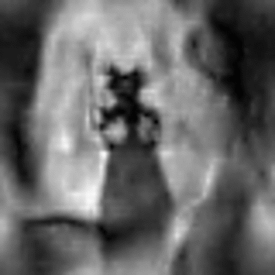 |
Sub-millisecond 2D MRI of the Vocal Fold Oscillation using Single Point Imaging with Rapid Encoding (SPIRE)
Johannes Fischer1, Ali Caglar Özen1,2, Matthias Echternach3, Bernhard Richter4, and Michael Bock1
1Dept. of Radiology, Medical Physics, Medical Center University of Freiburg, Faculty of Medicine, University of Freiburg, Freiburg, Germany, 2German Consortium for Translational Cancer Research Freiburg Site, German Cancer Research Center (DKFZ), Heidelberg, Germany, 3Division of Phoniatrics and Pediatric Audiology, Department of Otorhinolaryngology, Head and Neck Surgery, Ludwig-Maximilians-University, Munich, Germany, 4Institute of Musicians' Medicine, Medical Center University of Freiburg, Faculty of Medicine, University of Freiburg, Freiburg, Germany
We use single point imaging with rapid encoding (SPIRE) to image the vocal fold oscillations in the coronal plane. SPIRE is able to image fast, repetitive and two-dimensional motion, because the temporal resolution does not depend on TR but on the duration of the fast-switching phase encoding gradients, which is below one millisecond in this work. Data are gated using electroglottography and projection navigators are acquired during the sequence to detect shifts in larynx position which is corrected during reconstruction.
|
|
0616.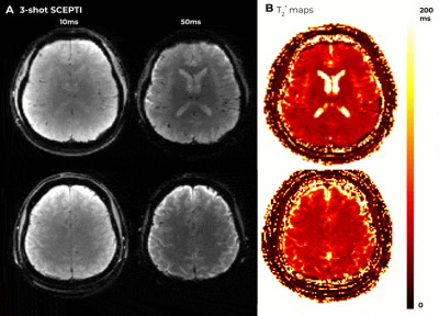 |
Spiral Crisscrossing Echo Planar Time-resolved Imaging (SCEPTI)
Gilad Liberman1, Fuyixue Wang1, Zijing Dong1, and Kawin Setsompop1
1A. A. Martinos Center for Biomedical Imaging, Department of Radiology, Massachusetts General Hospital, Charlestown, MA, United States
A new technique, termed Spiral Echo Planar Time-resolved Imaging (SKEPTIC), was developed to address both EPI’s geometric distortion and blurring and augment the recently introduced Echo Planar Time-resolved Imaging (EPTI). In SKEPTIC, the (2+1)-D k-t space is traversed using several matching out-in spirals within a single shot, and can benefit from additional rotated and time-jittered shots. The out-in multi-spiral trajectory is incoherent with field inhomogeneity phase evolution in both axes. This results in the ability to deliver single-shot 1.9mm2 in-plane resolution distortion-less, sharp multi-echo images with B0 and T2* mapping, and inherent motion, phase and B0-variation estimates for multi-shot imaging.
|
|
|
0617.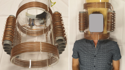 |
Dual axis gradient insert for supersonic MRI
Edwin Versteeg1, Tijl Van der Velden1, Jeroen Hendrikse1, Dennis Klomp1, and Jeroen Siero1,2
1Radiology, University Medical Center Utrecht, Utrecht, Netherlands, 2Spinoza Centre for Neuroimaging Amsterdam, Amsterdam, Netherlands
A silent gradient axis can be achieved by driving a gradient insert above 20 kHz. In this work, we investigate a prototype silent gradient insert that features two axes. Such a setup would enable both silent and fast imaging. The two axes were driven with an audio amplifier at 20 kHz and 22 kHz, and produced gradient amplitudes of 20.8 and 22 mT/m. We simulated the acceleration potential to be a factor of 9 and showed the feasibility of imaging with this setup on a phantom.
|
0618.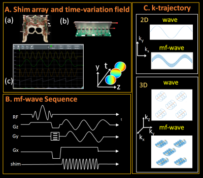 |
Multi-frequency wave-encoding (mf-wave) on gradients and multi-coil shim-array hardware for highly accelerated acquisition
Jinmin Xu1,2, Jason Stockmann2, Berkin Bilgic2, Thomas Witzel2,3, Jaejin Cho2, Congyu Liao2, Zijng Zhang1,2, Huafeng Liu1, and Kawin Setsompop2,3,4
1State Key Laboratory of Modern Optical Instrumentation, College of Optical Science and Engineering, Zhejiang University, Hangzhou, China, 2Department of Radiology, A.A. Martinos Center for Biomedical Imaging, Massachusetts General Hospital, Charlestown, MA, United States, 3Harvard Medical School, Boston, MA, United States, 4Massachusetts Institute of Technology, Harvard-MIT Health Sciences and Technology, Cambridge, MA, United States
Wave-CAIPI is a parallel imaging technique that can provide high accelerations with negligible g-factor and artifact penalties. However, gradient amplitude & slew rate limits impose a limitation on the amount of wave-encoding and hence the acceleration capability of this technique. In this study, we propose a multi-frequency wave-encoding method (mf-wave) that uses both the gradients and a combined RF and B0 shim array (32-channel) to perform wave-encoding simultaneously and synergistically at different wave frequencies during the acquisition. We demonstrate that mf-wave can enable ~20-fold acceleration with an acceptable g-factor noise penalty at 3T in vivo.
|
|
0619.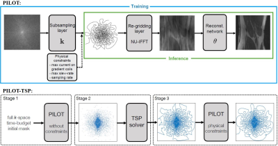 |
PILOT: Physics-Informed Learned Optimal Trajectories for Accelerated MRI
Tomer Weiss1, Ortal Senouf2, Sanketh Vedula2, Oleg Michailovich3, Michael Zibulevsky2, and Alex Bronstein2
1CS, Technion, Haifa, Israel, 2Technion, Haifa, Israel, 3University of Waterloo, Waterloo, ON, Canada
We propose a novel approach to the learning of conjoint acquisition and reconstruction of MRI scans. The acquisition is encoded in the form of general k-space trajectories, which constrained to obey the hardware requirements (peak currents and maximum slew rates of magnetic gradients). We demonstrate the effectiveness of the proposed solution in both image reconstruction and image segmentation, reporting substantial improvements in terms of acceleration factors and the quality of these end tasks. To the best of our knowledge, our proposed algorithm is the first to do data- and task-driven learning over the space of all physically feasible k-space trajectories.
|
|
0620.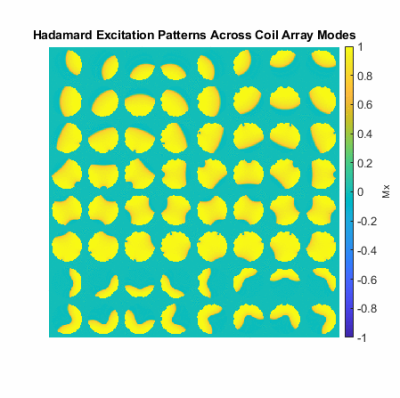 |
MR Barcoding: Gradient-Free MRI Using B1-Selective Parallel Transmission
Christopher E. Vaughn1,2, Mark A. Griswold3, and William A. Grissom1,2
1Vanderbilt University Institute of Imaging Science, Nashville, TN, United States, 2Department of Biomedical Engineering, Vanderbilt University, Nashville, TN, United States, 3Radiology, Case Western Reserve University, Cleveland, OH, United States
Conventional MR imaging uses linear B0 gradients for spatial encoding, which have high cost and bulk, and lead to patient discomfort via noise and PNS. We introduce MR Barcoding as a silent, low-profile, and low-cost replacement for B0 gradients. The technique is based on non-linear RF magnitude gradients synthesized by an array of conventional transmit coils, combined with B1+-selective Hadamard encoding pulses. A proof-of-principle simulation shows that a 64x64 image could be reconstructed using an MRF model and a sub-30s scan duration. Experimental results at 47.5 mT validate the encoding capabilities of the B1+-selective Hadamard encoding pulses.
|
|
 |
0621.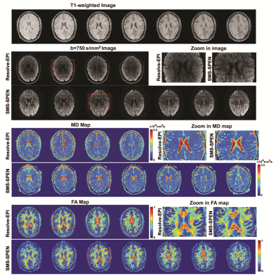 |
Rapid volumetric 3D MRI via simultaneous-multi-slab, multi-echo spatiotemporal encoding (SMS-ME SPEN)
Lingceng Ma1,2, Martins Otikovs1, Samuel F Cousin3, Gilad Liberman4, Qingjia Bao1, and Lucio Frydman1
1Department of Chemical and Biological Physics, Weizmann Institute of Science, Rehovot, Israel, 2College of Electronic science and technology, Xiamen University, Xiamen, China, 3Centre de RMN à Très Haut Champs, Lyon, France, 4Massachusetts General Hospital, Boston, MA, United States
SPatiotemporal ENcoding (SPEN) is a 2D single-shot MRI method with higher immunity to artifacts than EPI-based counterparts. The present study extends SPEN scans to 3D volumetric measurements, to achieve imaging over a 3rd dimension at higher resolution in minimal acquisition times. simultaneous multi-slab (SMS) and multi-echo (ME) kz-encoding procedures are here combined to cope with the SAR complications that would ensue from simply repeating 2D acquisitions over multiple slices. A framework to appropriately reconstruct and process 3D SMS-ME SPEN data to ensure the image quality by taking motion artifacts derived from different dimensions into account is also proposed, and demonstrated.
|
0622.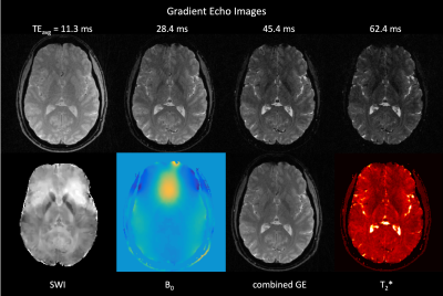 |
Simultaneous Multi-Slice Radial Echo Volumar Imaging for Fast Simultaneous Multi-Parametric Imaging
Christoph Alexander Rettenmeier1, Danilo Maziero2, Kai Tobias Block3, and V. Andrew Stenger1
1Medicine, University of Hawaii, Honolulu, HI, United States, 2University of Hawaii, Honolulu, HI, United States, 3New York University, New York, NY, United States
Echo Planar Imaging (EPI) is one of the most widely used fast acquisitions and has been shown to be useful for high-resolution Simultaneous Multi-Parametric (SMP) imaging. However, obtaining high spatial resolutions requires k-space segmentation which has a high sensitivity to motion. Radial sampling is promising because of its continuous k-space center update which can be used for self-navigation and its suitability for undersampling because of benign artifacts. We describe a new k-t sampling strategy based on a Radial Echo Volumar Imaging method for fast SMP imaging. Whole brain images including susceptibility, B0 and T2* maps acquired at 3T are presented.
|
 Watch the Video